Introduction
Hypoparathyroidism leading to hypocalcemia is an important treatable cause of recurrent seizures. Even though it is not an uncommon condition, primary hypoparathyroidism presenting for the first time as seizures in adulthood is quite infrequent. Patients may present with hypocalcemic seizures even in the absence of subtle hypocalcemic signs inclusive of tetany, Chvostek’s sign or carpopedal spasms. As this is an entirely treatable condition, a high index of suspicion for primary hypoparathyroism with hypocalcemic seizures should be maintained even in otherwise asymptomatic adults.
Case report
A 30 year old male, was presented to the emergency facility in an unconscious condition. He had been intubated on the way to the hospital as he had suffered from two episodes of ventricular tachycardia in the cardiac ambulance. He was being transported from a local hospital where he had been admitted for profuse diarrhea with dehydration.
He recovered during the hospital stay and on further inquiry it was discovered that he had a past history of recurrent seizures for the last 6 years inspite of being on multiple antiepileptic medications including phenytoin sodium, sodium valproate and leviteracitam. The seizure frequency had increased considerably in the last year, and he would have at least 5–6 episodes in a month, thereby creating a considerable toll on his personal and professional life. He had been evaluated with an MRI brain scan and an EEG at the onset of symptoms 6 years earlier and both were reported to be normal.
General physical examination was relatively normal, though he had a past history of being operated for bilateral cataracts six months ago. Also fundoscopic examination showed bilateral acute papillodema. There was no carpopedal spasm or any other signs of tetany like Chvostek’s or Trousseau’s sign.
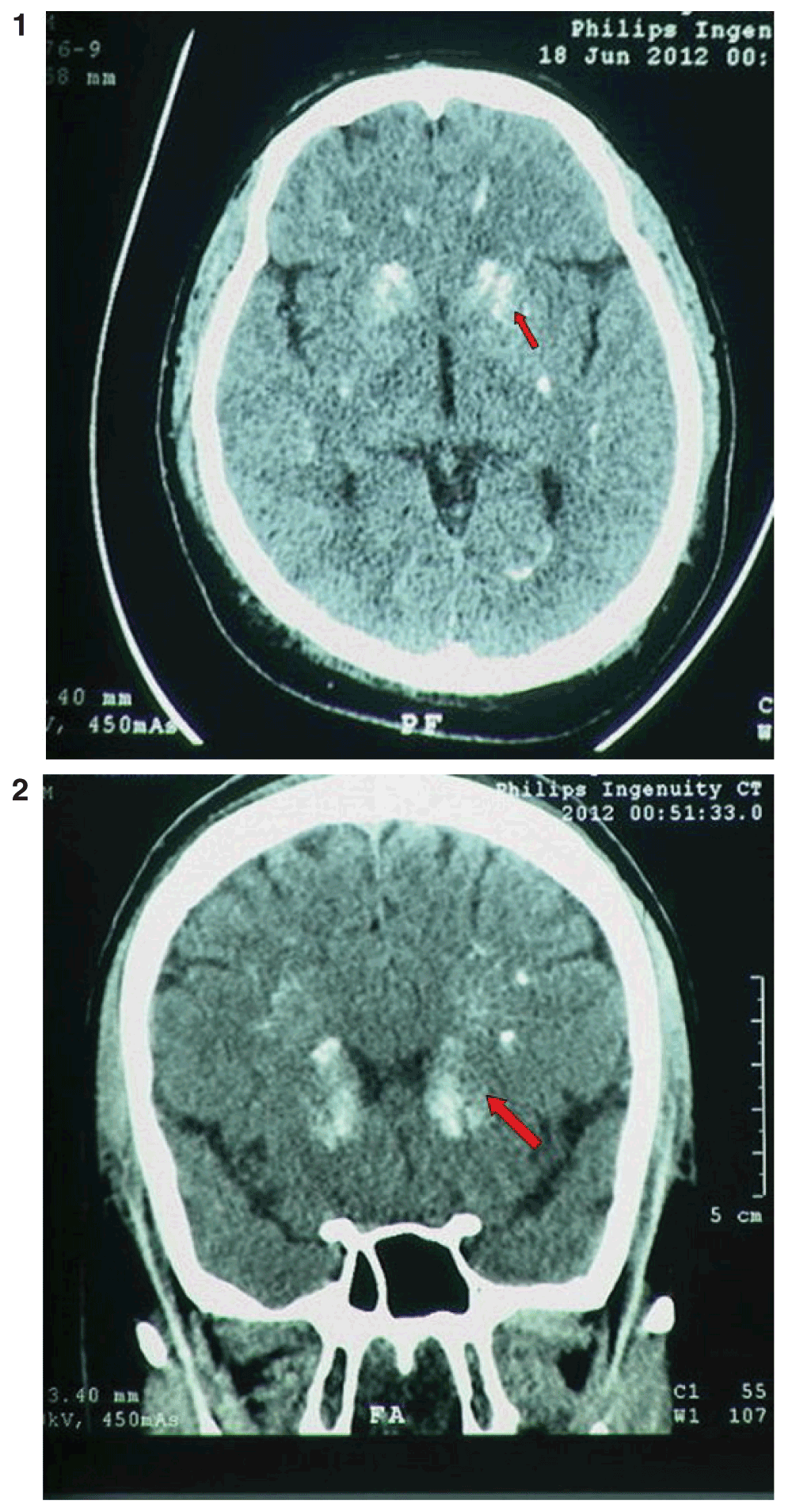
Investigations revealed normal hemoglobin and glucose level with normal sodium and potassium levels. TLC and DLC levels were also normal. He was found to have a serum calcium level of 3.3 mg% with a serum parathyroid hormone level of 1pg/ml, serum 25(OH) vitamin D levels of 6.6ng/ml and hypomagnesemia. NCCT head scan was done which showed bilateral basal ganglia calcification and deep white matter calcification. A 2D ECHO study was performed, and showed normal results (Figures 1 and 2).

Figures 1 and 2. NCCT head scan showing bilateral basal ganglia calcification and deep white matter calcification.
A diagnosis of primary hypoparathyroidism was made. He was treated with anticonvulsants, oral calcium, magnesium and vitamin D supplementation. During his hospital stay he did not have any other seizure events.
Discussion
Intracranial calcifications can be classified mainly into 6 groups based on their etiopathogenesis: age-related and physiologic, congenital, infectious, endocrine and metabolic, vascular, and neoplastic1
(Table 1). The function of the parathyroid hormone is primarily maintaining the plasma calcium levels. Hormonal disturbance of the parathyroid glands including hypoparathyroidism, hyperparathyroidism and pseudohypoparathyroidism may lead to intracranial calcifications. Calcium accumulation is demonstrated primarily in the bilateral basal ganglia, dentate nuclei, and peripheral subcortical white matter sites2.
Table 1. Causes of intracranial calcifications1.
| Classification | Cause |
|---|
Age-related
and physiologic | Pineal gland, habenula, choroid plexus,
falx cerebri, tentorium cerebelli, dura mater,
petroclinoid ligament, sagittal sinus |
| Congenital | Sturge-Weber syndrome, tuberous sclerosis,
neurofibromatosis, lipoma, Cockayne
syndrome, Gorlin syndrome |
| Infectious | TORCH diseases, granulomatous infections,
chronic viral encephalitis |
Endocrine &
Metabolic | Fahr disease, hypothyroidism,
hypoparathyroidism, hyperparathyroidism,
pseudohypoparathyroidism, postthyroidectomy |
| Vascular | Primary atherosclerosis, cavernous
malformation, arteriovenous malformation,
aneurysms, dystrophic in chronic infarction
and chronic vasculitis |
| Neoplastic | Oligodendroglioma, craniopharyngioma,
germ cell neoplasms, neurocytoma,
primitive neuroectodermal tumor (PNET),
ependymoma, ganglioglioma, dysembriyonic
neuroectodermal tumor (DNET), meningioma,
choroid plexus papilloma, medulloblastoma,
low grade astrocytoma, pilocytic
astrocytoma, pinealoma, pinealoblastoma,
schwannoma, dermoid, epidermoid, calcified
metastases (osteogenic sarcoma, mucinous
adenocarcinoma) |
The principal function of the parathyroid hormone (PTH) is the maintenance of calcium plasmatic levels, withdrawing the calcium from bone tissue, reabsorbing it from the glomerular filtrate, and indirectly increasing its intestinal absorption by stimulating active vitamin D (calcitriol) production. There are two mechanisms that may alter its function, limiting its control on calcium: 1) insufficient PTH production by the parathyroids (hypoparathyroidism), or 2) a resistance against its action in target tissues (pseudohypoparathyroidism). In both cases, there are significantly reduced levels of plasmatic calcium associated with hyperphosphatemia3.
In acute and/or severe symptomatic hypocalcemia there is a predominance of neuromuscular, neuropsychiatric, and cardiovascular abnormalities. There is an increase in neuromuscular excitability, latent or evident, with sensory and motor disruption. Perioral or extremity paresthesia, cramps, myalgia, and muscular weakness are mild to moderate symptoms. Neuropsychiatric manifestations include irritability, anxiety, psychosis, hallucinations, dementia, depression, mental confusion, and extrapyramidal abnormalities. Increased intracranial pressure, papilledema, and convulsions can also be present, and must be differentiated from severe tetany muscular spasms4,5. Typical clinical signs of neuromuscular irritability associated with latent tetany include hyperreflexia and Chvostek’s and Trousseau’s signs, respectively. Severe hypocalcemia may result in bradycardia or ventricular arrhythmias, cardiovascular collapse, and hypotension that is non-responsive to fluids and vasopressors3.
A decrease in myocardial contractility occurs, as well as a typical electrocardiographic abnormality, which is the rate-corrected QT interval (QTc) prolongation. Patients with chronic hypocalcemia may or may not have symptoms of discreet neuromuscular irritation, even with markedly low calcium levels. Asymptomatic cases may be detected by chance, by the dosage of calcium in routine exams, during periods of greater calcium demand (i.e. gestation, lactation, menstrual cycle and states of alkalosis), or during the use of hypocalcemic drugs (i.e. bisphosphonates)6.
Significant cognitive deficits, neuropsychiatric abnormalities, and extrapyramidal symptoms that resemble Parkinson’s disease or chorea are associated with the calcification of basal ganglia, which occurs in all forms of chronic hypocalcemia and may be detected with greater sensibility using computerized tomography7. Other findings of chronic hypocalcemia include sub-capsular cataracts, an increase in bone mineral density (BMD), and greater susceptibility to dystonic reactions induced by phenothiazines4.
Differential diagnosis of hypocalcemia will depend largely upon PTH and phosphorus levels, evaluated along with other clinical and laboratory data (Table 1). Cases presenting hypophosphatemia should include differential diagnosis of vitamin D, while cases associated with hyperphosphatemia are determined according to PTH levels. Hypoparathyroidism is an abnormality caused by a parathyroid hormone (PTH) secretion deficiency, and encompasses heterogeneous conditions (Table 2), which makes etiological differentiation crucial to the detection of abnormalities associated with some of these diseases beforehand, thereby preventing complications4. Signs and symptoms are caused by hypocalcemia.
Table 2. Causes of hypoparathyroidism3.
| Classification | Cause |
|---|
Parathyroid
Destruction | Surgery
Auto-immune (isolated or polyglandular)
Cervical irradiation
Infiltration by metastasis or systemic diseases
(Sarcoidosis, amyloidosis, hemochromatosis,
Wilson’s disease, thalassemia |
Reduced
parathyroid
function | Hypomagnesemia
PTH gene defects
Calcium sensing receptor mutations |
Parathyroid
agenesis | DiGeorge Syndrome
Isolated x-linked hypoparathyroidism
Kenny-Caffey syndrome
Mitochondrial neuropathies |
Laboratory measurements present hypocalcemia, hyperphosphatemia, and inappropriately low or undetectable PTH. Generally, levels of 1.25(OH) 2D are low and the alkaline phosphatase level is normal. In the majority of cases, hypoparathyroidism is sporadic, but there are familial cases in which transmission may be autosomic recessive, dominant, or X-linked.
Management of acute or severe symptomatic hypocalcemia must be made with intravenous calcium, with the goal of interrupting symptoms, preventing laryngeal spasm, and maintain total calcium levels above 7.0–7.5 mg/dL (ionized calcium greater than 0.7 mmol/L). Long-term treatment of patients with chronic hypocalcemia is done with 1 to 3 grams of elementary calcium per day in the various forms of salts available3.
All patients with hypoparathyroidism or pseudohypoparathyroidism who become hypocalcemic must use vitamin D or analogues in addition to calcium. The vast majority of patients obtain control with calcitriol in dosages of 0.25 μg, taken twice daily, up to 0.5 μg four times daily. Hypoparathyroidism causes increased excretion of urinary calcium in relation to serum calcium and predisposes hypercalciuria, nephrolithiasis, and nephrocalcinosis. The product of calcium × phosphate must be kept below 55. Patients must have their kidneys radiologically evaluated regularly in order to rule out nephrocalcinosis4.
Conclusion
1. Ironically, increasing reliance on high end investigations such as a MRI brain scan could lead to certain conditions being missed; conditions that could be easily identifiable by the humble CT scan.
2. All treatable metabolic conditions should be excluded at first before commencing with anticonvulsants; this will restrict patients from burdensome polytherapy and related side effects.
Consent
Written consent was obtained for publication of the patient’s clinical details and images obtained from the patient.

Comments on this article Comments (0)