ALL Metrics
-
Views
-
Downloads
Get PDF
Get XML
Cite
Export
Track
Data Note

The relationship between umbilical–portal–systemic venous shunt and outcome in 11 fetuses

[version 1; peer review: 2 approved with reservations]
PUBLISHED 18 Oct 2021
Author details Author details
OPEN PEER REVIEW
REVIEWER STATUS

Abstract

Objective: To investigate the relationship between congenital umbilical–portal–systemic venous shunt (UPSVs) and fetal outcome.
Methods: The ultrasonographic and genetic characteristics of 11 cases of UPSVS were retrospectively analyzed and followed up to postnatal.
Results: Four cases of ductus venosus -- systemic shunt (DVSS), one case of extrahepatic portal -- systemic shunt (EHPSS), and one case of umbilical systemic shunt (USS) combined with intrahepatic portal-systemic shunt (IHPSS), six cases of intrahepatic portal-systemic shunt (IHPSS) were observed. chromosomal abnormalities were observed in 9.1% (1/11), other ultrasonic abnormalities in 54.5% (6/11), cardiothoracic ratio increase in 45.5% (5/11), fetal growth restriction in 36.4% (4/11), edema was in 9.1% (1/11) and live birth was in 72.7% (8/11).
Conclusion: The incidence of IHPSS is the highest and the outcome is the best. Shunt of DVSS and IHPSS can be closed spontaneously after birth. When the prenatal diagnosis is congenital UPSVs, chromosomal abnormalities and other ultrasonic abnormalities should be required further examination, and the growth and development of the fetus should be closely monitored.

Keywords

umbilical-portal-systemic venous shunt; fetus; the outcome

Introduction

Congenital portosystemic shunt (CPSS) is associated with some complications, such as cholestasis, hyperammonemia, pulmonary arterial hypertension, hepatopulmonary syndrome, and benign and malignant liver tumors, in childhood. Some types of CPSS including extrahepatic, persistent intrahepatic shunt and ductus venosus must be inhibited by interventional radiology or surgery.1

Due to the low incidence, there is limited prenatal information on the relationship between umbilical-porta-systemic shunt and its prognosis. In the past, portal systemic shunts were divided into extrahepatic and intrahepatic shunts. They have been used to study fetal umbilical-portal system shunt and pediatric congenital portal systemic shunt.26 The recent systematic reclassification of the term “umbilical-porta-systemic venous shunt (UPSVS)” has been considered as the best choice for fetal prognosis analysis. This classification is based on the theory of UV-PV-DV as an intact structure.

According to Achiron and Kivilevitch,7 the UPSVs are divided into three types: Type I, umbilical–systemic shunt (USS), with the blood flow from the umbilical vein entered directly into the systemic veins; Type II, ductus venosus–systemic shunt (DVSS), with the DV blood flow shunted from its normal path into the systemic veins; Type III, portal–systemic shunt, which divided into two subgroups: Type IIIa, intrahepatic portal–systemic shunt (IHPSS), with an intrahepatic shunt between the IHPVS and the hepatic vein; and Type IIIb, extrahepatic portal–systemic shunt (EHPSS), with an extrahepatic shunt between the portal system and systemic veins (IVC, iliac vein, renal vein). According to previous studies, the incidence of trisomy 21 in UPSVS was 10.4%.8 Achiron R et al. observed that DVSS and IHPSS had the best prognosis, with spontaneous shunt closure after birth.7 Berg et al.9 observed that cases with extrahepatic shunt were more likely to develop into cardiac decompensation. Delle et al.10 observed a causal relationship between IHPSS and FGR.

The purpose of this study was to review our experience with fetal UPSVS and analyze its clinical and prognostic characteristics for better prenatal counseling with UPSVS.

Methods

Retrospective analysis was performed on 11 cases of UPSVSs admitted to the prenatal diagnosis center of our hospital from December 1, 2019, to December 1, 2020. According to the UPSVS classification criteria, four cases were DVSS, and six cases were IHPSS, including one case of USS combined with IHPSS and one case of EHPSS. All patients signed an informed consent form, performed routine ultrasound examinations of the fetus, placenta and amniotic fluid, and then performed detailed examinations and records of each fetus’s heart, celiac vessel and middle cerebral artery, focusing on observation of the umbilical vein and ductus venosus, main portal vein, left and right branch morphology, internal echo and surrounding structures, observed whether there are abnormal ducts between the umbilical vein, ductus venosus, portal vein, hepatic vein and other systemic veins, and follow up until the shunt is closed after birth. The karyotype and low-coverage massively parallel copy number variation sequencing (CNV-seq) of the fetus were further examined by amniocentesis. Statistical methods: Descriptive statistics were used to retrospectively analyze the clinical features and pregnancy outcomes of 11 cases. SPSS20.0 software was used for statistical analysis of data. Measurement data was expressed as mean (±SD), and the Bonferroni adjustment method in ANOVA was used for pairwise comparison. p < 0.05 indicates that the difference is statistically significant. Counting data are expressed as percentages.

Results

According to the UPSVS classification criteria, four cases of DVSS, six cases of IHPSS, one case of USS combined with IHPSS and one case of EHPSS were recorded.

The four cases of DVSS

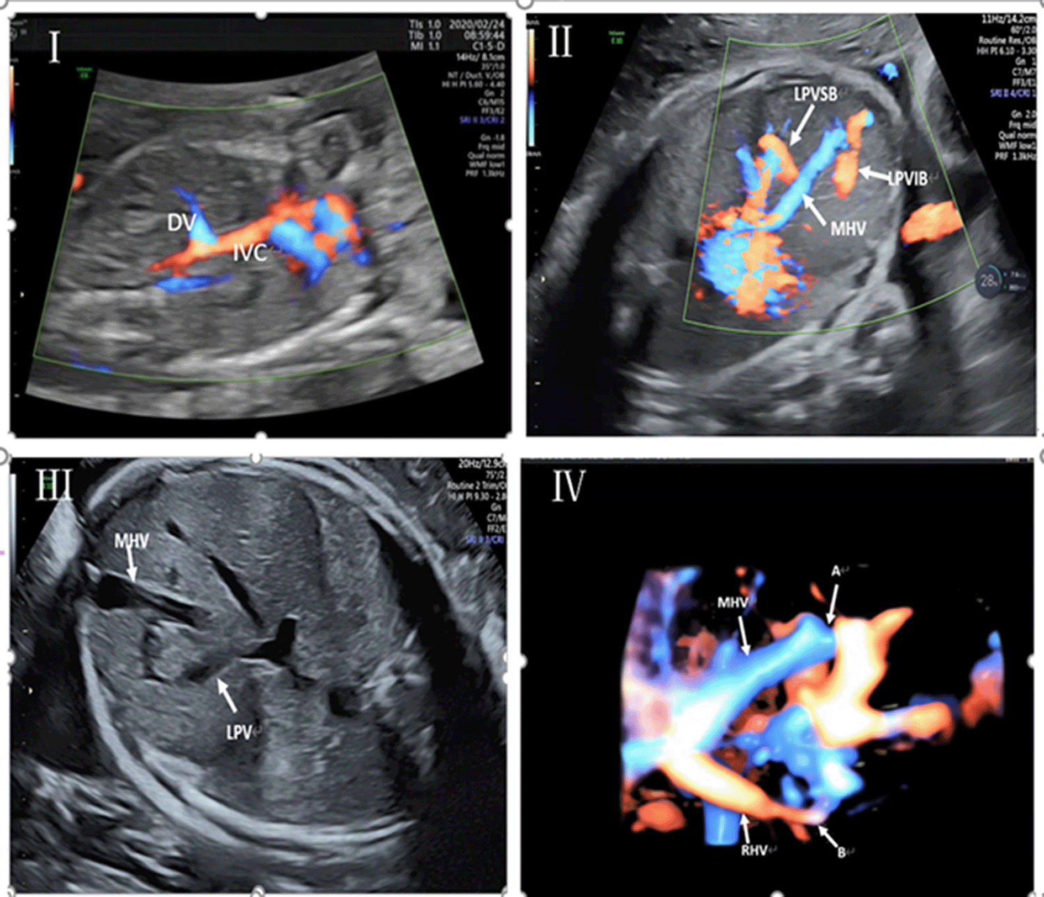
In two cases, the ductus venosus entered the middle of the inferior vena cava (Cases 2 and 4) and was observed at the gestational age of 23 weeks and 24+4 weeks, respectively. In one case, observed at 18 weeks of gestation, the ductus venosus was inserted into the hepatic segment of the inferior vena cava (Case 1, Figure 1). In one case, observed at 23+4 weeks, the ductus venosus was inserted into the middle hepatic vein (Case 3). Three cases received amniocentesis to do karyotype and CNV-seq test, and trisomy 21 was observed in Case 1; the other two cases had normal results. Two cases terminated their pregnancies: Case 1 terminated pregnancy because of trisomy 21, Case 3 terminated because of other associated structural abnormalities, including multiple hemivertebrae with scoliosis, and the left and right branches of the portal vein are not detected on ultrasound. Two cases were delivered prematurely (Cases 2 and 4), of which Case 4 had fetal growth restriction. The ductus venosus of the two cases had closed one month after birth, and the growth and development were normal.

  • One case of EHPSS

  • In Case 5, at 24+4 weeks of gestation, ultrasound showed fetal edema including abdominal effusion and skin thickening, widened hepatic veins, and increased cardiothoracic ratio, suggesting congestive heart failure. The umbilical vein was directly connected to the ductus venosus, the left and right branches of the portal vein and the main portal vein are not shown. The splenic vein was thin, and did not appear to enter the portal vein, the superior mesenteric vein was unclear. The distal end of the hepatic vein was widened and tortuous, seeming to be connected to the distal end of the hepatic artery, suggesting hepatic arteriovenous fistula. The pregnant woman finally chose to terminate the pregnancy without a chromosome examination.

f1bb4b0d-4d6c-455c-bba2-61c6d49f4a68_figure1.gif

Figure 1. I represents DV: ductus venosus, IVC: inferior vena cava, ductus venosus enters the hepatic segment of inferior vena cava. II represents: MHV: middle hepatic vein, LPVSb: left upper branch of portal vein; LPVLb: left lower branch of portal vein; the left upper branch and left lower branch of the portal vein enter into the middle hepatic vein. III indicates: MHV: middle hepatic vein, LPV: left portal vein, left portal vein enters into middle hepatic vein. IV (A) indicates: the confluence point of left outer lower branch of portal vein and middle hepatic vein, (B): the confluence point of left inner portal vein and the right hepatic vein.

One case of USS

Case 8 displayed USS combined with IHPSS, see IHPSS for details.

Six cases of IHPSS

The location of shunt in six cases is shown in Table 1. Amniocentesis was performed in two cases. The results showed no abnormality in karyotype or gene copy number variation. Two cases had fetal growth restriction (Cases 7 and 10). Ultrasound revealed cardiothoracic ratio increased in three cases (Cases 7, 8 and 10), congestive heart failure with possible cerebral edema in one case (Case 10). In Case 10, MRI revealed: cerebral vein, superior sagittal sinus, right transverse sinus and enlarged sigmoid sinus. Finally, one case underwent full-term delivery, five cases underwent premature delivery, due to congestive heart failure with possible cerebral edema (Case 10), increased cardiothoracic ratio (Cases 7 and 8), breech presentation combined with premature rupture of membranes (Case 9), fetal growth restriction with oligohydramnios (Case 11). All six cases had live births. Follow-up of those six cases showed that the shunts were closed within half a year after birth, and blood ammonia, liver function, growth and development were normal.

Results are shown in Tables 1 and 2.

Table 1. Clinical features and outcomes of the cases (n = 11).

ClassificationGAShuntFGRCardiac anomaliesEdemaAnomalies associatedKaryotypeCNV-seqPregnancy outcomeShunt closureGrowth and development after birth
Case1DVSS18WDV-IVCNoNoNoAbsence of nasal bone47, XN+21NormalTOP//
Case2DVSS23WDV-IVCNoNoNoNoNormalNormalPremature delivery by cesarean section1 month after birthNormal
Case3DVSS23+4WDV-MHVNoNoNoMultiple hemivertebrae with scoliosis//TOP//
Case4DVSS24+4WDV-IVCYesNoNoPolydactylismNormalNormalPremature delivery by cesarean section1 month after birthNormal
Case5EHPSS24+4WUV-DVNoCongestive heart failureYesNo//TOP//
Case6IHPSS26+4WLPV-LHVNoNoNoNoNormalNormalTerm delivery by cesarean section (breech presentation)4 months after birthNormal
Case7IHPSS35+4WLPV-MHV, LPV-RHVYesCTR: 0.42NoNo//Premature delivery by cesarean section4 months after birthNormal
Case8IHPSS36+5WLPV-MHV(2 branches), combined with UV-MHV (multiple branches)NoCTR: 0.46NoNo//Premature delivery by cesarean section1 month after birthNormal
Case9IHPSS30+1WLPV-MHVNoNoNoNo//Premature delivery via vaginal delivery (PPROM)6 months after birthNormal
Case10IHPSS35+1WLPV-MHVYesCongestive heart failureNoPossible encephaledemaNormalNormalPremature delivery by cesarean section6 months after birthNormal
LPV-RHV
LPV-LHV
Case11IHPSS34+4WLPV-LHVYesCTR: 0.42NoNoNormalNormalPremature delivery by cesarean section (oligoamnios)6 months after birthNormal

Table 2. Clinical features and outcomes of 11 cases.

Total (n = 11)DVSS (n = 4)IHPSS (n = 6)EHPSS (n = 1)p < 0.05
Age29.360 ± 5.00527.250 ± 4.78731.500 ± 4.84825p = 0.305
GA27.655 ± 6.15822.300 ± 2.94231.733 ± 5.08824.6p* = 0.028
Chromosome abnormality9.1% (1/11)33.33% (1/3)0%/
Other Ultrasonic Abnormalities54.5% (6/11)75% (3/4)33.3% (2/6)100%
CTR enlargement45.5% (5/11)0%66.7% (4/6)100%
Edema9.1% (1/11)0%0%100%
FGR36.4% (4/11)25% (1/4)50% (3/6)0%
Live birth72.7% (8/11)50% (2/4)100%0%

* Represents p-value: * p < 0.05.

  • What’s known/what’s new statements

  • What’s already known about this topic?

UPSVSs are divided into three types in recent research. Limited prenatal information is available on the relationship between umbilical–portal–systemic venous shunt and outcome due to the low incidence.

What does this study add?

We collected 11 cases of fetus from different UPVSV types, and analyze the clinical and prognostic characteristics in details to enable better prenatal counseling with UPSVS.

Ethics statement

The report was ethically approved by the institutional review board (Chongqing Health Center of Women and Children) and written informed consent was obtained from the mothers to publish this paper.

Data availability

Underlying data

Dryad: Underlying data for ‘The relationship between umbilical–portal–systemic venous shunt and outcome in 11 fetuses’.

https://doi.org/10.5061/dryad.crjdfn34g

Data are available under the terms of the Creative Commons Zero “No rights reserved” data waiver (CC0 1.0 Public domain dedication).

Comments on this article Comments (0)

Version 1
VERSION 1 PUBLISHED 18 Oct 2021
Comment
Author details Author details
Competing interests
Grant information
Copyright
Download
 
Export To
metrics
Views Downloads
F1000Research - -
PubMed Central
Data from PMC are received and updated monthly.
- -
Citations
CITE
how to cite this article
Fang L, Xueyan W, Yangxue X et al. The relationship between umbilical–portal–systemic venous shunt and outcome in 11 fetuses [version 1; peer review: 2 approved with reservations]. F1000Research 2021, 10:1058 (https://doi.org/10.12688/f1000research.55600.1)
NOTE: If applicable, it is important to ensure the information in square brackets after the title is included in all citations of this article.
track
receive updates on this article
Track an article to receive email alerts on any updates to this article.

Open Peer Review

Current Reviewer Status: ?
Key to Reviewer Statuses VIEW
ApprovedThe paper is scientifically sound in its current form and only minor, if any, improvements are suggested
Approved with reservations A number of small changes, sometimes more significant revisions are required to address specific details and improve the papers academic merit.
Not approvedFundamental flaws in the paper seriously undermine the findings and conclusions
Version 1
VERSION 1
PUBLISHED 18 Oct 2021
Views
5
Cite
Reviewer Report 27 Jun 2022
Johannes van der Merwe, University Hospitals Leuven, Leuven, Belgium 
Approved with Reservations
VIEWS 5
The manuscript describes the clinical outcomes of a small series of prenatal diagnosed UPSVS cases. The manuscript is overall sound and the topic is relevant. The biggest criticism of this manuscript is that it seems to add no new insights ... Continue reading
CITE
CITE
HOW TO CITE THIS REPORT
van der Merwe J. Reviewer Report For: The relationship between umbilical–portal–systemic venous shunt and outcome in 11 fetuses [version 1; peer review: 2 approved with reservations]. F1000Research 2021, 10:1058 (https://doi.org/10.5256/f1000research.59190.r140424)
NOTE: it is important to ensure the information in square brackets after the title is included in all citations of this article.
  • Author Response 07 Oct 2022
    Liu Fang, The department of prenatal diagnosis, Chongqing Health Center for Women and Children, Chongqing, 401147, China
    07 Oct 2022
    Author Response
    It's my great honor to receive your review. Thank you for your detailed review and the added references. Because the manuscript was submitted as datasets, discussion on this topic was ... Continue reading
COMMENTS ON THIS REPORT
  • Author Response 07 Oct 2022
    Liu Fang, The department of prenatal diagnosis, Chongqing Health Center for Women and Children, Chongqing, 401147, China
    07 Oct 2022
    Author Response
    It's my great honor to receive your review. Thank you for your detailed review and the added references. Because the manuscript was submitted as datasets, discussion on this topic was ... Continue reading
Views
6
Cite
Reviewer Report 03 Feb 2022
Bijoy K. Balakrishnan, Department of Feto-Maternal Medicine, CIMAR Cochin and Edappal Hospitals Pvt Ltd, Cochin, Kerala, India 
Approved with Reservations
VIEWS 6
Thank you for the opportunity to review this article. The cases that have been discussed are quite interesting and will provide necessary insight for clinicians while counselling their patients with UPVSS. However, it would have been more interesting if more ... Continue reading
CITE
CITE
HOW TO CITE THIS REPORT
Balakrishnan BK. Reviewer Report For: The relationship between umbilical–portal–systemic venous shunt and outcome in 11 fetuses [version 1; peer review: 2 approved with reservations]. F1000Research 2021, 10:1058 (https://doi.org/10.5256/f1000research.59190.r119590)
NOTE: it is important to ensure the information in square brackets after the title is included in all citations of this article.
  • Author Response 07 Oct 2022
    Liu Fang, The department of prenatal diagnosis, Chongqing Health Center for Women and Children, Chongqing, 401147, China
    07 Oct 2022
    Author Response
    Thank you for your review, we are still collecting cases. EHPSS cases are relatively rare, so it takes some time to summarize the cases. At present, A case of USS  ... Continue reading
COMMENTS ON THIS REPORT
  • Author Response 07 Oct 2022
    Liu Fang, The department of prenatal diagnosis, Chongqing Health Center for Women and Children, Chongqing, 401147, China
    07 Oct 2022
    Author Response
    Thank you for your review, we are still collecting cases. EHPSS cases are relatively rare, so it takes some time to summarize the cases. At present, A case of USS  ... Continue reading

Comments on this article Comments (0)

Version 1
VERSION 1 PUBLISHED 18 Oct 2021
Comment
Alongside their report, reviewers assign a status to the article:
Approved - the paper is scientifically sound in its current form and only minor, if any, improvements are suggested
Approved with reservations - A number of small changes, sometimes more significant revisions are required to address specific details and improve the papers academic merit.
Not approved - fundamental flaws in the paper seriously undermine the findings and conclusions
Sign In
If you've forgotten your password, please enter your email address below and we'll send you instructions on how to reset your password.

The email address should be the one you originally registered with F1000.

Email address not valid, please try again

You registered with F1000 via Google, so we cannot reset your password.

To sign in, please click here.

If you still need help with your Google account password, please click here.

You registered with F1000 via Facebook, so we cannot reset your password.

To sign in, please click here.

If you still need help with your Facebook account password, please click here.

Code not correct, please try again
Email us for further assistance.
Server error, please try again.