Keywords
Invasive mole – Hemoperitoneum – MRI – CT-scan
Invasive mole – Hemoperitoneum – MRI – CT-scan
Invasive mole, a rare subgroup of gestational trophoblastic desease, are much more common in women of productive age, and rarer in perimenopausal years.1 It usually occurs after molar pregnancy, and rarer, they can arise after any gestation even abortions.2
This disease is rarely documented in emergency medicine literature.3 It may present with a wide variety of symptoms depending on the extent of invasive growth and on the presence or not of metastases. The most common presentation is uterine bleeding,4 however, this abnormal bleeding may be masked by irregular menstrual cycles, commonly seen during perimenopausal period.
We are presenting the case of an unusual presentation of invasive mole in a perimenopause age woman, without history of molar pregnancy, complicated by a uterine rupture and hemoperitoneum, and occurring as acute abdominal pain.
A 48-year-old woman in perimenopause, G12A9P3 (gravida, abortion and living), without any past medical history, presented to the emergency department of Charles Nicolle Hospital Tunis, Tunisia in May 2021 with a two-day history of general abdominal pain, nausea and vomiting. The pain was dull and continuous with progressive worsening, resistant to analgesics. She had experienced amenorrhea for 8 weeks without any vaginal bleeding.
Physical examination revealed normal vital signs and no fever. The abdominal palpation showed a distended abdomen with generalized tenderness.
Laboratory results showed a highly elevated serum at 261 675.23 mIU/ml [normal range: <5 mIU/ml], normochromic normocytic anemia with Hemoglobin at 10.8g/dL [normal range: 12.3-15.3 g/dL], without biological inflammatory syndrome.
The patient was referred to the Gynecology Department where she was hospitalized. The examination by speculum and bimanual palpation revealed congested cervix with vaginal bleeding.
A complicated invasive mole was suspected and an abdominal Computed Tomography was performed.
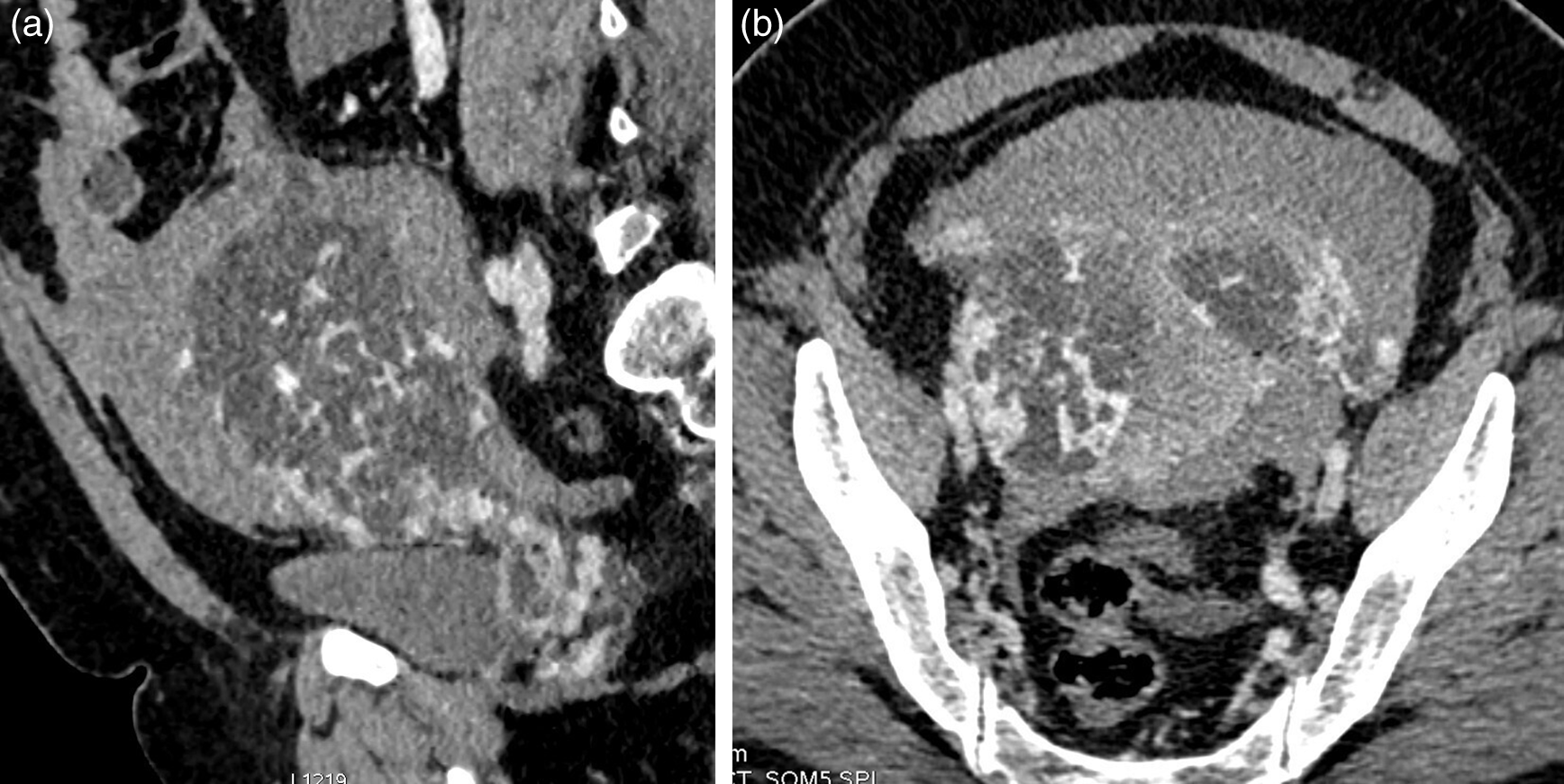
The CT-scan demonstrated a moderately abundant hemoperitoneum associated to a complex solid and cystic uterine mass of 13 cm. This mass was the seat of intense enhancement after injection of contrast, with thrombosis of the left ovarian vein (Figure 1). CT also showed a common left iliac adenomegaly.
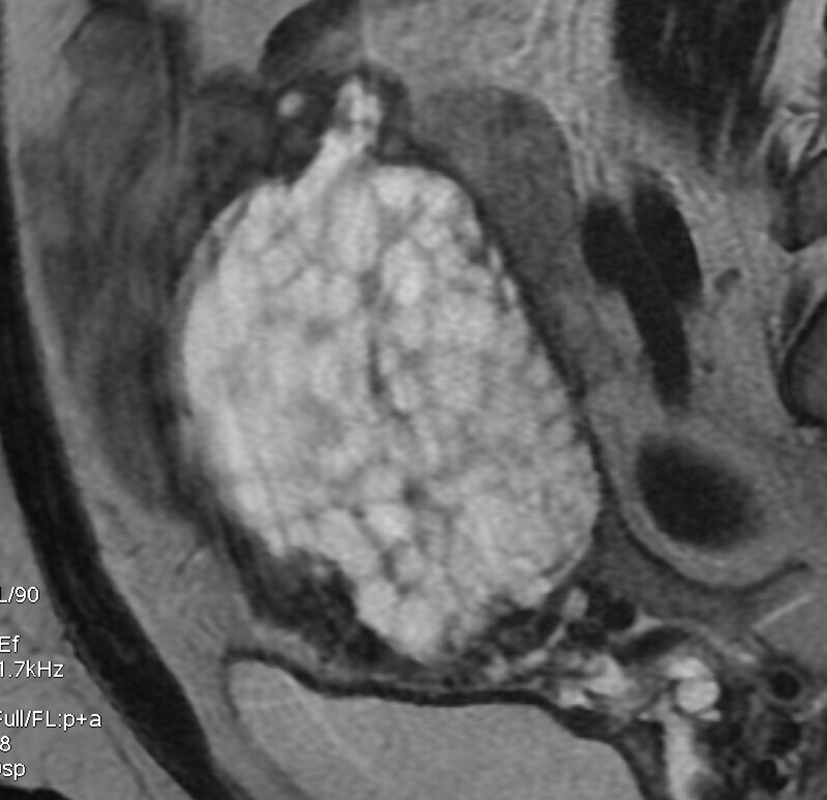
A pelvic MRI was performed and it showed a globular uterus, with a multiloculated cystic endocavitary mass occupying the fundal region, deeply thinning the myometrium. This mass was with high T2 and low T1 signal. Few areas of high T1 signal were present related to hemorrhage (Figure 2). A large zone of perforation in the posterior fundal uterine wall was noted (Figure 3), associated to a high abundance hemoperitoneum. Ovaries were normal.

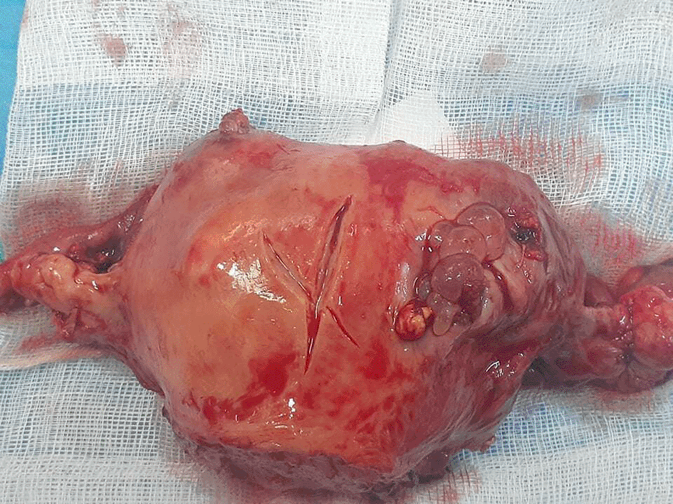
Intervention was decided immediately. Intra-operatively, one liter of hemoperitoneum was found. The uterus was globular, with perforation in its posterior wall, bringing chorionic villus (Figure 4). No macroscopic invasion of bilateral uterine parameters was noted.
A subtotal hysterectomy with bilateral annexectomy was done without any intra or postoperative incident.
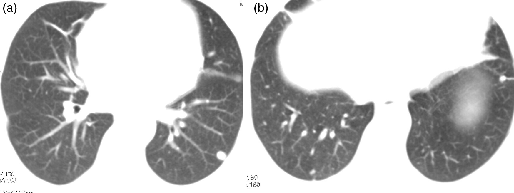
A thoracic computed tomography with a cerebral MRI were done as an assessment of extension showing multiple pulmonary ground glass and solid nodules with a metastatic aspect (Figure 5), and no cerebral lesion.
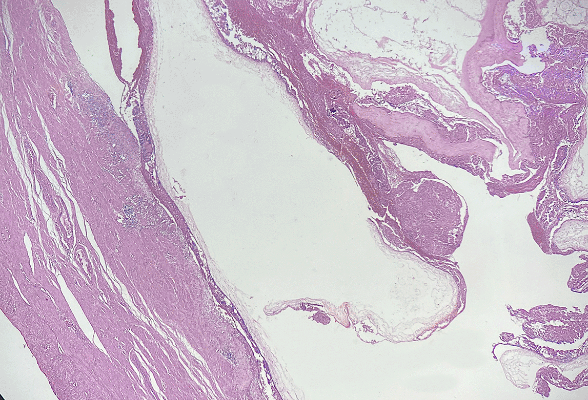
The pathologic examination of the specimen was positive for an invasive mole without parametrial invasion (Figure 6).
The patient was classified as high risk with a FIGO 2000 prognosis score to 15 [age >40 years (1 point); previous gestation: abortion (1 point); interval from index pregnancy more than 12 months (4 points); pretreatment hCG > 105 (4 points) ; largest tumor size >5 cm (2 points); site of metastases: lung (2 points); number of metastases identified between 5 and 8 (2 points); no previous failed chemotherapy (0 points)].5
Medical oncology consultation was obtained postoperatively, and the patient was proposed for adjuvant polychemotherapy including EMA-CO (Etoposide, Methotrexate, and Dactinomycin with Cyclophosphamide and Vincristine). The serum β-HCG level rapidly decreased after one cycle chemotherapy and dropped to normal values after the second cycle. Three additional courses of chemotherapy were given. Actually, four months after surgery, the patient is under periodic follow-up evaluations of serum β-HCG level which remained negative.
Gestational trophoblastic neoplasia refers to the aggressive subset of gestational trophoblastic disease (GTD) that has a capability for independent growth and metastases.6
It is a very rare group including invasive mole, choriocarcinoma, placental site trophoblastic tumor, and epithelioid trophoblastic tumor.7,8
Hydatidiform mole, accounts for 80% of all GTD, estimated to occur in 0.6 to 1.1 per 1000 pregnancies in North America.9 Invasive mole, by elsewhere is rarer and constitutes 5 to 8% of trophoblastic diseases.10 It may arise from any pregnancy event, although in most cases it is diagnosed after molar pregnancy.
The usual clinical presentation is vaginal bleedings, pelvic pain, abdominal distension due to enlargement of the uterus. Sometimes it can be accompanied with altered general state condition or pre-eclampsia.11,12
Acute abdomen without any past history of vaginal bleedings nor molar pregnancy is an unusual presentation that may mislead the diagnosis to an abdominal cause. Some authors such as Aminimoghadam et al. in 2017 and Bruner et al. in 2014 reported few cases of invasive mole presenting as acute hemoperitoneum.3,13
Development of trophoblastic tissue in uterine stroma and the invasion of myometrium by molar structures may lead to uterine perforation.14 In our case, this perforation was responsible for a heavy intraperitoneal hemorrhage.
Imaging aspects of gestational trophoblastic neoplasms reveal a great polymorphism. Distinction between invasive mole and choriocarcinoma is highly difficult in imaging.15
Ultrasound examination shows a heterogenous echogenic uterine structure due to zones of hemorrhage and necrosis. Infiltration of different tunics of the uterus with disorganization of their structure and irregularities is in favor of an aggressive tumor. Hypervascularization is usually seen in color doppler.16
Pelvic MRI is indicated essentially to check for local extension. It shows a poorly limited complex cystic mass, with high heterogenous enhancement after gadolinium. In fact, it shows the hyper vascularized character of these tumors and the dilation of adjacent myometrial vessels. MRI is performed for the study of mural uterine invasion and parametrial extension. Distinction between invasive mole and choriocarcinoma is not obvious in MRI despite a possible difference in enhancement which is peripheral in choriocarcinoma with large zones of central necrosis, and essentially central in invasive mole. These features are not always seen, and histological confirmation is necessary to confirm the precise diagnosis.15
These tumors have hematogenous dissemination, with pulmonary, hepatic, and cerebral metastatic sites. For extension assessment, a thoraco-abdominal Computed tomography is requested, completed eventually by cerebral MRI or CT scan. In our case an internal iliac adenomegaly with pulmonary nodules was discovered which is very rare in invasive mole unlike choriocarcinoma.
In summary, we reported a special case of an invasive mole, arising as a complication of hemoperitoneum and acute abdomen. This atypical presentation mislead the diagnosis initially. Imaging features and elevated rate of serum β-HCG permitted the proper diagnosis thereafter.
The absence of periodic follow-up after multiple abortions caused the delay in diagnosis, the degeneration and the distant extension of her molar pathology.
Moreover, the request for beta human chorionic gonadotropin (β-HCG) in case of acute abdomen presentation made it possible to quickly rectify the diagnosis and to take charge of the patient before the deterioration of her dynamic state.
| Views | Downloads | |
|---|---|---|
| F1000Research | - | - |
|
PubMed Central
Data from PMC are received and updated monthly.
|
- | - |
Provide sufficient details of any financial or non-financial competing interests to enable users to assess whether your comments might lead a reasonable person to question your impartiality. Consider the following examples, but note that this is not an exhaustive list:
Sign up for content alerts and receive a weekly or monthly email with all newly published articles
Already registered? Sign in
The email address should be the one you originally registered with F1000.
You registered with F1000 via Google, so we cannot reset your password.
To sign in, please click here.
If you still need help with your Google account password, please click here.
You registered with F1000 via Facebook, so we cannot reset your password.
To sign in, please click here.
If you still need help with your Facebook account password, please click here.
If your email address is registered with us, we will email you instructions to reset your password.
If you think you should have received this email but it has not arrived, please check your spam filters and/or contact for further assistance.
Comments on this article Comments (0)