Keywords
airway obstruction, ENT emergency, Ludwig’s angina, oral hygiene
airway obstruction, ENT emergency, Ludwig’s angina, oral hygiene
Ludwig’s angina (LA) was first reported and published by a German physician Wilhelm Frederick von Ludwig in 1836. He described it as a throat infection that was different from other typical throat infections with specific characteristics; peculiar hardness of the involved tissue, swelling beneath the tongue, edema of the neck, and sparing of the glands.1 Modernization in medical care has made this entity rare and of low mortality at the present. But it is still seen in emergency settings and is still feared as a lethal entity due to the rapidly progressive airway obstruction that follows.2,3 To highlight this most feared complication of LA, the word ‘angina’ is added as its origin is a Latin word ‘angere’, which means ‘to strangle’.4
Odontogenic infection, especially pre-apical infection of mandibular molars, is the most common origin of Ludwig’s angina and this predominance of dental etiology has been well backed up by evidence.2,3 Other less common etiologies include peritonsillar abscess, mandibular fracture, or other facial trauma. Although males and age groups from 20 to 40 years are commonly affected by LA,4 it has also been reported in neonates, children, and the elderly.3 Causative agents are usually a mix of both aerobic and anaerobic bacteria, including mouth organisms like streptococci or staphylococci.5
Here, we report a case of a 15-year-old male who developed LA, was overwhelmed by its complications and later recovered from it. Also, there was an incidental finding of a roundworm in his respiratory tract. This case report is presented in line with the CARE guidelines.6
A 15-year-old male presented to the emergency department with concerns of swelling over his submandibular region (for nine days), fever (for four days), dysphagia, dysphonia, and drooling (for two days). Meticulous history taking revealed that the patient had experienced pain in his mandibular teeth recently, but he did not seek any medical attention for it. He had no known history of allergy, no history of trauma nor surgical or dental procedures. When asked about his personal history, he stated that he inconsistently brushed his teeth, and he had a habit of eating sugary items and drinking carbonated drinks. There was also no practice of visiting a dentist regularly in the patient’s family. On the day of the presentation, physical examination showed a fever of 102 degrees Fahrenheit, pulse rate of 143 beats per minute, respiratory rate of 37 breaths per minute, and blood pressure of 133/90 mmHg. Remarkable findings on examination were halitosis, firm and tender swelling which extended from one angle of the mandible to the other with bilateral involvement of submandibular space and sublingual space, restriction of mouth opening with an inter-incisor gap of 2 cm, swollen tongue, which was lifted up, and dental carries over both his mandibular first molars. However, examination for stridor, headache, cyanosis and regional lymph node involvement were negative (Figure 1).

The diagnosis of LA was made by judging the clinical picture: firm, tender, bilaterally symmetric swelling of sublingual space and submandibular space, presence of carious teeth, and history of poor oral hygiene. The patient was immediately admitted to the intensive care unit (ICU) and management was started promptly due to the possible impending airway obstruction. A vertical midline incision just above the sternal notch was given to perform an emergency tracheostomy, but couldn’t be performed successfully due to the purulent discharge coming out of the incision site. Then emergency endotracheal (ET) intubation was performed with the help of direct rigid laryngoscopy under general anesthesia. Multiple incisions along the angle of the mandibles were performed for drainage of the neck swelling. The patient was put on clindamycin (300 mg) and meropenem (1 gm) intravenously for 12 days and carious teeth (both mandibular first molars) were extracted. Elective tracheostomy was performed eight days later (Figure 2).
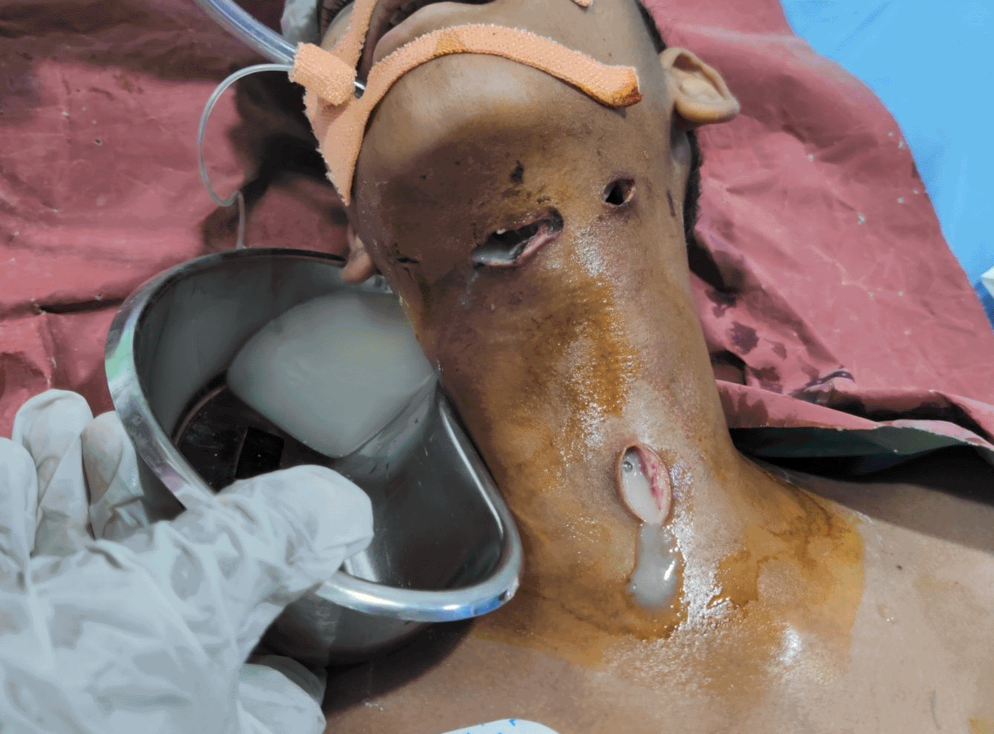
Discharge can be seen coming from the vertical incision that was carried out to perform emergency tracheostomy.
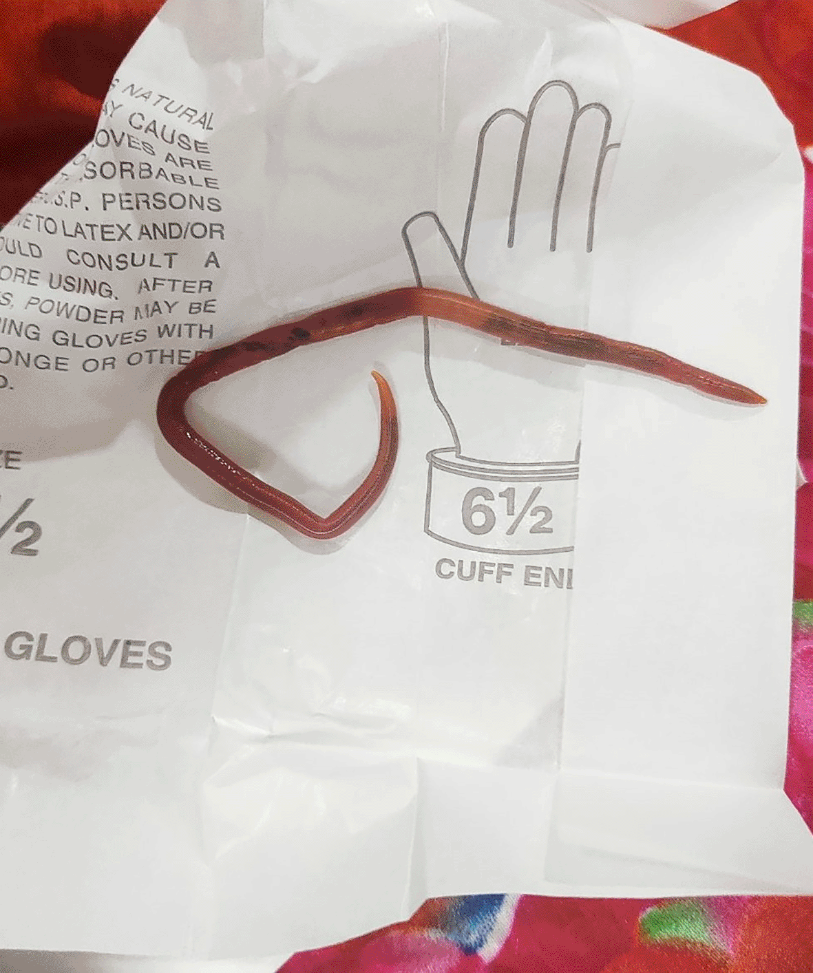
On the third day of admission, while suctioning and caring for secretion from the ET tube, a roundworm came out of the ET tube. Adult worm identification examination confirmed that it was Ascaris lumbricoides. However, eosinophils were not raised and there was no trace of other worms and their eggs on the X-ray. For deworming, oral albendazole 400 mg was administered for three days. As the patient was being monitored regularly for possible complications of LA, empyema thoracic and loculated effusions were noticed on the fourth day of admission. Chest-tube insertion was carried out for its management. Our patient was also prescribed vancomycin (40 mg/kg) for 14 days as urine culture tested positive for Enterococcus species on the twelfth day of ICU admission (Figures 3 and 4).

After successfully managing a series of complications and close monitoring for up to 26 days, the patient was transferred to the pediatric ward from the ICU. The patient recovered very well and faced no new issues. The tracheostomy was closed, and discharge was planned for the patient. Our patient and his parents were counseled about his clinical condition and the interventions carried out. Our patient was prescribed mupirocin (2%) ointment for application over the closed incision sites and prednisone tablets (5 mg, once a day, for five days) along with pantoprazole tablets (40 mg, once a day, for one month). He was called for a follow-up two weeks after discharge (Figures 5 and 6).


We report a case of LA that originated from an odontogenic infection. Prabhu et al.7 concluded that odontogenic infections with poor oral hygiene are consistent in the majority of cases. Firstly, infection originates in the subgingival pocket then spreads to the musculature of the floor of the mouth. This infection now progresses below the mylohyoid line gaining access to sublingual space. Since the roots of the second and third mandibular molars lie below the mylohyoid line, infection of these teeth are most frequently involved in the development of LA. The infection then spreads to the submandibular space from the sublingual space. Okoje et al.8 pointed out in their study that the first mandibular molar can be involved in the development of LA but at low frequency. In our patient, there was the presence of unattended carious mandibular molars which acted as the source of infection.
Diagnosis of LA is made based upon the clinical picture, and laboratory tests are of little immediate value.9 Grodinaky’s criteria for diagnosis of LA includes the involvement of more than one space usually bilaterally, the presence of gangrene with serosanguineous, putrid infiltration with very little or no frank pus, sparing of glandular structures and spread is through continuity not by lymphatics.3 As soon as we diagnosed the patient, he was intubated and admitted to the ICU due to the possible impending airway obstruction. This is the most feared complication of LA and this was to be considered with the utmost emergency. After securing the airway, early treatment of the underlying cause was important to avoid a lethal outcome. We removed the patient’s carious teeth, and he was put under broad-spectrum antibiotics. Since causative agents are usually a mix of both aerobic and anaerobic bacteria, the aim while using antimicrobials should be to cover all possibilities.4,5 Besides airway complications, our patient also showed empyema and pleural effusion on radiological examination. Empyema and pleural effusion are some of the noted complications of LA.2 This was only noticed later when radiological examinations were carried out as the priority was to intubate the patient and secure the airway first. Enterococcus species were isolated from the urine of the patient and this is thought to be hospital-acquired as Enterococcus species are a common cause of nosocomial urinary tract infection.10
LA is not only a subject of concern of dental, otorhinolaryngology, and respiratory disciplines but a matter that raises multidisciplinary concern due to its other complications: mediastinitis, aspiration pneumonia, rupture of the carotid artery, thrombophlebitis of the internal jugular vein, necrotizing fasciitis, and osteomyelitis.2
Although LA is a rare entity, it should be considered for differential diagnosis in cases of neck swelling especially in those having a history of poor oral hygiene and recent dental procedures. This condition spreads aggressively and demands aggressive management with the priority of securing the airways first and the use of broad-spectrum antimicrobials. Complications of LA require special attention from multiple disciplines. The practice of good oral hygiene is not just about the oral cavity but is also about life-threatening conditions like LA.
Our 15-year-old patient has developed a positive outlook towards the condition he went through. He understands the clinical state he was in and is thankful to the entire hospital, especially the nurses who took care of him during his hospital stay. The patient understands the importance of maintaining good oral hygiene.
| Views | Downloads | |
|---|---|---|
| F1000Research | - | - |
|
PubMed Central
Data from PMC are received and updated monthly.
|
- | - |
Provide sufficient details of any financial or non-financial competing interests to enable users to assess whether your comments might lead a reasonable person to question your impartiality. Consider the following examples, but note that this is not an exhaustive list:
Sign up for content alerts and receive a weekly or monthly email with all newly published articles
Already registered? Sign in
The email address should be the one you originally registered with F1000.
You registered with F1000 via Google, so we cannot reset your password.
To sign in, please click here.
If you still need help with your Google account password, please click here.
You registered with F1000 via Facebook, so we cannot reset your password.
To sign in, please click here.
If you still need help with your Facebook account password, please click here.
If your email address is registered with us, we will email you instructions to reset your password.
If you think you should have received this email but it has not arrived, please check your spam filters and/or contact for further assistance.
Comments on this article Comments (0)