Keywords
penetrating brain injury, PBI surgical management, transorbital approach wooden foreign body
penetrating brain injury, PBI surgical management, transorbital approach wooden foreign body
Transorbital penetrating brain injury (PBI) secondary to a non-projectile foreign body is rare and potentially life-threatening. The orbital wall is thin; therefore, access to the cranial cavity through this region can be swift. It can cause severe damage to the eye, optic nerves, brain parenchyma, and neurovascular structures.1 Prevalence studies have reported that transorbital PBI accounts for 45% and 24% of all traumatic brain injury in children and adults, respectively, and accounts for 0.04% of all traumatic brain injury and 4.5% of all orbital pathologies.2,3
Transorbital PBI at the skull base region can cause severe neurovascular damage. Vital structures, such as cranial nerves, arteries, and the cavernous sinus, are of particular concern during preoperative planning for surgery in patients with transorbital PBI in the skull base region.4 Immediate complications (that can occur within a week of injury) include haemorrhage, vascular damage (such as carotid-cavernous fistula [CCF], traumatic aneurysm, and intravascular thrombosis), ischemic brain injury, brain oedema, and cerebral contusion.5 Delayed complications (that can occur after more than a week of injury) include infections (meningitis, encephalitis, and orbital cellulitis), foreign body migration, hydrocephalus, and cerebrospinal fluid (CSF) leakage.5 Therefore, it is essential to understand the mechanism of injury, reconstruction of preoperative planning surgery, and postoperative care.4 The timing of surgery also needs to be considered in patients with a wooden transorbital PBI, especially when it involves a rough wooden surface, as it can cause more severe tissue damage.4 Adequate debridement, administration of antibiotics, and anti-seizure prophylaxis are also required to achieve a satisfactory outcome.6
Surgery is the primary treatment for most cases of transorbital PBI. Available approaches include the transorbital approach, right or left frontal craniotomy applying the subfrontal approach, right or left frontotemporal craniotomy, bifrontal craniotomy using the anterior interhemispheric approach, lobectomy, and the percutaneous lumbar intrathecal approach.7–12 In our case, we considered using the transorbital approach in view of the clinical and radiological features that are associated with such cases of wooden transorbital PBI.
An 18-year-old male carpenter sustained a wooden PBI at two locations—the right frontoparietal and left orbito-temporal areas, and the base of the skull—while cutting wood with a machine. The wood in the frontoparietal area was extracted, and bone decompression along with evisceration of his left eye were performed six hours after the incident at the district general hospital. Afterwards, the patient was referred to our hospital for further management. He complained of pain and loss of sight in the left eye. He had a Glasgow Coma Scale (GCS) score E4V5M6, trigeminal nerve palsy (V2 and V3), facial nerve palsy of the peripheral type (House Brackmenn grade IV) and left-sided hemiparesis. There was no sign of CSF leakage.
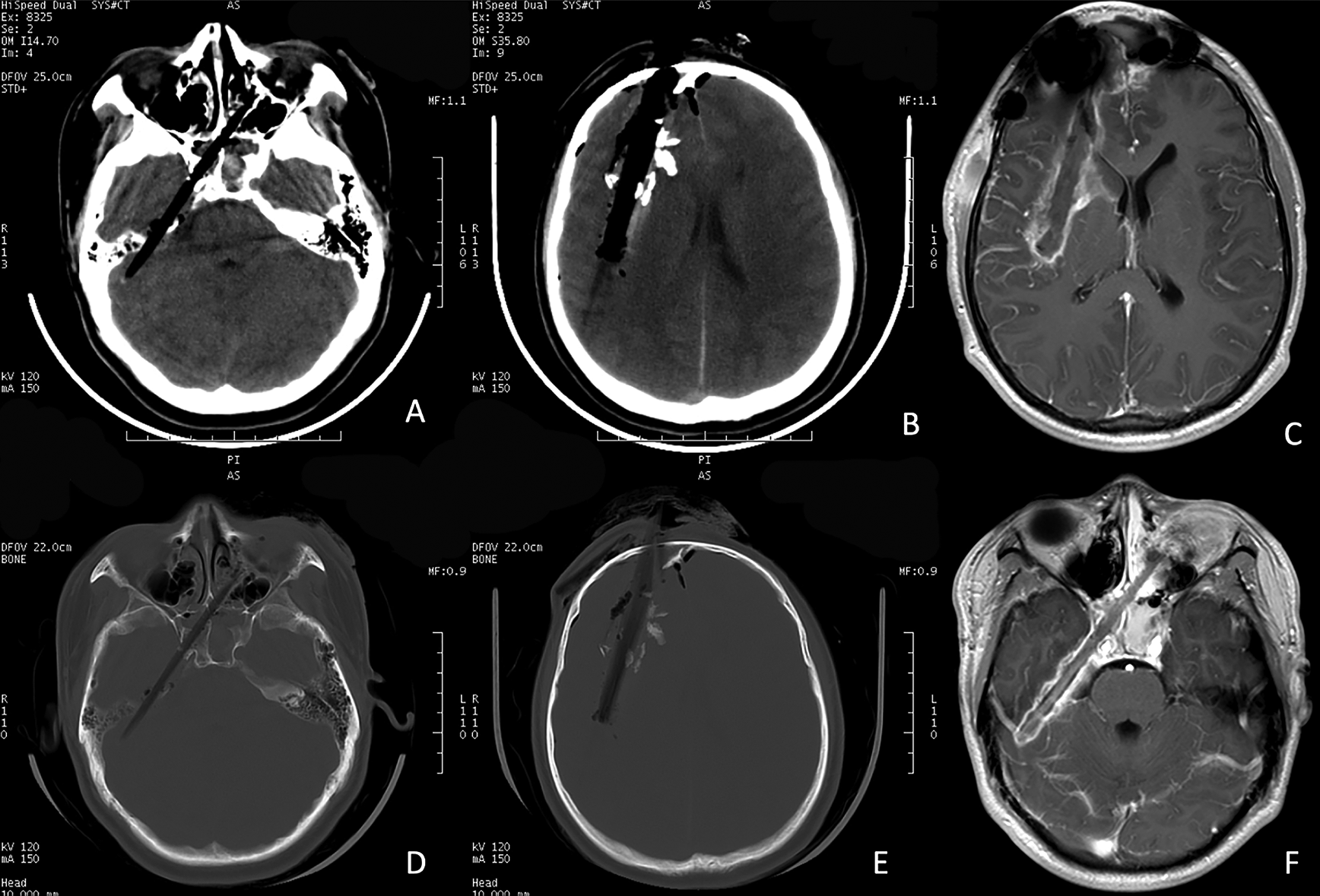
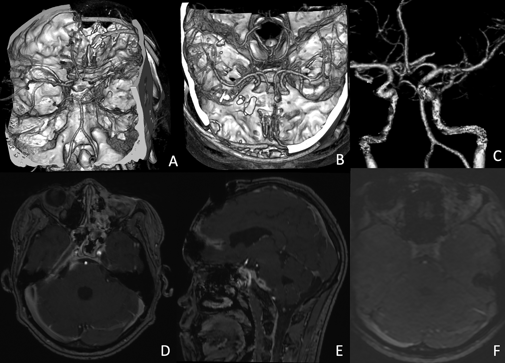
Laboratory examinations revealed leucocytosis and increased C-reactive protein (CRP) and procalcitonin levels. Computed tomography (CT), magnetic resonance imaging (MRI), magnetic resonance angiography (MRA), and a 3D-printed model were used preoperatively to plan for surgery (Figure 1). There were no lesions in the internal carotid artery, cavernous sinus, and transverse sinuses (Figure 2). We surgically extracted the wood 14 days after the accident. Empirical antibiotics were administered for six weeks while awaiting definitive antibiotic treatment based on microbiological and antibiotic susceptibility tests. Phenytoin was administered for a week after the accident for post-traumatic seizure prophylaxis.

(A) The wood is seen to extend from the left medial orbital region, ethmoid sinus, across the contralateral sphenoid sinus, and reach the superior part of the posterolateral surface of the petrous bone before crossing to the right side. (B) Another piece of wood that extends from the right frontal to the right parietal region (about 10 cm long) is also observed. (D) and (E) show bone window segments. (C) and (F) T1-weighted contrast magnetic resonance imaging (MRI) using gadolinium shows that the foreign body had formed a fibrous capsule.

(A), (B), and (C) computed tomography (CT) angiography and reconstruction of the cerebral arteries shows no rupture in the internal carotid artery (ICA). (D) magnetic resonance angiography (MRA) reveals narrowing of the right ICA that is compressed by the one of the pieces of wood. (E) magnetic resonance venography (MRV) shows no lesion in the cavernous sinus.
| Step 1: | The patient was placed in the supine position, facing the right side. Following surgical draping, the operating area around his left eye was disinfected. He was placed perpendicular to the operator's view. |
| Step 2: | A linear incision was made on the conjunctiva. |
| Step 3: | The superior and inferior palpebrae were gently retracted using the Langenbeck retractor. Blunt dissection of the left orbital soft tissue was then performed. |
| Step 4: | The tip of the wooden foreign body was identified on the medial orbital wall. |
| Step 5: | Osteotomy was performed using Kerrison punch forceps until the hole at the penetration area became wider. |
| Step 6: | The wood was extracted gently using a bone rongeur. Extraction was performed perpendicular to the operator’s view. |
| Step 7: | Haemorrhage was evaluated and treated after extracting the wood. Further haemostasis was achieved using Gelfoam and Surgicel. |
| Step 8: | Suturing in layers and canthorrhaphy were performed (Figure 3). |
Postoperative CT scan evaluation revealed gross total extraction without any haemorrhagic lesion along with a GCS of E4V5M6 (Figure 4). The patient was discharged six weeks after the pieces of wood were extracted, and no apparent clinical complications and abnormal infectious parameters (leucocyte, CRP, and procalcitonin) were found. Six months postoperatively, there was no feature of trigeminal nor facial palsy. The patient could return to his usual activities, and a customized ocular prosthesis was made for his left eye.
The goals of surgery in cases of wooden transorbital PBI include: (1) debridement of non-vital tissues, considering the extensive damage the wood may have caused; (2) evacuation of hematomas, such as extradural, subdural, and intraparenchymal hematoma; (3) removal of as much bone fragments as possible; (4) retrieval of foreign body fragments (larger fragments should be meticulously sought out, as they tend to migrate); (5) securing of adequate haemostasis; and (6) watertight closure of the dura mater, which usually requires the use of grafts. Turbin anatomical criteria refer to zone 3b (medial canthus) as the area where the orbital area exits at the superior orbital fissure and sphenoid wing.1 The aspects of the central nervous system at risk of damage in that area include the cavernous sinus, temporal lobe, brain stem, or basal cistern.1
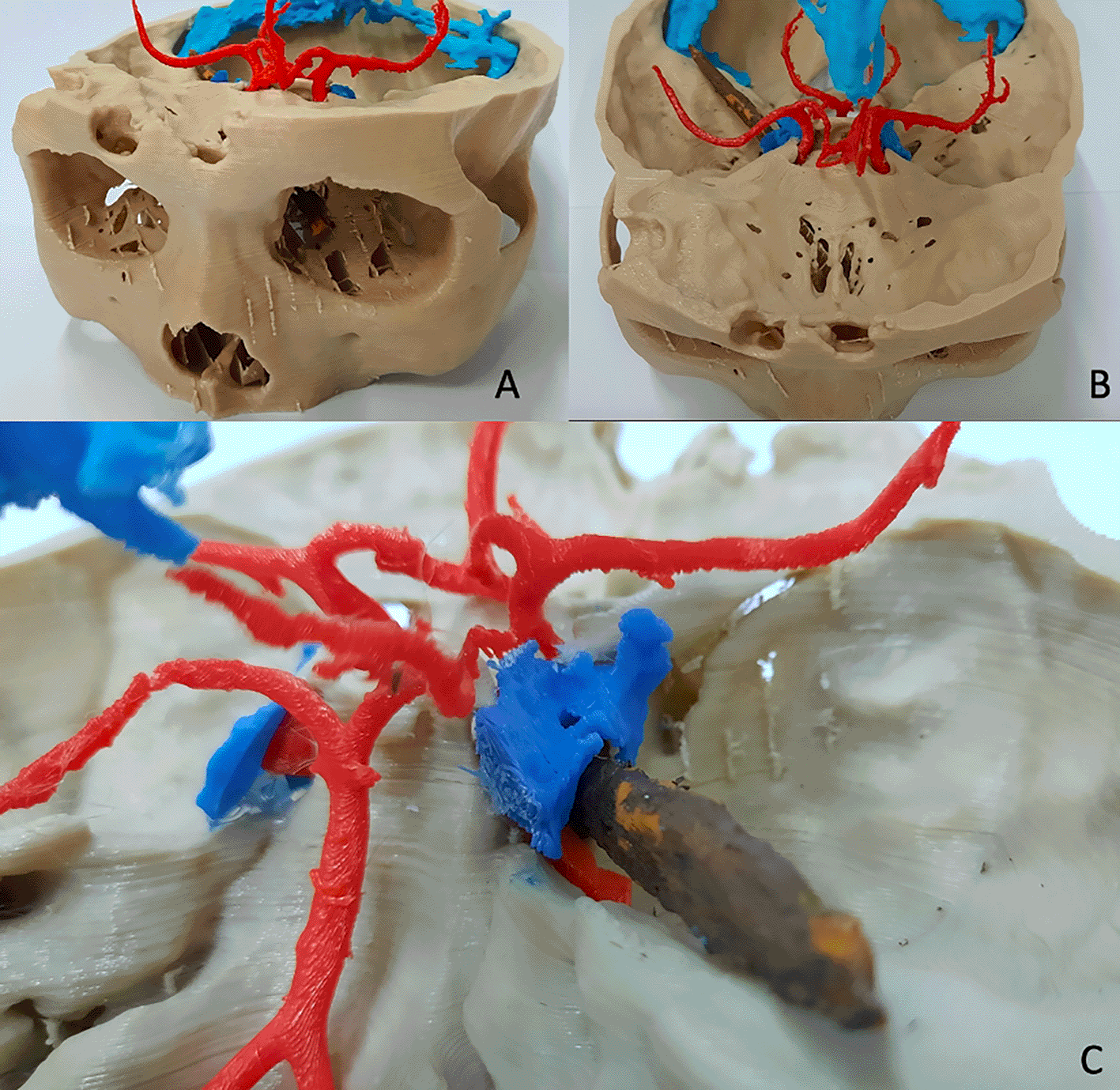
In our case, the patient underwent a two-stage surgery. The first stage of surgery was debridement, surgical extraction of the right frontoparietal wooden object, bone decompression, and left ocular evisceration, which were done six hours after the incident at the district general hospital. The bone flap removed from the injured side was placed on the contralateral subgaleal layer of the injured side. The patient was referred to our hospital because we were more equipped to manage the extraction of the wood from the orbito-temporal part of the base of the skull. In this case we also performed the 3D-printed reconstruction for the surgery plan because 3D-printed model reconstruction can help neurosurgeons for surgical strategies, and it enhances the anatomical visualization of the location and trajectory of the foreign body, which is not completely visible from the outside (Figure 5).13

(A). Tip of the wooden foreign body at the medial orbital wall. (B) and (C) Show that there is no lesion in the internal carotid artery (red) and cavernous sinus (dark blue).
Based on details obtained from CT angiography (CTA), magnetic resonance venography (MRV), MRA, and the 3D-printed reconstruction, no serious neurovascular lesions were seen. Therefore, we decided to perform the second stage of surgery in the orbito-temporal part of the base of the skull base. The patient was positioned supine facing to the right for a transorbital approach, perpendicular to the wooden foreign body’s entry zone. The eyelid was retracted gently, and the flap was sutured using silk 3.0. The conjunctiva was incised horizontally from the medial to the lateral canthus. We performed soft-tissue exploration of the orbit using a Langenbeck retractor until the medial wall of the orbit (ethmoid bone) was visible. When performing soft tissue retraction of the orbit using a Langenbeck retractor, it is essential to pay attention to the presence of an oculocardiac reflex due to stretching of the afferent stretch receptors that are transmitted through the ciliary nerve, Gasserian ganglion, and efferent vagal fibres, which are capable of increasing the parasympathetic tone. This could cause bradycardia in the patient.
After exploring the soft tissue of the orbit medially, the superficial tip of the wood was revealed in line with its trajectory. Osteotomy of the interlocking bone around the tip of the wooden foreign body was performed using a Kerrison punch, then the bone around the tip of the wood was meticulously removed. The wooden foreign body was gently extracted, perpendicular to the operator’s view, using a bone rongeur. There was no brisk bleeding from the internal carotid artery nor from the cavernous sinus. Adequate debridement (irrigation for dilution) and haemostasis (using Gelfoam and Surgicel) were achieved after the wood was extracted. The wooden foreign body and the fibrous capsule formed were examined for infection via microbiological culture and antibiotic susceptibility testing. Finally, we sutured the skin in layers and performed tarsorrhaphy. The wood was most probably contaminated; therefore, we debrided any easily accessible impacted bone and other extracranial tissues along the track.
Regarding the timing of surgery, for our patient surgery was performed 14 days after the accident. The purpose of this was to ensure that a consistently thick fibrous capsule had formed over the entire body of the wood. This is a physiological foreign body reaction that protected the brain from the rough surface of the wood. Ibrahim et al., in their experimental study in 2017, reported that the immune response to foreign body implantation began on day 14.14 Mast cells (MC) are mobilized to the site of the wound, where they mature and are activated early in the inflammatory process through chemotactic inflammatory signaling. The activated MC secrete mediators, such as IL-4 and IL-13, attracting macrophages through chemotactic signals. To improve their effectiveness, macrophages fuse to form foreign body massive cells that reside at the tissue interface.15 Foreign-body giant cells are still incapable of digesting large implants but can secrete cytokines that cause fibroblasts to produce a collagen capsule that embraces the foreign body, and this effectively isolates it from surrounding tissue.16 On day 14, the thickest fibrous capsule forms, followed by those on days 60, 90, and 180 (Figure 6).14 Therefore, watchful waiting until day 14 needs to be considered for a hemodynamically stable patient without severe neurovascular complication. Allograft cranioplasty was performed six months after the accident. This was done to reduce the risk of infection.
Many surgical approaches exist for managing transorbital PBIs. In this case, we decided to perform transorbital approach techniques perpendicular to the entry zone, 14 days after the injury. This enabled us to perform a minimally invasive procedure based on the patient’s clinical and radiological features.
All data underlying the results are available as part of the article and no additional source data are required.
The authors would like to thank all those who contributed to the process and completion of this report, including all neurosurgery consultant and fellow residents of the Department of Neurosurgery, Faculty of Medicine, Universitas Airlangga, Surabaya.
| Views | Downloads | |
|---|---|---|
| F1000Research | - | - |
|
PubMed Central
Data from PMC are received and updated monthly.
|
- | - |
Is the background of the case’s history and progression described in sufficient detail?
Yes
Are enough details provided of any physical examination and diagnostic tests, treatment given and outcomes?
Yes
Is sufficient discussion included of the importance of the findings and their relevance to future understanding of disease processes, diagnosis or treatment?
Yes
Is the case presented with sufficient detail to be useful for other practitioners?
Yes
Competing Interests: No competing interests were disclosed.
Reviewer Expertise: Neroradiology & Neurointervention
Is the background of the case’s history and progression described in sufficient detail?
Yes
Are enough details provided of any physical examination and diagnostic tests, treatment given and outcomes?
Yes
Is sufficient discussion included of the importance of the findings and their relevance to future understanding of disease processes, diagnosis or treatment?
Partly
Is the case presented with sufficient detail to be useful for other practitioners?
Yes
Competing Interests: No competing interests were disclosed.
Reviewer Expertise: neurosurgery
Alongside their report, reviewers assign a status to the article:
| Invited Reviewers | ||
|---|---|---|
| 1 | 2 | |
|
Version 1 15 Dec 21 |
read | read |
Provide sufficient details of any financial or non-financial competing interests to enable users to assess whether your comments might lead a reasonable person to question your impartiality. Consider the following examples, but note that this is not an exhaustive list:
Sign up for content alerts and receive a weekly or monthly email with all newly published articles
Already registered? Sign in
The email address should be the one you originally registered with F1000.
You registered with F1000 via Google, so we cannot reset your password.
To sign in, please click here.
If you still need help with your Google account password, please click here.
You registered with F1000 via Facebook, so we cannot reset your password.
To sign in, please click here.
If you still need help with your Facebook account password, please click here.
If your email address is registered with us, we will email you instructions to reset your password.
If you think you should have received this email but it has not arrived, please check your spam filters and/or contact for further assistance.
Comments on this article Comments (0)