Keywords
collagen scaffold, jellyfish Aurelia aurita, alveolar bone, regeneration, hMSCs
collagen scaffold, jellyfish Aurelia aurita, alveolar bone, regeneration, hMSCs
The alveolar bone is a part of the maxilla and mandible bones that support the formation of dental sockets (alveoli)1. The alveolar bone is formed during an eruption of a tooth that serves to provide a mounting place for the formation of periodontal ligaments. Etiology of alveolar bone damage can occur in two categories, namely infectious and non-infectious. Damage to the alveolar bone that occurs due to infection is usually caused by anaerobic gram-negative bacteria such as Porphyromonas gingivalis, Prevotella intermedia, Agregatibacter actinomycetemcomitans. Infectious diseases of dental support tissue are commonly called periodontitis. In addition to periodontitis, tooth bone damage can be caused by mechanical trauma in the form of occlusion trauma, post-tooth extraction, and post-use orthodontic wire due to excessive pressure. The result of alveolar bone damage is the onset of a defect in the alveolar bone that results in teeth faltering and eventually loss2,3.
Regeneration therapy is needed to prevent tooth loss by regrowing alveolar bone. Regenerative periodontal tissue healing occurs through the formation of new periodontal tissues, i.e. the formation of alveolar bones, the functional periodontal ligaments, and the new cementum. The indicators of bone regeneration process are increased of the differentiation and the number of bone cells (osteoblast cells)4.
One of the critical stages in the development of artificial bone tissue is the design and the manufacture of a scaffold. A scaffold is a porous biodegradable material that has a 3-dimensional structure. Scaffold serves as a structural supporter of cells, and it becomes an extracellular matrix during the natural process of bone regeneration and development, which will be naturally degraded later as new tissues grow. Scaffold material can be obtained from synthetic and natural sources5. Protein as collagen or fibrin and polysaccharides (such as chitosan and glycosaminoglycan (GAG) are proven to match the biocompatibility properties of cells but are problematic to the immunogenicity6,7.
Collagen is the most commonly used polymer in the development of a scaffold. Collagen is a very low soluble protein that has typical triple helix characteristics and consists of two cross-linked a1(I) and a2(II) chains with hydrogen bonds between protein hydroxy and other residues. The triple helix structure consists of three dominant amino acids in which each parts contains amino acid ((-Gly-X-Y)n)8. Since Collagen naturally is able to activate immunogenic response when penetrated into the scaffold, it is then more often to be used in scaffold development9. The type of collagen that is often used is type I10,11, because its biocompatibility, chemical and physical properties are suitable for scaffold development. Type I collagen is classified as the main structural protein of the original extracellular matrix (ECM), which is beneficial for cell growth10. The scaffold can be applied as bone healing12,13 because of its ability to induce osteogenesis, osteoinduction, and osteoconduction phenomena, so that mesenchymal stem cells require scaffold as the attachment media, to proliferate, and to differentiate into osteoblast cells.
During this time, the source of collagen used for the manufacture of scaffold comes from various organisms. The source of collagen that is usually used comes from the skin and tendons of bovine and porcine. However, collagen derived from bovine poses a risk of bovine spongiform encephalopathy infection while collagen derived from porcine sometime has become prohibited due to religious reasons14–17.
Sources of collagen from marine animals are more preferred nowadays because they are safer than land animals. One of the aquatic biota that has the potential to be a scaffold in Indonesia is jellyfish18. Based on the statistical data of the jellyfish production value in Indonesia in 2008, jellyfish production reached 500,000 tons per year19 and experienced an increase in 2011 up to 674,000 tons per year20. The increase of the jellyfish production value in Indonesia from 1998 to 2002 has an average of 2.46 % per year. Of all the types of jellyfish, A.aurita is the most numerous and consumed species in Indonesia that can be found in all the seawaters of Kalimantan, Sumatra, and Java Islands. Although the presence of jellyfish in Indonesia is relatively abundant, jellyfish is one group that receives less attention, so information about jellyfish biota resources is relatively limited. In addition, jellyfish not only contain collagen but are also rich in fatty acids that are useful for the human body21. The content of amino acids in the jellyfish is high so that almost all the needs of amino acids of the human body can be fulfilled by consuming jellyfish, except for methionine and histidine which are less than the provisions provided by FAO / WHO. Collagen in jellyfish has also become an available and relevant source for use as a component of matrices in tissue engineering7,14,22.
According to previous studies, collagen contained in jellyfish has been used for a wide range of biomedical purposes. Hoyer et al.14 have used all parts of Rhopilema esculentum to extract its collagen and produce scaffold based on the principle of biometric mineralization. Collagen extracted from jellyfish was then cross-linked with 1-ethyl-3-(3 dimethyl aminopropyl) carbodiimide hydrochloride (EDC) and performed by dehydrothermal treatment (DHT)23. Collagen is considered as the gold standard for this field due to its high biocompatibility9,24. Collagen-based biomaterials are widely used for the manufacture of scaffolds intended for bone regeneration. In addition to having elastic mechanical properties, scaffold with the base material of jellyfish collagen has interconnection characteristics between its pores that allow mesenchymal stem cells to live and multiply so that it can be a good basis for osteogenic differentiation or osteogenesis14. Rhopilema esculentum was found to contain 16 types of amino acids with the most compositional sequences being glutamic acid (Glu), lysine (Lys), glycine (Gly), aspartic acid (Asp), leucine (Leu)25,26. In contrast, A. aurita was found to contain 15 types of amino acids with the most compositional sequences being glycine (Gly), glutamic acid (Glu), phenylalanine, (Phe), threonine (Thr), tyrosine (Tyr), lysine (Lys), serine (Ser). Of the entire A. aurita tissue, it has 57 mg of protein per gram of dry weight basis. The most composition in collagen content consists of glycine and other amino acids, including proline and hydroxyproline27.
In this present study, we create and develop a new biomaterial scaffold of jellyfish A. aurita collagen that is biocompatible, biodegradable, pore interconnected, non-toxic, and can support cell viability to help regenerate alveolar bone defects.
Dried A. aurita was obtained from Prigi Beach, Trenggalek Regency, East Java, Indonesia. Dried jellyfish were cut into small pieces then washed using sterile ddH2O. After that, the sample was rewashed using NaOH. Then it was dissolved in the sterile ddH2O, then blended using a laboratory blender (Blender Philips) until crushed. The sample was then carried out for sonification and further centrifuged. A pellet was taken and put in a petri dish then freeze-dried to obtain the collagen in membrane form.
Membrane and porous collagen scaffold was obtained after the freeze-drying process, it was then cross-linked in solution of N-(3-dimethyl aminopropyl)-N’-ethyl-carbodiimide hydrochloride (EDC). The scaffold was carefully rinsed in the sterile water then diluted in glycine solution. After that, the scaffold was rewashed with sterile water before freeze-dried again”
Scaffold membranes were affixed with carbon tape and coated with Aurum-Palladium (Au-Pd) prior to SEM and EDX Spectroscopy. The size of the samples used in SEM was 1 cm × 1 cm. Each sample was observed using SEM (FEI inspect S50) with 500x and 2000x magnifications to characterize the topography (surface, texture) and the morphology (pore size and pore interconnectivity). Meanwhile for EDX Spectroscopy, it was performed to measure the element combinations contained in the scaffold.
The scaffold membrane sample was crushed with mortar until it became a powder (± 2 mg) then further mixed with Kalium-bromide (KBr) until it formed pellets. Sample was observed using FTIR spectroscopy (Shimadzu IR-prestige 21).
A scaffold membrane weighing 15 mg (W0) was then soaked into 4 mL of Phosphate Buffer Saline (PBS) solution in a Petri dish and incubated in a water bath at 37°C. PBS was replaced every two days. The scaffold was rinsed from PBS after nine days and then washed with water, lyophilized, and weighted (Wt). This assay was done in triplicate.
Percentage water uptake = [(Wt – W0) / W0] × 100
W0 and Wt – initial weight and weight after incubation for time ‘t’ respectively
Human mesenchymal stem cells (hMSCs), isolated from adipose cells, were kindly provided by Biomedic Laboratory, Brawijaya University. The treatment of hMSCs for in vitro experiments was approved by The Ethics Committee of Brawijaya University (1052-KEP-UB). The cells were maintained in 25 cm2 culture flask in complete medium, Dulbecco's Modified Eagle's Medium (DMEM) high glucose (Gibco) containing 10% fetal calf serum and 1.25% penicillin, streptomycin, ampicillin at 37°C in a humidified 5% CO2 incubator. The medium was replaced every 2–3 days and the culture process was completed at passage 5. The scaffold membrane was dissolved in a 1xPBS buffer (pH 7.4) with concentration of 5mg/ml and incubated in a shaker incubator (BJPX-103B, Biobase-USA) at 37°C for ± 60 days. When the scaffold membrane has dissolved completely, the mixture then centrifuged at 5000 rpm for 20 seconds to remove the undissolved material. The supernatant was then filtered using 0.45 μm millipore and diluted with complete medium to generate a treatment concentrations of 10; 100; and 1000 μg/ml. The cells were seeded in 96 well plate with density of 1.0 × 10⁴ cells per well total volume of 100 μL. The cells were incubated at 37°C and 5% CO2 for 24 hours. The medium was replaced with treatment medium and incubated for another 24h at 37°C and 5% CO2. Then, the treatment medium was replaced with medium containing 10% WST-1 reagent and incubated at 37°C and 5% CO2 for 180 minutes. Absorbance was measured by a microplate reader (ELx808, BioTek-USA) at 450 nm wavelength. Percent viability was calculated based on the percentage of absorbance of treated cells to untreated cells (control).
The use of scaffold as a brace of alveolar bones was inseparable from the topography and morphology, which includes pore size and interconnectivity of the scaffold. The topography of this scaffold shows a porous surface with a slightly rough texture. SEM observations on the scaffold membrane of A. aurita collagen obtained pore size ranged between 159.9 μm – 325.6 μm (Figure 1). The presence of porosity in the scaffold gives room for new cells to attach, proliferate, and differentiate into the new alveolar bone. This topology of jellyfish collagen scaffold is potential as a support on the development process of new cells in the alveolar bone.
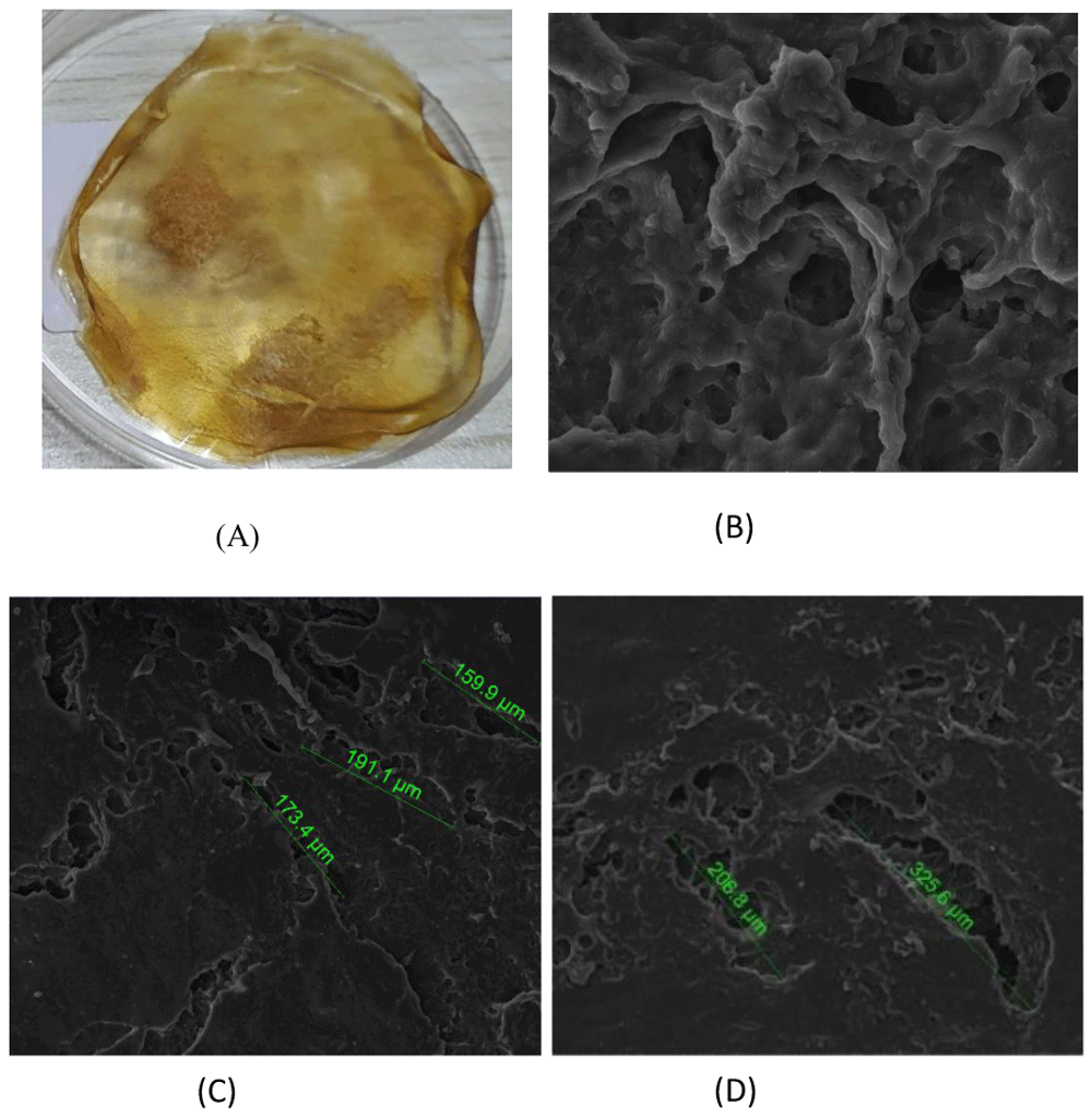
(A) Collagen Scaffold Membrane; The Porosity of scaffold by SEM Analysis with (B) 2000x magnification; (C) and (D) 500x magnification.
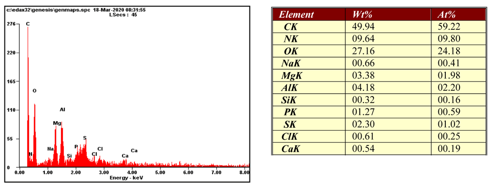
The microstructure elements contained in collagen scaffold were measured using EDX spectroscopy. As a result, microstructure elements found in the scaffold membrane are carbon (C), nitrogen (N), oxygen (O), sodium (Na), magnesium (Mg), aluminium (Al), silicon (Si), phosphor (P), sulfur (S) and calcium (Ca). The highest element found in the scaffold membrane was carbon (C) and the second highest was oxygen (O), 49.94 % and 27.16% of weight, respectively (Figure 2).
Amide structure has been found in jellyfish A. aurita scaffold based on FTIR analysis (Figure 3.). In scaffold treated with EDC, there is Amide A found in 3426.10 cm-1, Amide B in 2924.66 cm-1, while Amide I, Amide II, and Amide III were found in 1655.57 cm-1, 1547.57 cm-1 and 1240.91 cm-1, respectively. In addition, in collagen scaffold without EDC, Amide A found in 3451.18 cm-1, Amide B in 2961.29 cm-1, while Amide I, Amide II and Amide III were found in 1651.72 cm-1, 1553,35 cm-1 and 1238.98 cm-1, respectively. Amide A, Amide B, and Amide II of the treated scaffold are lower than the untreated scaffold. On the other hand, Amide I and Amide III in the treated scaffold are stronger than untreated scaffold (Table 1).
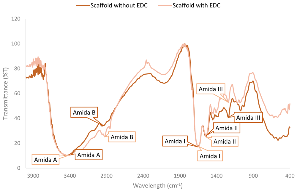
| Wavelength (cm-1) | Prediction of functional groups28,29 | ||
|---|---|---|---|
| Treated Scaffold | Untreated Scaffold | ||
| Amide A | 3426.10 | 3451.18 | N-H binding hydrogen |
| Amide B | 2924.66 | 2961.29 | Asymmetrical stretch of CH2 |
| Amide I | 1655.57 | 1651.72 | C=O |
| Amide II | 1547.57 | 1553.35 | N-H, C-N, C-C |
| Amide III | 1240.91 | 1238.98 | C-N, N-H |
Biodegradablity is one of the properties that the scaffold must possess. Biodegradability in the scaffold is very important because the scaffold has a function as a temporary supporter, in which it will be naturally degraded by the time the alveolar bone has been regenerated. Scaffold natural degradation is expected to be in line with the growth of new bone cells so that it will not be degraded too fast or too late. The result of this study has showed that collagen scaffold treated with EDC was 53% degraded after the 9-days incubation period, while in the same incubation time, the untreated scaffold was 72% degraded after 9-days (Figure 4).
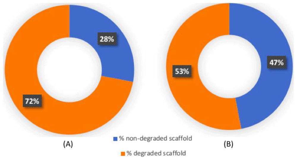
Biodegradability of scaffold membrane (A) untreated scaffold; (B) Scaffold treated with EDC.
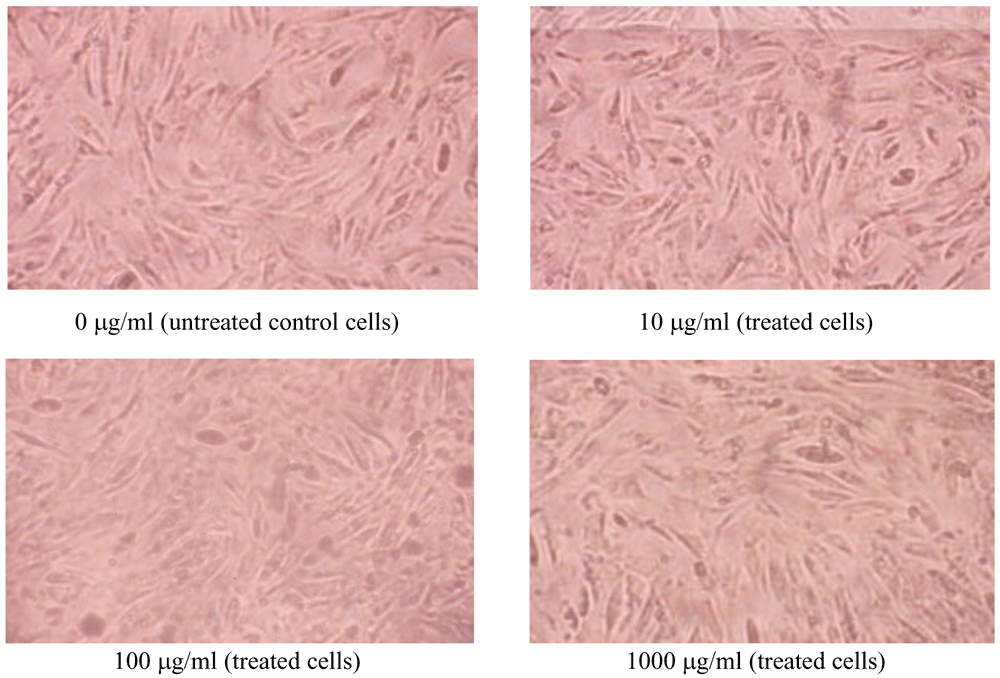
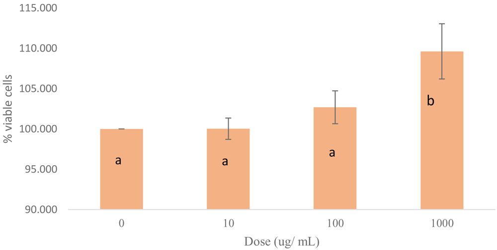
Cell cytoxicity test aims to determine whether collagen can support the cells growth. Human mesenchymal stem cells (hMSCs) were treated with collagen scaffold concentrations of 10; 100; and 1000 μg/ml, respectively (Figure 5). The viability cells were shown as a percentage of treated cells toward untreated control cells. The untreated control group with 0 μg/ml collagen-EDC showed 100% viable cells, while the treated groups with 10 μg/ml, 100 μg/ml and 1000 μg/ml collagen-edc treatment showed 100.03% ±1.32%, 102,686 ± %, and 109,623 ±3.42% viable cells respectively.
On the bar chart, × shows the dosage of each treatment while Y shows the %viable cells of each dose group (Figure 6). Statistical results using one-way ANOVA and Tamhane showed significant differences in concentrations between all groups (0 μg/ml, 10 μg/ml and 100μg/ml) to 1000 μg/ml (p ≤ 0.05), while each concentrations of 0 μg/ml to 10 μg/ml, 0 μg/ml to 100 μg/ml and 10 μg/ml to 100 μg/ml showed no significant difference (p>0.05) (Figure 6).
In the medical sector, the scaffold has an important function especially for the treatment of new cell formation. Scaffold creation must have specific characteristics to support the quality during its application. Biocompatible, bioresorbable, and non-immunogenic properties become important requirements for scaffold manufacturing30. A scaffold of collagen jellyfish is expected to be applied as one of the options for eco-friendly and good quality treatment. Advantages of using collagen as a scaffold material are due to its biocompatible and bioactivity properties. The function of collagen as new cell growth container and to be able to facilitate cell differentiation is expected to enhance cell regeneration in alveolar bones.
The topography of the scaffold from jellyfish collagen with EDC as a cross-linker in this study shows a porous and a slightly rough surface. This topography is different from the type 1 collagen structure that usually has fibrillar structure. Adding EDC gives the binding effect of collagen structure and reconstruct it to porous form. Regarding the cell to material interaction, it is important to increase cell attachment and this depends on the surface topography of material. The topography of scaffold surface has a significant role in supporting cell activities, such as initiation of cells attachment, the proliferation of cells, and differentiation of cells. The morphology of jellyfish collagen scaffold has as a characteristic a porosity rate of ± 159.9 – 325.6 μm. According to Zhang et al.31, collagen scaffold, which has good interconnectivity, generally has a porosity rate of 45 – 800 μm. Interconnectivity of a good collagen scaffold for tissue engineering usually has a collagen scaffold with a porosity of 100–200 μm32. Proportional porosity and adequate pore size are necessary to facilitate nutrient diffusion of the entire structure of the cell so that cells can be proliferated and differentiated correctly33.
Elements of collagen scaffold found in this research have a higher carbon compound. Based on the research, carbon can be used to increase the value of scaffold material. Carbon in some treatment will be good in strength and stability. As scaffold material, carbon has been classified as a non-degradable scaffold. So, it can not be degraded over bone growth, but it can be a real bone substitute34. Oxygen is also having an important role in cell survival. Oxygen is needed to avoid cell hypoxia condition during the cell migration in the scaffold. Oxygen will be diffused in the cells during the respiration activity and it will also help the cells to produce ATP. The lower oxygen in tissue, namely hypoxia condition, can damage the tissue. The lower oxygen in environments will cause the production of reactive oxygen species (ROS) which can further damage the DNA by causing mutation so that it will be harmful to regenerate new cells35. In bone generation, calcium (Ca) and phosphor (P) have a close interaction. P and Ca will bind with each other and form a CaP that has a function to help the proliferation and differentiation of cells. Interaction between Ca and P will induce osteoclast and osteoblast in bone generations. While calcium phosphate is widely used in bone regeneration material like scaffold, cement, and coating36.
Based on FTIR results, the collagen scaffold plus EDC has a lower infrared absorption rate compared to untreated collagen scaffolds. In this research, Amide A, Amide B, and Amide II of treated scaffold absorb lower infrared waves compared to the untreated scaffold. The differences in absorption of group function is due to the addition of EDC, indicating an interaction between the functional group in collagen (scaffold) and the functional group in EDC. EDC is an agent of imide-based zero-length cross-linker that has a functional group RN=C=NR which is known to be effective in inhibiting the enzyme degradation process of denude collagen fibrils and increasing bonds in the collagen. Besides, the functional group of EDC is known to be able to act with carboxyl ionized clusters in proteins forming O-acylisourea that can bind to protein components so that covalent amino acid chain bonds form in two more stable proteins and produce products in the form of urea. The addition of EDC to stop exposed matrix metalloproteinases (MMPs) bound to the collagen matrix is likely to occur due to the changes from the catalytic domain to allosteric23.
Besides giving an effect on IR absorption in FTIR analysis, EDC also gives an effect in biodegradability of the scaffold. Biodegradability is also an important factor as the scaffold is supposed to be absorbed by surrounding tissues, without repeating the surgery process. The time needed by the scaffold to be degraded must be the same with tissue development period. So that, when the cells develop the collagen matrix, the scaffold can be provided as a structural support and then degraded after the tissue has been developed32. The addition of EDC to the scaffold is known to increase the stabilization of the collagen matrix and to lengthen the degradation period37,38. This is proven in this study where scaffold with EDC showed lower degradation compared to the untreated scaffold i.e. 53% to 72% after the 9th day of incubation, respectively (Figure 4).
WST-1 (2-(4-iodophenyl)-3-(4-nitrophenyl)-5-(2,4-disulphonelyl)-2H tetrazolium monosodium salt) can be converted into a water-soluble formazan by mitochondrial dehydrogenase enzymes of alive cells. The water-soluble salt is then released into the culture medium producing a color change which shows the amount of mitochondrial dehydrogenase in cell culture. Thus, the assay represents the viable cells through their metabolic activity. The result showed that collagen scaffold that had been combined with EDC was not toxic to hMSCs and even able to increase the cells growth (Figure 5 and Figure 6). This result is in accordance with reports of EDC which have showed it to be not toxic in fibroblastic L929 cell38 and increasing cells growth without being toxic39. The statistical analysis showed to be not toxic on 10 μg/ml concentration but the cell growth was insignificant compared with the ones on 100 μg/ml and 1000 μg/ml concentrations. On 1000 μg/ml concentration, the cell growth has the highest result.
As a conclusion, we assumed that collagen scaffold of jellyfish A. aurita which use EDC as a cross-linker has a good quality for scaffold manufacturing. The collagen scaffold from jellyfish A. aurita is appropriate with the requirements as the biomaterial in supporting the alveolar bone regeneration process, by fulfilling the following criteria, such as biocompatible, biodegradable, having ideal porous interconnection, being non-toxic, and able to support cell viability. In order to get more optimal scaffold development results, further research focusing on in vivo models will be useful in the future.
Zenodo: Jellyfish (Aurelia aurita) collagen scaffolds potential in alveolar bone regeneration, https://doi.org/10.5281/zenodo.445004040.
This project contains the following underlying data:
- Uncropped/unedited electron microscopy images showing morphology of jellyfish A.aurita collagen scaffold (500x magnification)
- Uncropped/unedited electron microscopy images showing morphology of jellyfish A.aurita collagen scaffold (2000x magnification)
- Raw data of Energy Dispersive X-ray (EDX) spectroscopy showing elements contained in a jellyfish A.aurita collagen scaffold
- Unedited Fourier Transformed Infrared (FTIR) images showing wavelength of jellyfish A.aurita collagen scaffold without EDC (untreated scaffold)
- Unedited Fourier Transformed Infrared (FTIR) images showing wavelength of jellyfish A.aurita collagen scaffold with EDC (treated scaffold)
- Raw data of biodegradable test result
- Raw data of absorbance reading for WST assay for hMSCs
Zenodo: Jellyfish (Aurelia aurita) collagen scaffolds potential in alveolar bone regeneration, http://doi.org/10.5281/zenodo.443116041.
This project contains the following extended data:
- Uncropped/unedited electron microscopy images showing morphology of jellyfish A.aurita collagen scaffold (500x magnification)
- Uncropped/unedited electron microscopy images showing morphology of jellyfish A.aurita collagen scaffold (2000x magnification)
- Uncropped/unedited electron microscopy images showing morphology of jellyfish A.aurita collagen scaffold (20.000x magnification)
Data are available under the terms of the Creative Commons Attribution 4.0 International license (CC-BY 4.0).
| Views | Downloads | |
|---|---|---|
| F1000Research | - | - |
|
PubMed Central
Data from PMC are received and updated monthly.
|
- | - |
Provide sufficient details of any financial or non-financial competing interests to enable users to assess whether your comments might lead a reasonable person to question your impartiality. Consider the following examples, but note that this is not an exhaustive list:
Sign up for content alerts and receive a weekly or monthly email with all newly published articles
Already registered? Sign in
The email address should be the one you originally registered with F1000.
You registered with F1000 via Google, so we cannot reset your password.
To sign in, please click here.
If you still need help with your Google account password, please click here.
You registered with F1000 via Facebook, so we cannot reset your password.
To sign in, please click here.
If you still need help with your Facebook account password, please click here.
If your email address is registered with us, we will email you instructions to reset your password.
If you think you should have received this email but it has not arrived, please check your spam filters and/or contact for further assistance.
Comments on this article Comments (0)