Keywords
Post-partum, pubic symphysitis, case report.
Post-partum, pubic symphysitis, case report.
Septic arthritis of the pubic symphysis is a rare infection, often occurring in particular patients including: athletes with chronic pubic inflammation, women in the postpartum period, drug addicts, patients following a surgical treatment for urinary incontinence or patients with pelvic cancer.1,2
To date, it is an often unknown infection because it is sometimes difficult to diagnose especially as the nosology remains unclear in the literature. Indeed, it must be suspected in the presence of pelvic girdle pain with fever and sometimes lameness or painful irradiation towards the lower limbs, but the symptomatology can be misleading.3
This clinical presentation helps guide additional studies that are not always specific with many differential diagnoses. CT and MRI are the two most sensitive exams.3,4
As for treatment, there are several therapeutic options whose pillar is antibiotic therapy.5,6
We present here a case of pubic symphysitis complicating the postpartum period.
This was a 31-year-old female Caucasian patient with no relevant personal or family history who was re-admitted at day 25 postpartum for septic shock. The patient was primigravid, with no significant pathological history.
The pregnancy was normal. The patient had a low-birth-weight newborn baby of 3750 g. The delivery was without incident. The day after the delivery, the physical examination was strictly normal and the patient was discharged.
At day 15 postpartum, the patient described a gait lameness due to bilateral inguinal pain, in the context of unencrypted fever. The patient took this to be common postpartum joint pain and self-medicated with analgesics. However, the evolution was marked by the aggravation of functional impotence becoming disabling with the appearance of a fever which did not respond to medical treatment; thus, the patient consulted us.
Clinical examination at admission found a confused patient, febrile at 39°C, heart rate of 160 bpm, with blood pressure at 80/60 mmHg, hypogastric defense and sensitivity to palpation of the pubic symphysis and hip mobilization. The diagnosis of septic shock was retained. After conditioning, a pelvic ultrasound was performed showing multiple hyperechogenic heterogeneous compartmentalized collections occupying almost the entire pelvis without visualization of the uterus or ovaries.
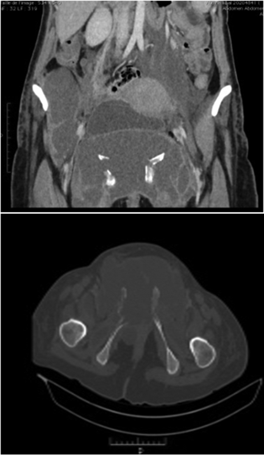
We then performed an abdomino-pelvic CT that showed multiple compartmentalized collections, communicating with peripheral enhancement and water contents, distributed in the form of cubicles of variable size and shape occupying almost the entire perineum (intra-muscular), fusing within the root muscles of the lower limbs, coxo–femoral joints, rectus muscles and along the iliacus and psoas muscles. Calcifications were found within these collections (osteolysis). The most voluminous cubicle was located opposite the the 15-mm pubic symphysis. Osteolysis of the ilio- and ischio-pubic branches responsible for total disjunction with pubic diastasis. Floating, marginal and total thrombus in places at the level of the iliac bifurcation, the left common and external iliac veins, the right external iliac vein and bilateral common femoral veins (Figure 1).
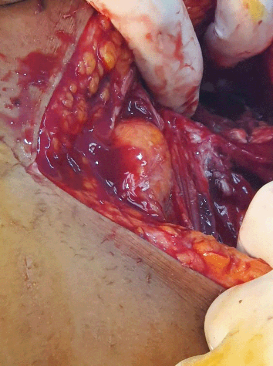
A few hours after admission, the patient’s condition quickly deteriorated with a severe collapse and recourse to noradrenaline. After conditioning, a laparotomy was made with exploration resulting from the collection of two liters of frank pus from Retzius’ space, with the presence of free bone fragments and a significant osteolysis of the pubic symphysis.
The initial investigations were bacteriological sampling, removal of the bone sequester with slight bone and excision of the false membranes, an access to the cubicles at the perineal muscles and iliopsosas and cleaning with physiological serum. Then we entered into exploration of the peritoneal cavity, which was without anomalies. At the end of the operation, two undulating blades were installed at the root of the lower limbs and a Salem probe between the bladder and the uterus (Figure 2).
Biologically there was moderate leukocytosis at 10,000 cells/mm3, associated with an inflammatory syndrome (CRP = 30 mg/L).
The diagnosis of septic arthritis of the complicated pubis of a state of septic shock with abscesses in adjacent muscles and extensive iliac venous thrombosis was therefore retained. The patient was placed on broad-spectrum probabilistic antibiotic therapy using vancomycin (500 mg four times per day) and Tienam (1 g four times per day) parenteral antibiotics and on a curative dose low-molecular-weight heparin. The bacteriological sampling done per-operatively and the blood cultures came back negative. The evolution was marked by progressive clinical improvement.
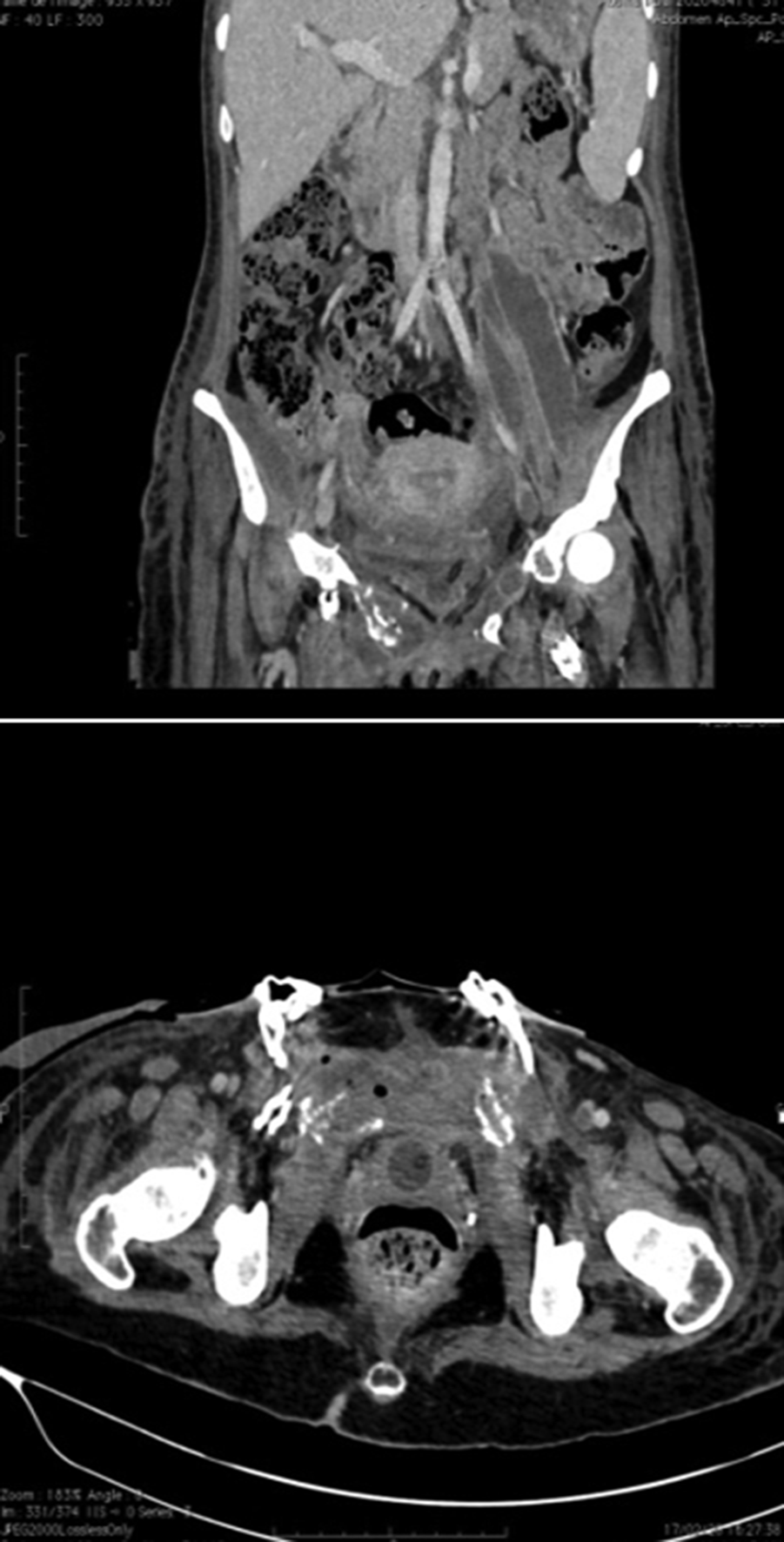
A control CT was performed on day 13 of the treatment objectifying: 1) a right retroperitoneal collection of 5×7 cm; 2) a partial regression of the collections of the left iliopsoas muscle; 3) a raised wall delimiting multiple communicating cubicles; 4) a near total regression of the perineal collections, the right abdominal muscle and the roots of the lower limbs; 5) a stable aspect of venous thrombosis; and 6) as well as bilateral anterior osteolysis of the ilio and ischio pubic branches. Following these CT findings, a CT-guided puncture was then used in the retroperitoneal collection bringing back a thick greenish liquid with the installation of a drain (Figure 3).
Dual therapy was maintained for 6 weeks with an apyrexia as of day 3. Biological inflammatory syndrome disappeared after one month. The removal of the two initially placed corrugated blades was done after 3 weeks.
The patient was asymptomatic during the last check-up with a recoil of 8 months.
Pubic symphysis represents less than 1% of all hematogenous osteomyelitis.7 They are exceptional in post-partum women.6 This is the first case encountered in our service. Pubic symphysis often occurs in a particular field causing symphysis lesions or locoregional infection.3
Indeed, in post-partum women, in addition to the risk of lesions of the symphysis in peripartum (even in the absence of traumatic childbirth), the immunodeficiency due to pregnancy and delivery promotes the pillulation of germs. Concerning the pathophysiology, it is still poorly elucidated by Mynors in 1974 suggested that it is mainly pubic thrombosis due to the dramatic response to heparinization of resistant cases of pubic osteitis.8
The pubic symphysitis in post-partum is difficult and often late diagnosis because pain is often related to the circumstances of childbirth with recourse to self medication that will alleviate the symptomatology. According to a literature review of 100 cases of pubic symphysitis, published by Ross et al.6 In 2003, most patients reported pubic pain (68%). Pain is also localized to the groin (41%), thigh (15%) and hip (12%), probably due to radiation along the hip adductors, which insert onto the pubic symphysis.
Reaching the hip adductors also explains the pain during walking (59%) and hip movement (45%). Long delays between symptom onset and diagnosis were typical (mean, 29 days), due to insidious presentation and low clinical suspicion of this rare disease.6 In our patient the diagnosis was made 10 days after the onset of symptoms.
Indeed, the secondary appearance of a pain that did not exist in early post-partum, its increasing intensity, or its unfavourable and unconventional evolution, the presence of a fever or a functional impotence associated especially after non-dystocic delivery are elements that can guide the diagnosis.9
To support the diagnosis or guide the samples two essential examinations to be requested are CT and MRI of the pelvis.3,9
These examinations typically show bone erosions, a bank abscess, enlargement or effusion of the pubic symphysis. MRI provides early detection of edema and inflammation of the bone and muscles, intra-articular effusion or abscess.4
It will then be necessary to search for the responsible germ. Staphylococci aureus (34%) and Pseudomonas aeruginosa (24%) are the bacteria most often incriminated.3 Pubic symphysis with group B streptococcus are exceptional.6 This bacteria can be isolated in blood cultures or post-partum lochia. If this investigation is negative, a local biopsy sample by trocar or surgical biopsy may be proposed. The low virulence of these bacteria, their long culture time, or prior antibiotic therapy may lead to the culture being wrongly considered sterile3 as is the case with our observation.
In this pathology, the biological signs are neither specific nor constant. In the majority of cases, there is an inflammatory syndrome with increased sedimentation rate, but leukocytosis is inconstant, exceeding 11 000 cells per mm3 in only 35% of cases according to the same series.6 For our case, the inflammatory syndrome is moderate with leukocytosis at 10000 and CRP at 30 despite the severity of the clinical picture and per-operative findings.
The treatment is based, classically, on antibiotic therapy of at least 6 weeks sometimes associated with the drainage of a collection.6 But there is no clear recommendation in the literature about which molecule to use.
The evolution is generally favorable in case of early diagnosis and treatment; however, 55% of patients required surgery either for debridement or for abscess evacuation.10 In the absence of treatment, the progression can be towards chronicity with the possibility of appearance of fistulas, bone sequesters and the possibility of pelvic cellulitis.11
For our patient, although she presented with septic shock and the per-operative findings were serious, the evolution was still favorable.
The control check is based mainly on the clinical evolution and the surveillance of the inflammatory syndrome because the imaging takes a long time to show a healing para port to the clinical cure. But for our patient, we asked early for a control scan of extensive associated lesions (venous thrombosis and abscess of the soft parts).
Post-partum pubic symphysitis is a very rare condition with a difficult diagnosis. Two essential tests that should not be delayed to confirm the diagnosis: CT and pelvic MRI are required to confirm the diagnosis and eliminate complications. A treatment based on antibiotic therapy is to start from the diagnosis because the earlier treatment, the more favorable the evolution.
| Views | Downloads | |
|---|---|---|
| F1000Research | - | - |
|
PubMed Central
Data from PMC are received and updated monthly.
|
- | - |
Provide sufficient details of any financial or non-financial competing interests to enable users to assess whether your comments might lead a reasonable person to question your impartiality. Consider the following examples, but note that this is not an exhaustive list:
Sign up for content alerts and receive a weekly or monthly email with all newly published articles
Already registered? Sign in
The email address should be the one you originally registered with F1000.
You registered with F1000 via Google, so we cannot reset your password.
To sign in, please click here.
If you still need help with your Google account password, please click here.
You registered with F1000 via Facebook, so we cannot reset your password.
To sign in, please click here.
If you still need help with your Facebook account password, please click here.
If your email address is registered with us, we will email you instructions to reset your password.
If you think you should have received this email but it has not arrived, please check your spam filters and/or contact for further assistance.
Comments on this article Comments (0)