Keywords
Case Report, Biliary, Hepatitis, Autoimmune, Steroids
Case Report, Biliary, Hepatitis, Autoimmune, Steroids
Autoimmune hepatitis (AIH) is a major immune-mediated and female-dominant chronic liver disease.1,2 The association of primary biliary cholangitis (PBC) and AIH, known as overlap syndrome (OS) is a rare condition. The diagnosis of OS is generally complex and difficult to establish. It is usually defined by the presence of at least two of the three recognized biochemical, serological, and histological criteria of each disease.3 A new scoring classification has been proposed for the diagnosis of OS, with a high sensitivity and specificity, where the cut-off score is 21.4
Clinical manifestations of AIH are varied; however, the majority of patients present with a subclinical or chronic disease.5 The development of acute liver failure (ALF) during the initial presentation of AIH alone is uncommon.6 Few cases of ALF have been reported in relation to PBC- AIH OS,3,7 but no cases have been reported which relate to PBC alone. There are very limited published data on AIH presenting with ALF in male patients.8,9
Herein, we report a case of acute presentation of AIH with features of PBC, suggesting PBC-AIH OS, in a male patient with no previously known liver disease. The patient was successfully treated with corticosteroids and ursodeoxycholic acid.
A 40-year-old male Caucasian patient, working as a salesman, was admitted to our department with the diagnosis of ALF. He had a two-week history of jaundice, dark urine, nausea, poor appetite, and pruritus that had worsened over the previous two days. His past medical history was unremarkable. There was no history of excessive alcohol or any hepatotoxic drug consumption. His family history was negative for liver diseases and autoimmune disorders.
On admission, the patient’s vital signs were stable. The body temperature was 37.4°C, pulse rate was 80 beats/minute, blood pressure was 110/70 mmHg, and jaundice was present. He was conscious, and no flapping tremor was observed. There was no abdominal distension, palpable mass, ascites, or any other clinical features of advanced cirrhosis.
The laboratory workup showed: serum alanine aminotransferase 1238 IU/L (normal value: 40 IU/ L), aspartate aminotransferase 1049 IU/L (normal value: 40 IU/L), alkaline phosphatase (ALP) 192 IU/ L (normal range: 50 to 130 IU/ L), gamma glutamyl transferase (GGT) 164 IU/ L (normal value: 30 IU/ L), total bilirubin 211 umol/L, prothrombin time (PT) 30% and international normalized ratio (INR) 2,87.
Serological markers for hepatotropic viruses such as A, B, C, D and E were all negative. The patient had no clinical or serological signs of infection with other viruses, such as cytomegalovirus, Epstein-Barr virus, herpes simplex virus or human immunodeficiency virus.
All the autoantibodies (anti-nuclear antibody, smooth muscle antibody, liver-kidney-microsomal antibody, and liver soluble antibody) were negative, except for anti-mitochondrial antibody (AMA) M2, which was positive. It was detected using indirect immunofluorescence (IIF), but no quantitative analysis was carried out. Quantitative serum immunoglobulins were within normal levels: IgG: 12,4 g/L (normal range: 6,9 to 16,2 g/L) and IgM: 1, 43 g/L (0.6-2.60 g/L).
Abdominal ultrasonography showed normal liver parenchyma, and normal-size spleen. Color Doppler ultrasound imaging showed normal venous flow. There were no collateral circulations or hepatic vein thrombosis. Endoscopic findings did not reveal any esophageal or gastric varices. Percutaneous liver biopsy could not initially be performed because of the significantly elevated INR. Transjugular liver biopsy would have been preferable in this case, but was unavailable at our institution. As the main potential etiologies were excluded (viral, drug-induced hepatitis, toxins, herbal medication, ischemic hepatitis, or alcohol), the underlying cause was still indeterminate. We then suspected him of having ALF due to autoimmune liver disease, namely AIH. The positivity of AMA M2 in this case implied a probable diagnosis of PBC- AIH OS.
Given the acute, severe, and life-threatening presentation, corticosteroids were promptly initiated. He was first treated with 1 mg/kg of body weight of equivalent prednisolone daily for 10 days, and then with 60 mg prednisolone, daily for approximately 60 days. Five days after the initiation of corticosteroids, his hepatic function tests started to improve. The cytolysis decreased significantly, whereas ALP and GGT levels rose slightly. PT increased to 50%. Although the diagnosis of OS was uncertain, we decided to start ursodeoxycholic acid on day seven, at the dosage of 13 mg/kg body weight daily.
That patient’s jaundice gradually subsided and we noticed an overall clinical improvement especially in fatigue. He was released from our hospital three weeks after admission. Later, during follow up, we introduced azathioprine at the dose of 100 mg daily and prednisolone was progressively tapered to a dose of 10 mg daily, without any sign of exacerbation.
Percutaneous liver biopsy was performed three months after his discharge. Histopathological assessment of the liver biopsy showed a fibrous expansion of the portal triad, with severe portal inflammation consisting of lymphocytes and plasma cells and periportal piecemeal necrosis. Apoptotic bodies were found in lobules.
No evidence of lymphocytic cholangitis or ductopenia was found. These findings were compatible with AIH (Figure 1).
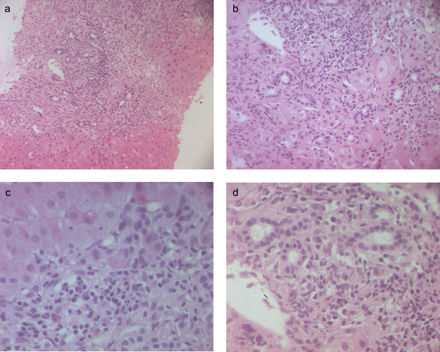
Severe interface chronic hepatitis with apoptotic bodies associated with a dense inflammatory infiltrate (lymphocytes, plasmocytes).
(d) H. E x 400: Cholangiolar proliferation, without biliary duct lesions.
We retrospectively applied the New Scoring Classification for PBC-AIH OS,4 and our patient who presented a score of 20, was identified as potentially having the diagnosis of PBC-AIH OS.
This diagnosis remained questionable as IgM level was normal, AMA M2 titer was not quantified and may have been a false positive, and the characteristic lesions of PBC were not found.
The patient was maintained on10 mg of prednisolone, 100 mg of azathioprine and 800 mg of ursodeoxycholic acid (UDCA), daily.
During the two-year follow-up, the patient remained asymptomatic, and his hepatic function tests became utterly normal.
The present case illustrated a rare presentation of autoimmune hepatitis in a male patient, with acute onset and unexpected features of PBC. The diagnosis was challenging as we have excluded the main potential etiologies of ALF, and serological markers supporting the diagnosis of AIH were absent. Little is known about the management of AIH–induced ALF as there are very limited published data.
AIH is an immune-mediated chronic liver disease, with variable clinical presentations. The chronic, insidious presentation of the disease is commonly revealed by asymptomatic elevation of liver enzymes. However, acute onset hepatitis, including ALF, is rare.10 It defines a syndrome characterized by markers of liver damage (elevated serum transaminases) and impaired liver function (jaundice and INR >1.5).11 This pattern, insufficiently described in the literature, is challenging for physicians as it can mimic other types of acute hepatitis.
Although diagnostic criteria of the acute and fulminant forms of AIH were codified by the International Autoimmune Hepatitis Group in 1992,12 and who removed the condition of six months of disease activity to establish the diagnosis, it is still difficult to diagnose and treat these forms.12 Additionally, autoantibodies can be absent or weakly positive and serum γ-globulin levels are lower than in chronic classical presentations.
Consequently, AIH should be considered in all patients with acute and chronic hepatitis of undetermined cause.13
Women are four times more likely to have AIH than men.14 The rarity of AIH in men is demonstrated by epidemiological studies.2 The acute onset of AIH is a rare condition in the male gender, thus the circumstances of our case are very unique. Similarly, PBC is an autoimmune, cholestatic liver disease, characterized by chronic cholestasis, circulating AMA, and, typically, histological lesions of nonsuppurative destructive cholangitis of the small interlobular bile duct.15 PBC is a slowly progressive disease,16 and there have been no cases reported in the literature of PBC patients presenting with an ALF.11
When PBC and AIH simultaneously coexist in the same patients, it is classified as PBC-AIH OS. Nevertheless, in the absence of clearly established diagnostic criteria, the OS of PBC- AIH is generally evoked by the association of cytolysis, hepatic cholestasis, AMA, and other autoantibodies, as well as histological lesions. Although liver biopsy is mandatory for diagnosis and to guide treatment, detailed histological criteria of AIH with clinically acute presentation have not been well-established.17
Therefore, the diagnosis of AIH-PBC OS is accepted when two or three Chazouillères criteria for PBC and AIH are fulfilled.17,18 With regard to these definitions, we suspected the diagnosis of an acute presentation of AIH. Although our patient presented PBC features, OS could not be confirmed as AMA positivity is only observed in approximately 20% of AIH patients, and was not observed in this case.19 The histology of AIH with acute presentation may reveal heterogeneous patterns of hepatic injury, and typical histological findings of classic AIH can be absent or poorly demonstrated.20
In our case, the liver biopsy specimen demonstrated the absence of cirrhosis with evidence of acute damage including confluent periportal necrosis, plasma cell infiltration in portal areas, perivenular necroinflammatory activity and lymphocytic cholangitis. However, typical histological lesions of PBC, particularly nonsuppurative destructive cholangitis of the small interlobular bile duct, were not found.
It is very important that patients with ALF, such as the case presented here, are treated as soon as possible, to avoid the need for urgent liver transplantation.
Immunosuppressive therapy, namely corticosteroids with or without azathioprine, can achieve sustained remission in more than 80% of patients with AIH. However, drug therapy management in severe forms of AIH remains a subject of debate, and the usefulness of corticosteroid therapy is not clear.21,22 Another crucial question that remained unanswered was whether the optimal dose of corticosteroid should be weight-based or higher doses should be given in these cases, and about the optimal route of administration, oral or intravenous hydrocortisone administration. The benefits of a high-dose regimen of corticosteroids, i.e. higher response rates and, consequently, a reduced need for liver transplantation, should be considered alongside the higher risk of septic complications.23 The decision to initiate corticosteroids in our patient who did not fulfill conventional diagnostic criteria for AIH was made because of the very severe and immediately life-threatening presentation of the illness. In addition, no underlying etiology had been determined. In such situations, the decision should be made on an individual basis, and remains the prerogative of the treating hepatologist. Although it was not evident whether the introduction of corticosteroid therapy would be beneficial, it was the only therapeutic option available, considering that urgent liver transplantation is not available in Tunisia.10 The steroid dose that we decided to prescribe to our patient was based on similar cases previously reported in the literature which had favorable outcomes.21,22 The only predictor of outcome has been the treatment response, which according to Ichai et al,21 has to be assessed over two weeks. The absence of improvement within two weeks of treatment initiation, or the deterioration of any clinical or laboratory feature during this interval, defines refractory disease and justifies the need for an alternative therapeutic option.24 For our patient, although the diagnosis of PBC- AIH OS was uncertain, we associated UDCA with prednisolone and got a favorable response. UDCA can markedly decrease serum bilirubin, ALP, and GGT levels, improve histological damage and fibrosis in patients with PBC, and long-term treatment can delay the histological progression of the disease, particularly in patients with early histological stages. Some authors might prefer to prescribe the initial therapy according to the predominant component of the OS, and to change or add other therapies during clinical follow up.
We described a case of ALF with overlap features of both PBC and AIH, which was successfully treated with corticosteroids.
Acute onset of PBC-AIH OS during the initial presentation is uncommonly reported, and the diagnosis of this entity remains challenging.
In our case, corticosteroids were an effective and lifesaving therapeutic option that prevented urgent liver transplantation. However, identifying early predictors of corticosteroid treatment failure is very important in preventing clinical deterioration in non-responders and in selecting patients for liver transplantation.
All data underlying the results are available as part of the article and no additional source data are required.
| Views | Downloads | |
|---|---|---|
| F1000Research | - | - |
|
PubMed Central
Data from PMC are received and updated monthly.
|
- | - |
Provide sufficient details of any financial or non-financial competing interests to enable users to assess whether your comments might lead a reasonable person to question your impartiality. Consider the following examples, but note that this is not an exhaustive list:
Sign up for content alerts and receive a weekly or monthly email with all newly published articles
Already registered? Sign in
The email address should be the one you originally registered with F1000.
You registered with F1000 via Google, so we cannot reset your password.
To sign in, please click here.
If you still need help with your Google account password, please click here.
You registered with F1000 via Facebook, so we cannot reset your password.
To sign in, please click here.
If you still need help with your Facebook account password, please click here.
If your email address is registered with us, we will email you instructions to reset your password.
If you think you should have received this email but it has not arrived, please check your spam filters and/or contact for further assistance.
Comments on this article Comments (0)