Keywords
Duodenoscopy, aorto-duodenal fistula
Duodenoscopy, aorto-duodenal fistula
Secondary aorto-enteric fistula (SAEF) is an abnormal communication between a prosthetic graft and the gastrointestinal tract that happens after an abdominal infrarenal aortic graft implant to treat aortic aneurysm or occlusive disease.1 SAEF is a rare life-threatening complication with an incidence lower than 2% after aortic prosthetic reconstruction.1 Its diagnosis is sometimes challenging and delayed because of a lack of specific signs.2 Upper endoscopy should be done primarily to eliminate another cause of digestive bleeding. In rare cases it can also confirm the diagnosis by showing the protrusion of the aortic graft in the distal duodenum.3 We report in this paper a case of secondary aorto-duodenal fistula revealed by a severe anemia and confirmed with exceptional duodenoscopy images showing the Dacron prothesis protruding into the lumen of the duodenum. It is only the second report in the medical literature where we clearly and directly see a Dacron vascular graft protruding into the duodenum.
A 75-year-old North African man was referred to our department for management and exploration of severe anemia, symptoms of dyspnea, vertigo, and asthenia. Four years previously, he had undergone an aorto-bifemoral bypass by a Dacron prosthesis for aorto-iliac Leriche occlusive disease.
The patient did not report any history of gastric ulcers or other gastro-duodenal pathologies.
Upon physical examination, the patient was pale but awake and alert. The hemodynamic state was stable, his blood pressure was 130/70 mmHg and his heart rate was 92 bpm. He was febrile (temp = 38 EC). Cardiac and pulmonary examinations were normal. Neurological and otorhinolaryngological examinations were normal also.
The digital rectal examination did not show any bleeding.
Complete blood count revealed microcytic hypochromic anemia with a hemoglobin level of 5,5 g/dL and hyperleukocytosis with a leukocyte count of 12.1 × 10⁹/L. The C-reactive protein level was 17 mg/dL.
The patient received blood transfusion by four units of packed red blood cells and the hemoglobin level reached 9 g/dL.
In order to determine the cause of this severe anemia, the patient had a colonoscopy and upper gastrointestinal endoscopy under general anesthesia. Both exams were normal, not showing an obvious cause of intestinal bleeding.
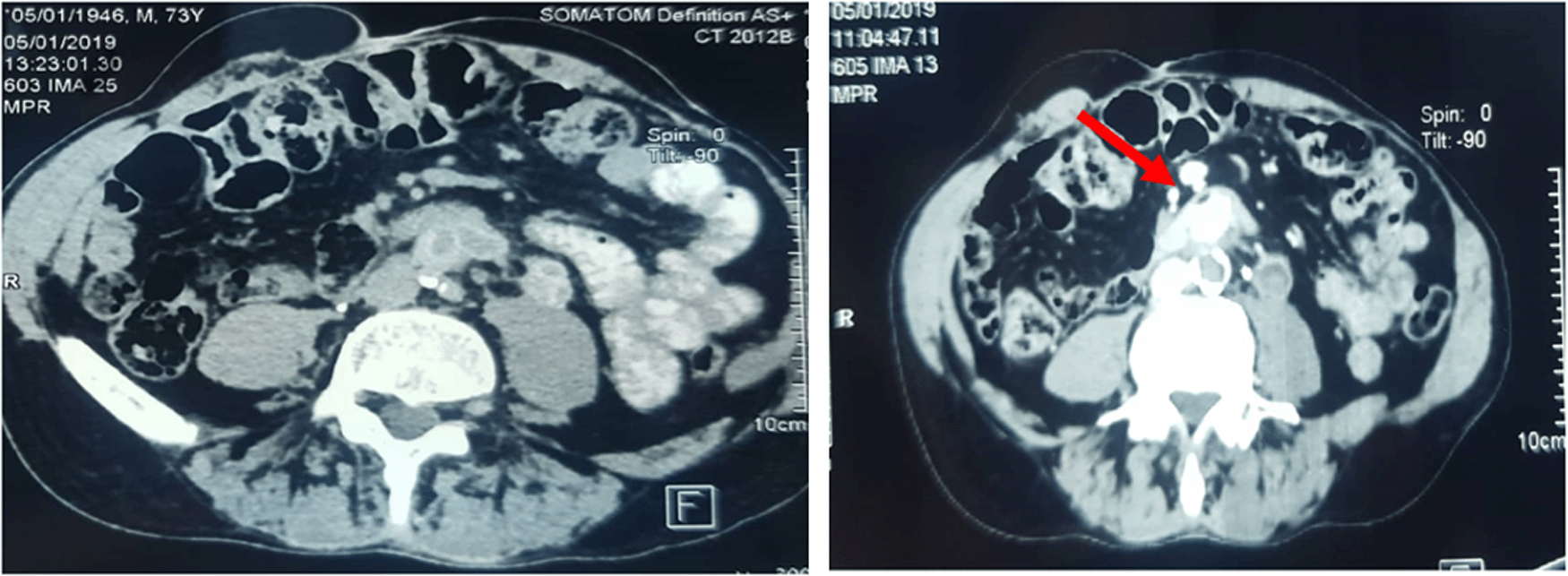
Contrast Computed Tomography (CT scan) was performed urgently, showing a closed contact between the third duodenum and the proximal part of the aorto-bifemoral graft with air bubbles present in the aortic graft associated with a fluid collection of 8,8 × 3,9 mm in diameter in contact with the Dacron prothesis (Figure 1). At this stage the diagnosis of SAEF was strongly suspected and we decide to perform another endoscopic exploration of the duodenum.

Contrast Computed Tomography revealing a closed contact between the third duodenum and the proximal edge of aorto-bifemoral graft with air bubbles in the aorta (red arrow) without extravasation of the aortic contrast material into the duodenum lumen associated with a little collection of 8,8 × 3,9 mm in diameter.
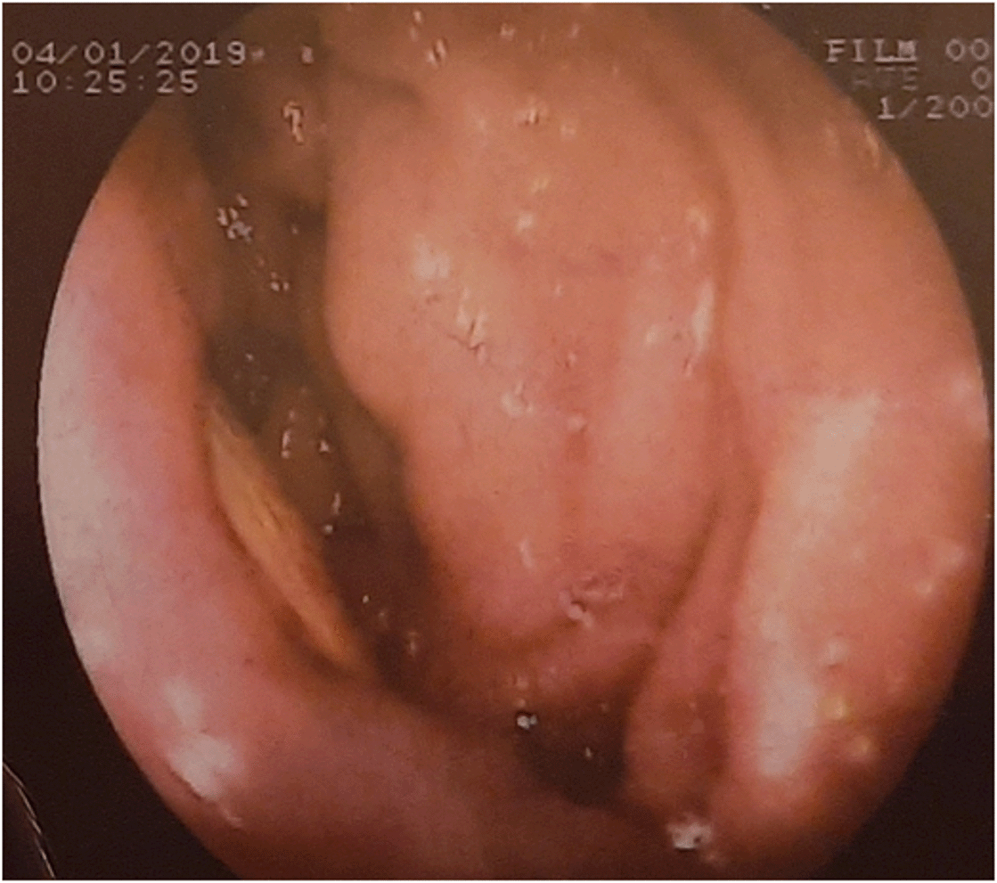
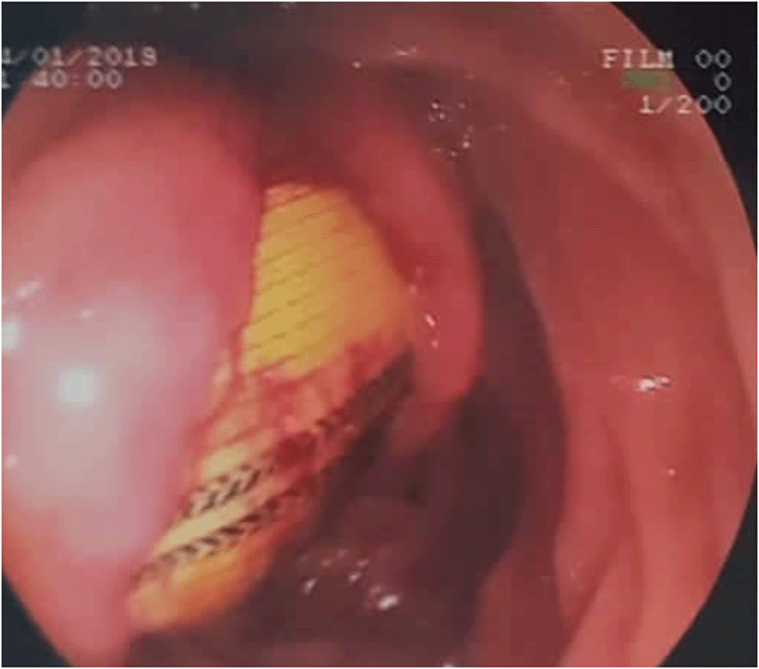
The second esophagogastroduodenoscopy showed a large duodenal fold beside a white foreign body evoking a synthetic material (Figure 2). In order to better elucidate this finding, a duodenoscopy was performed, demonstrating a large parietal defect >3 cm in diameter involving the third duodenum with visualization of Dacron mesh through the mucosal defect (Figure 3).


The patient received a broad-spectrum intravenous probabilistic antibiotic therapy based on Teicoplanin, Imipenem, and Amikacin. The patient was transferred to the vascular surgery department for surgical intervention.
Unfortunately, the day before his operation, the patient presented a cataclysmic hemorrhage and died from hemorrhagic shock.
In 1956, Claytor reported the first case of SAEF after an aortic prosthetic graft.4 More than 75% of SAEF occur between the aorta and the 3rd or 4th part of the duodenum.5 This localization is explained by the retroperitoneal fixation and the close proximity of the duodenum to the aorta.6–8 Rarely the fistula is located in the ascending, transverse, sigmoid colon, or rectum.6–8
The SAEF pathogenesis is controversial. Several hypotheses have been postulated. The most significant mechanisms for its development are graft infection and inflammation, and mechanical factors.2,6–9 Mechanical effect is explained by the presence of a continuous pulsatile movement between the graft and intestine, leading to the erosion of the aortic prosthetic material into the adjacent digestive structure.6,9 This hypothesis is supported by the fact that most SAEFs involve, like our case, the third and the fourth duodenum.6 Another possible mechanism of SAEF is infection introduced at the time of the surgery or secondary to the prosthetic graft colonization by the bowel flora, passed through the suture line.6,9
The clinical presentation varies significantly. A patient with SAEF may present with gastrointestinal bleeding, sepsis, or weight loss. Gastrointestinal hemorrhage, as manifested by hematemesis, hematochezia, melena, hemorrhagic shock, or chronic anemia is the most frequent presentation, encountered in 18-100% of cases.5,6,10 After GI bleeding, sepsis or fever is the commonest presentation in patients with SAEF accounting to 30-87% of cases.5,6 Signs of infection such as fever and leukocytosis sometimes accompany gastrointestinal bleeding.7,9 Our patient presented an occult gastrointestinal bleeding revealed by a severe anemia associated with fever and hyperleukocytosis.
The SAEF diagnosis must be suspected first in every patient who has gastrointestinal bleeding and a history of abdominal graft. Diagnosis is aided by morphological exams such as computerized tomography (CT scan) and digestive endoscopy.6,8,9 The CT scan is the most frequent initial diagnostic test.6,8,9 It allows the visualization of the fistula conduit and its location and may aid in identification of an infection or abscess if present. Its sensibility and its specificity are around 94% and 85% respectively.11 The most specific CT scan features suggestive of SAEF are gas shadow in or around the graft (sensitivity 40%; specificity 100%) and visible graft (sensitivity 22%, specificity 100%).5,12,13 In our case, CT scan showed the presence of air bubbles around the aortic graft. Esophagogastroduodenoscopy is recommended in suspected SAEF patients presenting with upper gastrointestinal bleeding.6,9 Sometimes, the diagnosis should be suspected when we discover stigmata of arterial bleeding, adherent clot, a pulsatile blood clot, or a pulsatile structure on the lumen of the distal duodenum.14
When the synthetic graft is visible in the intestinal lumen, the diagnosis is confirmed. The accuracy of EGD is however limited beyond the second portion of the duodenum while most of SAEFs occur in the third and fourth parts of the duodenum. This explains the scarcity of reports in the medical literature that have been able to highlight by duodenoscopy this aortoduodenal fistula.3,15–19 Compared with these reports, the duodenoscopy findings in our case are the more relevant allowing for the first time to clearly see the characteristic cutmarks of the Dacron prothesis.
The endoscopic findings of prosthetic graft within the duodenum allows the certain diagnosis and surgical treatment should be performed immediately to avoid fatal hemorrhage, as seen in our patient.19
The main objectives of SAEF treatment are maintenance of hemodynamic stability, surgical repair of the underlying defect, infection control via empiric intravenous antibiotics and perfusion of the lower limbs.3,5,6,20 The optimal SAEF repair technique is currently not well defined. The conventional vascular treatment consists of an extra-anatomic bypass grafting with aortic ligation and subsequent graft removal associated with bowel repair.7,21
Management of sepsis consists of early administration of empiric antibiotics covering the majority of identified organisms.6,22 The prognosis of patients with SAEF remains uncertain, and depends on hemodynamic status of the patient at presentation, the operative technique performed and the time of surgical exploration.6,9 A delay in surgical exploration is the main predictive factor of mortality.23
SAEF is a rare life threatening complication. The diagnosis must be suspected in front of a patient having a gastrointestinal bleeding or severe anemia with the history of previous aortic prosthetic reconstruction. Unfortunately, the lack of specific signs is responsible for a diagnostic delay. The diagnosis depends on clinical and biological examinations and CT scan findings. In rare cases, meticulous duodenoscopy exploring the third and the fourth segment of duodenum can confirm the diagnosis by showing the prosthetic graft into the duodenal lumen. The management of SAEF must be done urgently to avoid fatal cataclysmic bleeding.
Written informed consent for publication of their clinical details and/or clinical images was obtained from the family of the patient.
This paper would not have been possible without the exceptional support of our friend Jack Hukill, especially for his help in verifying English this article.
Elloumi Hanen: Writing – Original Draft Preparation
Ben Mrad Melek: Writing – Review and Editing
Ganzoui Imen: Writing – Original Draft Preparation
Ben Hmida Sonia: Resources
Triki Wissem: Writing – Original Draft Preparation
Mchirgui Ilhem: Review and Editing
Ben Hmida Makrem: Review and Editing
Derbel Bilel: Supervision
Cheikh Imed: Validation
All data underlying the results are available as part of the article and no additional source data are required.
| Views | Downloads | |
|---|---|---|
| F1000Research | - | - |
|
PubMed Central
Data from PMC are received and updated monthly.
|
- | - |
Is the background of the case’s history and progression described in sufficient detail?
Yes
Are enough details provided of any physical examination and diagnostic tests, treatment given and outcomes?
Yes
Is sufficient discussion included of the importance of the findings and their relevance to future understanding of disease processes, diagnosis or treatment?
Partly
Is the case presented with sufficient detail to be useful for other practitioners?
Partly
Competing Interests: No competing interests were disclosed.
Reviewer Expertise: Gastrointestinal surgery
Is the background of the case’s history and progression described in sufficient detail?
Yes
Are enough details provided of any physical examination and diagnostic tests, treatment given and outcomes?
Partly
Is sufficient discussion included of the importance of the findings and their relevance to future understanding of disease processes, diagnosis or treatment?
Partly
Is the case presented with sufficient detail to be useful for other practitioners?
Partly
Competing Interests: No competing interests were disclosed.
Reviewer Expertise: Vascular surgeon
Is the background of the case’s history and progression described in sufficient detail?
Yes
Are enough details provided of any physical examination and diagnostic tests, treatment given and outcomes?
Yes
Is sufficient discussion included of the importance of the findings and their relevance to future understanding of disease processes, diagnosis or treatment?
Partly
Is the case presented with sufficient detail to be useful for other practitioners?
Yes
Competing Interests: No competing interests were disclosed.
Alongside their report, reviewers assign a status to the article:
| Invited Reviewers | |||
|---|---|---|---|
| 1 | 2 | 3 | |
|
Version 1 19 Jul 21 |
read | read | read |
Provide sufficient details of any financial or non-financial competing interests to enable users to assess whether your comments might lead a reasonable person to question your impartiality. Consider the following examples, but note that this is not an exhaustive list:
Sign up for content alerts and receive a weekly or monthly email with all newly published articles
Already registered? Sign in
The email address should be the one you originally registered with F1000.
You registered with F1000 via Google, so we cannot reset your password.
To sign in, please click here.
If you still need help with your Google account password, please click here.
You registered with F1000 via Facebook, so we cannot reset your password.
To sign in, please click here.
If you still need help with your Facebook account password, please click here.
If your email address is registered with us, we will email you instructions to reset your password.
If you think you should have received this email but it has not arrived, please check your spam filters and/or contact for further assistance.
Comments on this article Comments (0)