Keywords
Adrenal cortical adenoma, adrenal tumor, pheochromocytoma, adrenal myelolipoma
Adrenal cortical adenoma, adrenal tumor, pheochromocytoma, adrenal myelolipoma
The adrenal gland is a unique retroperitoneal endocrine organ that is different if compared with other retroperitoneal structures in embryological, anatomical, and their role in homeostasis. Tumors of the adrenal gland can be classified as functional or non-functional. The tumors may arise from the adrenal gland or could be secondary lesions. They may be benign or malignant.1,2 Tumors of the adrenal gland are relatively common. The prevalence of adrenal gland tumors is three to ten percent of the human population.3 Adrenal tumor sometimes found as an incidentaloma, which means the tumor more than 1 cm in size was found during an imaging study with indications other than the adrenal condition, but this excludes the patients who were done an imaging study during cancer work-up. Adrenal tumors are known to be one of the most commonly found incidentaloma.4–8
Adrenal masses could be classified into functional, malignant, or benign masses. In functional terms, adenoma causing Conn’s of Cushing’s syndrome, pheochromocytoma, aldosteronoma, and adrenal carcinoma were included. While malignant were comes from metastases, carcinoma, lymphoma, or neuroblastoma. Last but not least, benign masses consisted of non-functioning adenoma, angiomyolipoma, cysts, and hemorrhage.9 Pheochromocytomas are neural crest cell tumors and linked with catecholamine production.10 The initial examination, usually using ultrasound as the diagnostic modality, could be detected in 90% of cases as an enlargement of the adrenal gland.11 The founding could be variable from solid to mixed cystic and solid to cystic.12 Pheochromocytomas patients often present with hypertension, tachycardia, headaches, palpitations, diaphoresis, chest pain, anxiety, and even weight loss. Around 10% of the patients do not have hypertension as their primary symptom.13
An adrenal cortical adenoma is one of the most common incidentalomas found.13 An adenoma mass should be grouped into functioning and non-functioning adrenal adenomas. It should be noted that functioning and non-functioning adenomas could not be classified using imaging techniques.14 Adenomas show a uniform hypoechoic mass relative to the fat. Adenomas are relatively small mass made them be difficult to be found during an ultrasound examination.9
The treatment for adrenal tumor requiring surgery usually because of the treatment failure in functional adrenal masses or a malignant tumor for either primary adrenal cortical carcinoma or solitary metastasis from nonadrenal sources, such as lungs, breasts, kidneys, and melanomas.15 Today, laparoscopic adrenalectomy is the first choice for adrenal disorder surgical procedures. But, there are some conditions that are requiring open adrenalectomies, such as patients with adrenal carcinoma, large pheochromocytoma with blood pressure that may be hard to control, and patients that are requiring a simultaneous abdominal procedure.16 But the absolute contraindications for adrenalectomy are extensive metastatic disease, uncorrected coagulopathy, and severe cardiopulmonary disease that precludes anesthesia.14 The authors report two cases of adrenal glands tumors requiring open adrenalectomy. This study was conducted with surgical CARE checklist guideline as a guidance.15
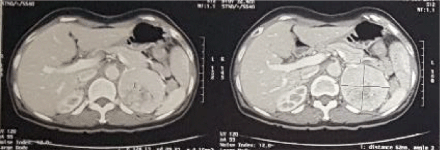
A 52-year-old Indonesian female presented with pain on the left flank. Pain has been felt for six months. Hematuria was found. History of diabetes was found with the highest fasting glucose reported was 159 mg/dL, hypertension was found with a regular blood pressure of about 170/100 mmHg—patient with a history of percutaneous cardiac intervention four months ago. The patient was referred to our Regional Referral Hospital with a left adrenal tumor. The patient also has depression diagnosed by a psychiatrist having 23-score in Beck Depression Inventory-II (BDI II) questionnaire. It has been experienced by the patient two years ago, initiated by the death of her husband. The patient also felt a problem in her marriage, as she never felt intimate with her husband from the first day of their marriage. The patient was diagnosed with an adrenal tumor for the last five years, and since then, she has felt useless in her life. From the radiologic examination, we found a mass from the suprarenal organ, suggestive of an adrenal tumor as a conclusion (Figure 1). Thus, we used the left adrenal tumor as a working diagnosis.
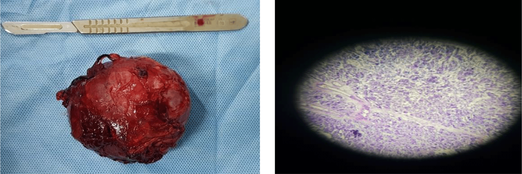
Adrenalectomy was performed for the left adrenal tumor. During the operation, unstable blood pressure was noted and could increase over 200 mmHg. Then the anesthesiologist used several medications to lower the blood pressure immediately. The problems with her blood pressure did not persist after the surgery. The highest blood pressure after adrenalectomy was 130/80 mmHg. Post adrenalectomy mass was sent to the pathology department in order to perform a pathology examination (Figure 2). The result came within one week after the procedures and revealed a picture of tumor cells forming nests is largely separated by fibrous fibers by an invasion of blood vessels. Tumor cells with rounded and oval nuclei are enlarged, rough chromatin, protruding nuclei, cytoplasm partly eosinophilic, partly clear, and bubbly. Abnormal mitosis is easy to find. The stroma consists of infiltrated fibrous connective tissue, with a conclusion of adrenal cortical adenoma.
During the follow-up, the patient shows a normal condition, with normal blood pressure (maximum 125/80 mmHg) but the fasting glucose still not yet within the normal value.
We were reporting an Indonesian male 27-year-old with pain in the upper right abdomen. The pain has been experienced by the patient since one month ago and aggravating within this week. A mass was felt by the patient on the upper right abdomen. No hematuria was reported, no passing stone, no history of hypertension and diabetes. From physical diagnosis, we found a positive ballottement test on the right flank. Other physical examinations were within normal limits.
During the preoperative preparation, blood pressure was within the normal limit. An initial complete blood test showed normal hemoglobin (12.3 g/dL), white blood cells (9,800/μL), and thrombocyte (319,000/μL). Normal glucose analysis was found, different from our first case.
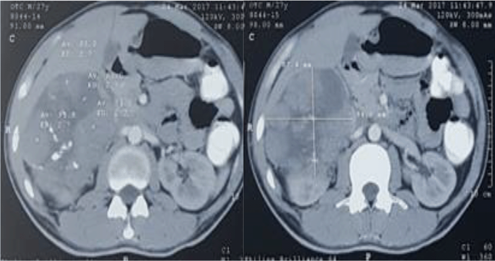
The radiological examination found an adrenal tumor (Figure 3). Based on the radiological examination, the right adrenal tumor was suspected as our diagnosis.
Right adrenalectomy was performed on December, 7th 2017. We found a bulging mass from the posterior. Then we opened the white line of Toldt. The next step was releasing the mass from lateral, superior, inferior, and the last, media. Anterior pedicels and branches from the vena cava and aorta towards the right adrenal were then ligated and cut. Intraoperatively, several antihypertensive drugs were used by the anesthesiologist to maintain the blood pressure at a lower level. Normal blood pressure was preserved after removal of the adrenal gland.
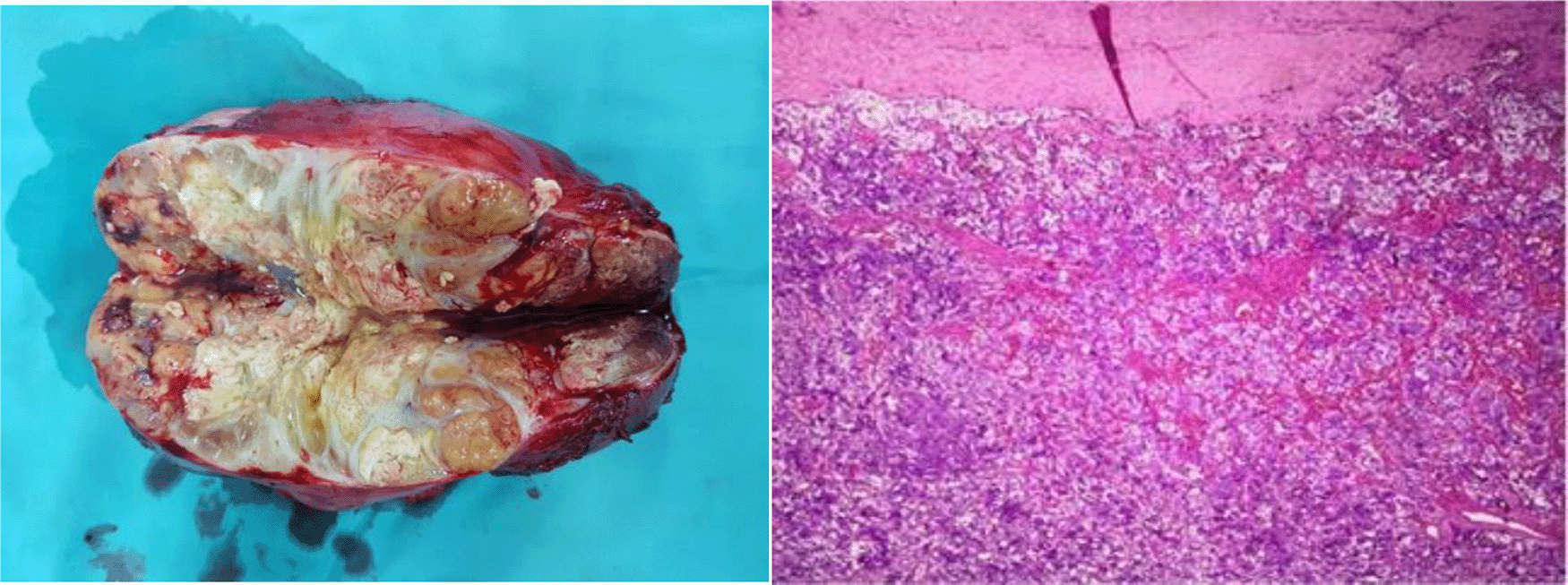
The mass on the right upper abdomen was inspected by the pathology department on December, 8th 2017, and after the next five days, a result was reported, which was a pheochromocytoma (Figure 4). The follow-up was done, and the patient did not show any problem with the blood pressure, blood glucose, and the complete blood count was normal.
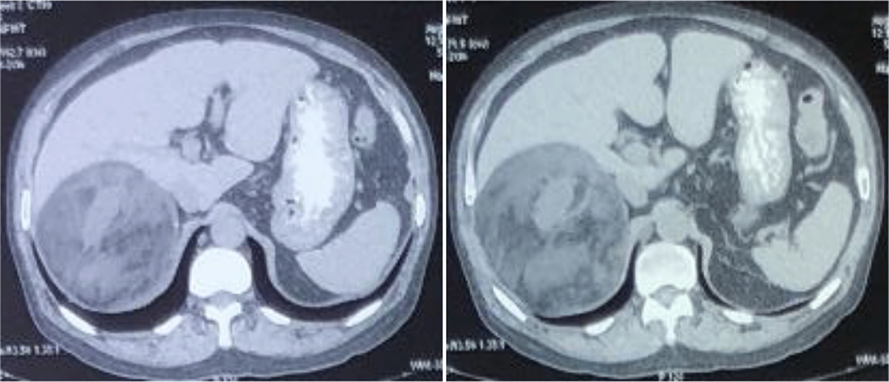
We were reporting a 48-year-old male who came to urology clonic referred from digestive surgery consultant with an intraabdominal tumor suspected with adrenal myelolipoma. The mass was felt to increase in size for the last one month. No pain on palpation, no history of hematuria, cloudy urine, or passing stone. The patient also complained of fatigue and shortness of breath. On examination, hemodynamic was within normal limits. On urological examination, ballotement was found on the right flank without any tenderness. From computed tomography scan, an upper right abdomen mass was found, pushing liver to the superior and kidney to the inferior (Figure 5). The mass sized 12.8×10×10 cm, suspected of adrenal myelolipoma.
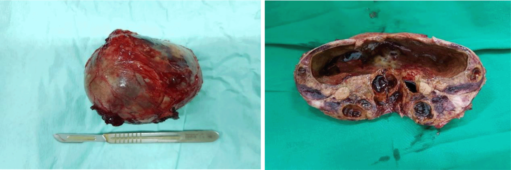
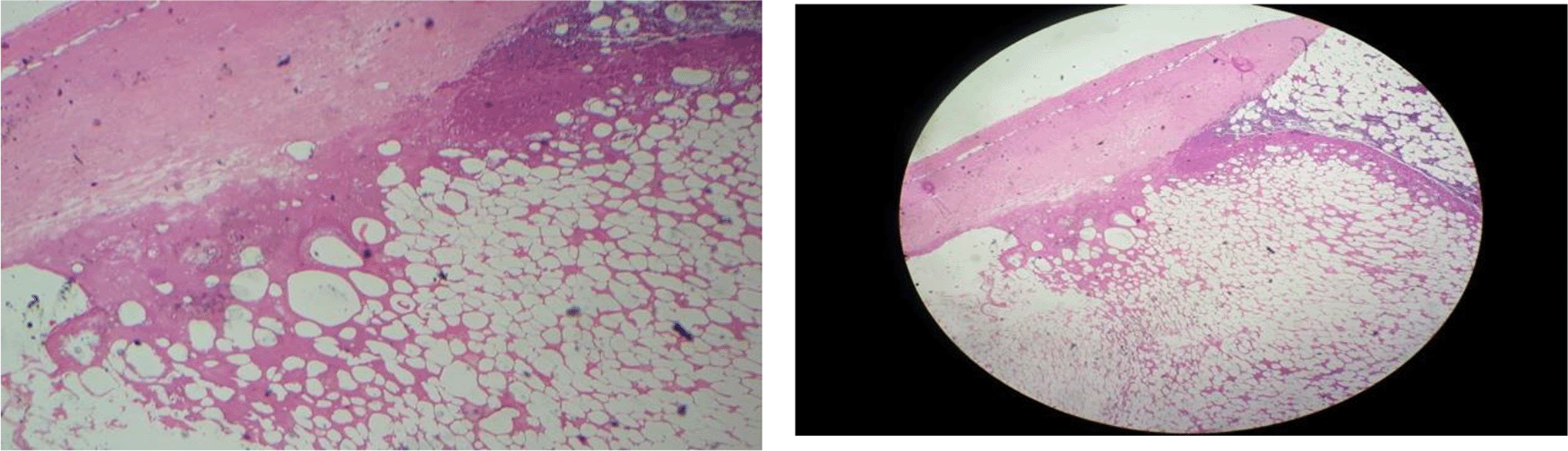
We performed a tumor removal for this patient (Figure 6). Intraoperatively, the hemodynamic remained stable. Based on this finding, we could rule out pheochromocytoma. Later, a histopathological examination revealed adrenal preparations appear to be the proliferation of mature fat cells with a round, oval, eccentric, smooth chromatin nucleus, numerous cytoplasm and clear, between groups of fat cells appear to group hematopoietic cells, at certain focus looks interstitial bleeding (Figure 7). No signs of malignancy were found in this preparation, the impression of Myelolipoma of Adrenal.
From this case series, we reported two cases of adrenal tumors, with one case of adrenal cortical adenoma and one case of pheochromocytoma. Adenomas are the most common benign tumors from the adrenal gland. From our first case, an adrenal cortical adenoma was found associated with diabetes, hypertension, and depression. This case is suggestive of a functional adrenal cortical adenoma. The glucocorticoids’ production from the adenoma could increase hypercortisolism, or known as Cushing syndrome.17
Cortisol production from benign adenomas often results from a unilateral hyperplastic mass, although a bilateral one might also happen. Systemic manifestations from hypercortisolism are central obesity, dyslipidemia, or hypertension.17 In our patient, we found hypertension without dyslipidemia or central obesity. On the other hand, one patient may come to clinical practice with a high blood glucose level, and when screened for hypercortisolism, he/she may have a subclinical Cushing syndrome.18 In our patient, we have a high blood glucose level, which becomes normal after adrenalectomy. Similar results were stated by Midorikawa and Mitchel. They found that an adrenalectomy procedure may improve glucose control together with hypertension.19,20 Cushing syndrome may also be related to depression. Labeur et al. also stated this manifestation in their study.21
Pheochromocytoma is a tumor of the catecholamine-producing cells of the adrenal medulla. The classic presenting sign of pheochromocytoma patients is paroxysmal hypertension, while the other could demonstrate persistent high blood pressure and normotensive.17 There is a classic hallmark triad of pheochromocytoma, headache, sudden episodic perspiration, and tachycardia.22 In our case, we failed to find any clinical manifestation, which has been stated earlier. Our patient has normal blood pressure without headache, perspiration, or even tachycardia. This might be similar to a study from Adler et al. They said as many as 20% of pheochromocytoma patients could present with no symptom at all.23
Intraoperatively, both of our cases showed similar symptoms with a spike in blood pressure. This manifestation has also been reported by Kakoki et al.24 However, after the adrenalectomy procedure, the patients did not sustain any elevated blood pressure. This also happened during the follow-up.
Adrenal myelolipoma is a rare, non-functional, and benign neoplasm of the adrenal gland.25 Because of the rarity, in the past adrenal myelolipomas, were found during the autopsy. Nowadays, because of radiological studies such as ultrasonography, computed tomography, and magnetic resonance imaging, incidentaloma has become more commonly found.26 The management of this tumor is usually conservative because most of them are asymptomatic. However, surgical intervention is suggested in a large tumor (larger than six cm).27 So, the management is based on the size and the symptoms of the tumor.28
We report a case series with three cases of adrenal tumor. The first case is an adrenal cortical adenoma suggestive a functional type. The second case is a pheochromocytoma without the classic hallmark. Both cases have unstable intraoperative blood pressure and have to be stabilized using several medications. During the follow-up, normotensive was achieved in both patients. The third cases have no hemodynamic disturbance in preoperative, intraoperative, and postoperative. In case of adrenal tumor, management tailoring should be based on individual patient.
| Views | Downloads | |
|---|---|---|
| F1000Research | - | - |
|
PubMed Central
Data from PMC are received and updated monthly.
|
- | - |
Provide sufficient details of any financial or non-financial competing interests to enable users to assess whether your comments might lead a reasonable person to question your impartiality. Consider the following examples, but note that this is not an exhaustive list:
Sign up for content alerts and receive a weekly or monthly email with all newly published articles
Already registered? Sign in
The email address should be the one you originally registered with F1000.
You registered with F1000 via Google, so we cannot reset your password.
To sign in, please click here.
If you still need help with your Google account password, please click here.
You registered with F1000 via Facebook, so we cannot reset your password.
To sign in, please click here.
If you still need help with your Facebook account password, please click here.
If your email address is registered with us, we will email you instructions to reset your password.
If you think you should have received this email but it has not arrived, please check your spam filters and/or contact for further assistance.
Comments on this article Comments (0)