Keywords
Gastric tuberculosis, abdominal tuberculosis, Subepithelial tumor, Tuberculous Lymphadenitis, case report
Gastric tuberculosis, abdominal tuberculosis, Subepithelial tumor, Tuberculous Lymphadenitis, case report
Abdominal tuberculosis (TBC) can affect many organs in the peritoneal cavity such as the gastrointestinal tract, peritoneum, lymph nodes, spleen, and liver. It can affect one organ or many in combination.1 Gastrointestinal TBC’s presentation is varied, depending on the site that is involved.2 Its diagnosis is especially difficult. This particularity is explained by differential diagnoses that can mimic the various manifestations of gastrointestinal TBC, including infectious and noninfectious causes.3 TBC of the stomach is the rarest form and is generally misdiagnosed because it may mimic a gastric tumor.4
Here, we report a case of mesenteric tuberculous lymphadenitis that had involved the gastric wall and mimicked a gastric submucosal tumor with no evidence of tuberculosis elsewhere.
A 52-year-old woman, with a history of hypertension, was hospitalized in our department of surgery following three months of epigastric pain and discomfort with weight loss. She had neither fever nor respiratory symptoms. Physical examination revealed mild tenderness in the upper abdomen associated with an palpable and painful epigastric mass measuring 4 cm. There was no cervical lymphadenopathy or hepatosplenomegaly, and laboratory data were normal. There were no abnormalities on the chest X-ray.
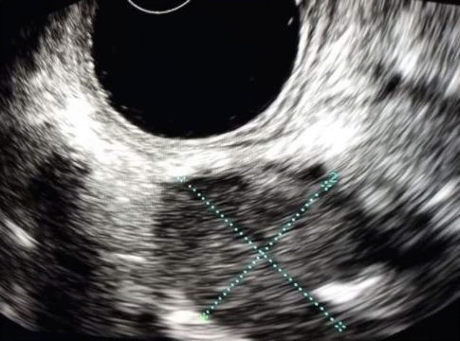
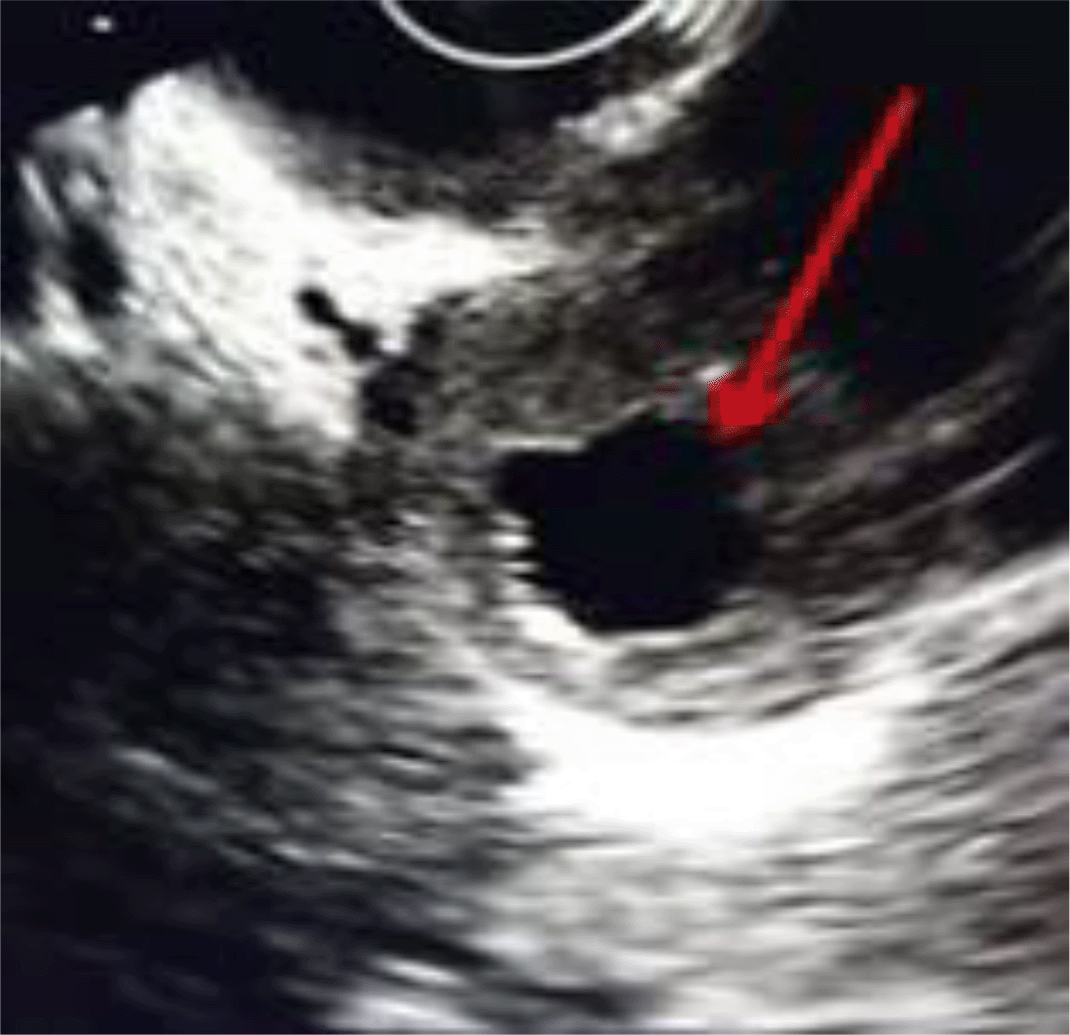
The upper endoscopy was inconclusive and showed either submucosal compression or an anterior submucosal lesion with erosive anterior gastropathy and a fistulous orifice located in the bulb (Figures 1 & 2). A biopsy was performed and concluded there was no malignancy and no evidence of tuberculosis. The endoscopic ultrasound revealed a rounded lesion approximately 30*26 mm located in the antrum. The lesion was hypoechogenic and discretely heterogeneous, not vascularized, and distant from the gastric wall whose five layers appeared of a normal aspect (Figure 3). By positioning the probe next to the bulbar fistulous orifice, it was found that there was a second lesion with a hypoechogenic center (Figure 4) with a hypoechoic fistulous path.
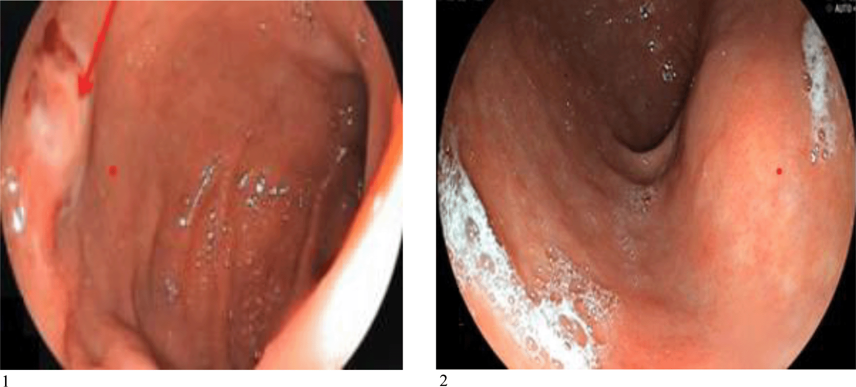
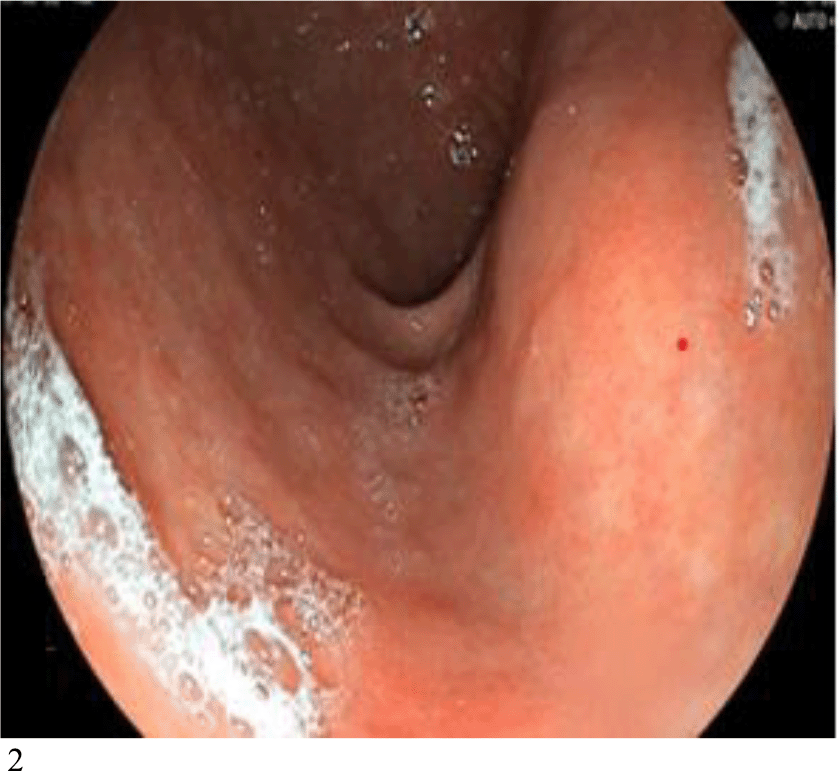
Esophagogastroduodenoscopy shows an extrinsic compression of the gastric wall and an anterior submucosal lesion, with a fistulous orifice located in the bulb (images were edited in Microsoft PowerPoint 2016 to remove patient’s scan data).
An abdominal computed tomography (CT) scan was performed and showed an exophytic, heterogenous gastric formation with an axial necrotic center measuring 44*24 mm. After injection of contrast agent evoking peritoneal carcinosis nodules, the formation was found to be 24 mm and associated with multiple tissue nodules of enhanced infra centimeter. There were hepatic hilum and coeliomesenteric lymph nodes, one of which had a necrotic center measuring 9 mm in diameter corresponding to the one described on the endoscopic ultrasound (Figures 5 & 6).


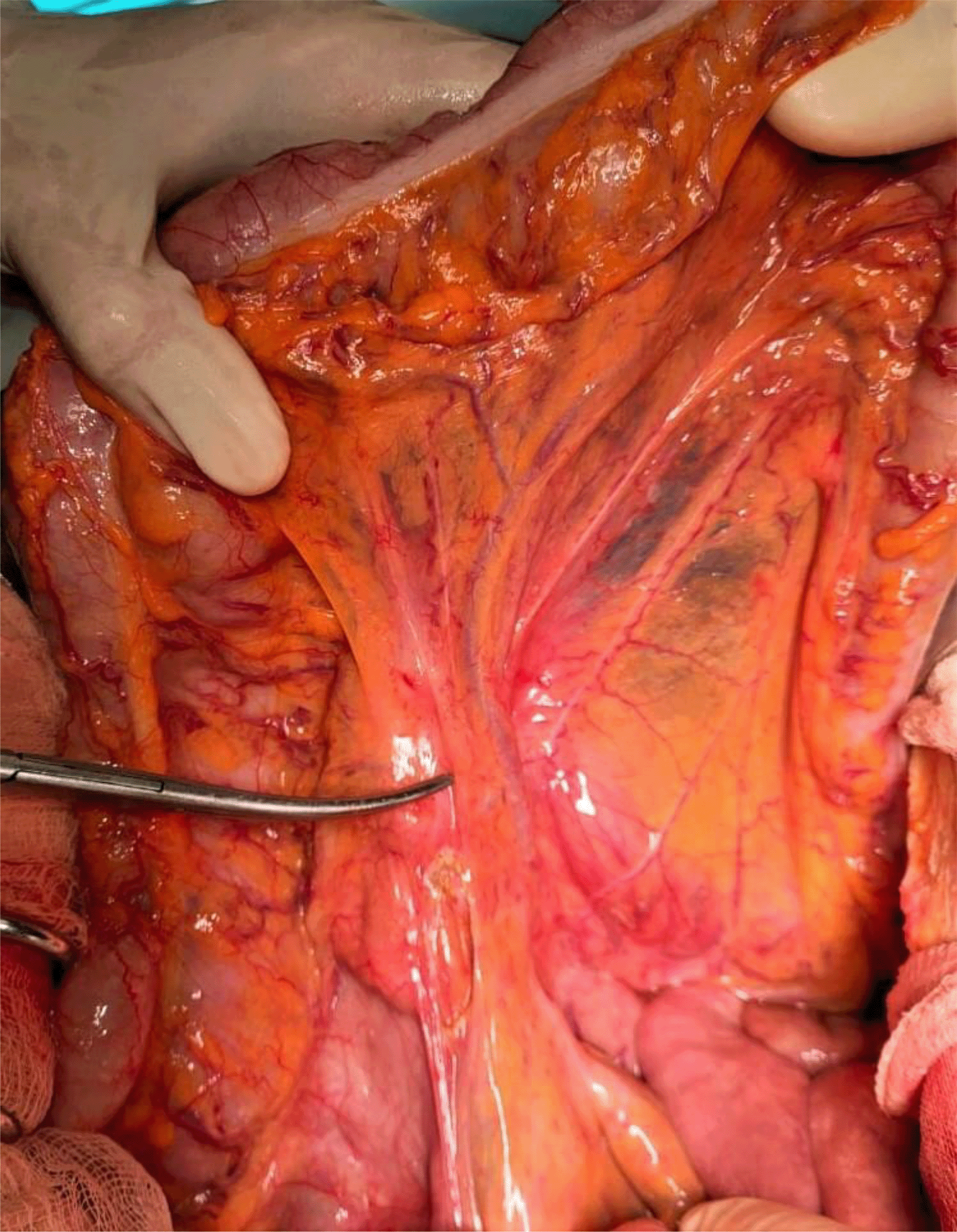
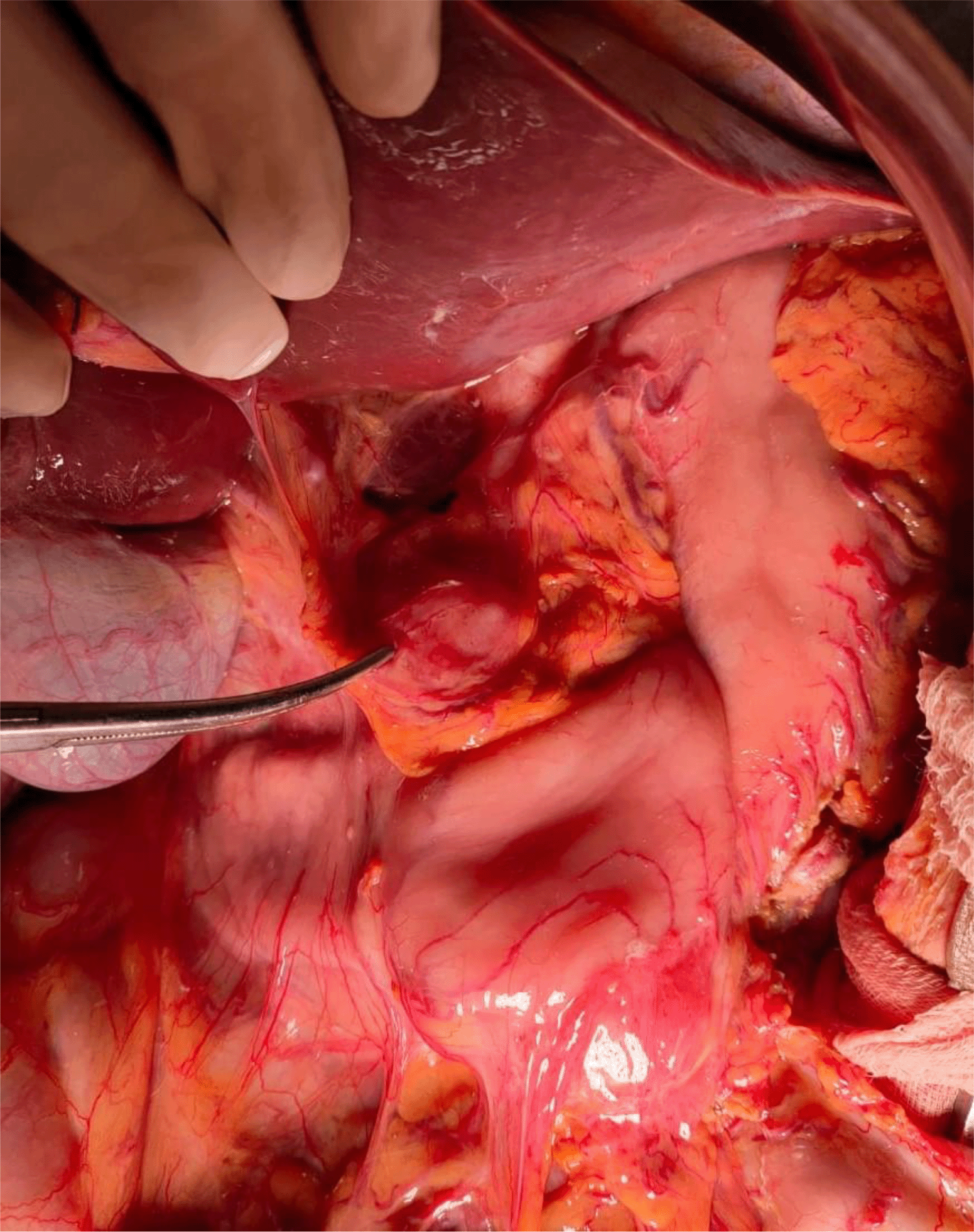
The patient underwent an exploratory laparotomy with a prediagnosis of suspected gastric cancer. The surgical findings indicated a bulky mass adjacent to the antrum with posterior development invading the transverse mesocolon (Figures 7A & 7B), associated with multiple adenopathies of the mesentery, the transverse mesocolon, and the greater omentum, and organized inflows. There was a second mass of 3 cm located in the small omentum in contact with the left gastric artery, probably corresponding to a voluminous adenopathy (Figure 7C). A biopsy was taken and sent for frozen section examination, which found numerous epithelioid and giganto-cellular granuloma and central caseous necrosis, confirming the diagnosis of tuberculous intraperitoneal lymphadenitis (Figure 8). Indeed, gastrotomy was not performed because of the benign nature of the pathology. The patient was administered anti-tuberculosis treatment and was closely monitored. A CT scan taken six months after surgery revealed a total regression of all the gastric lesions and nectrotic lymph nodes previously described, and the disease was fully controlled.

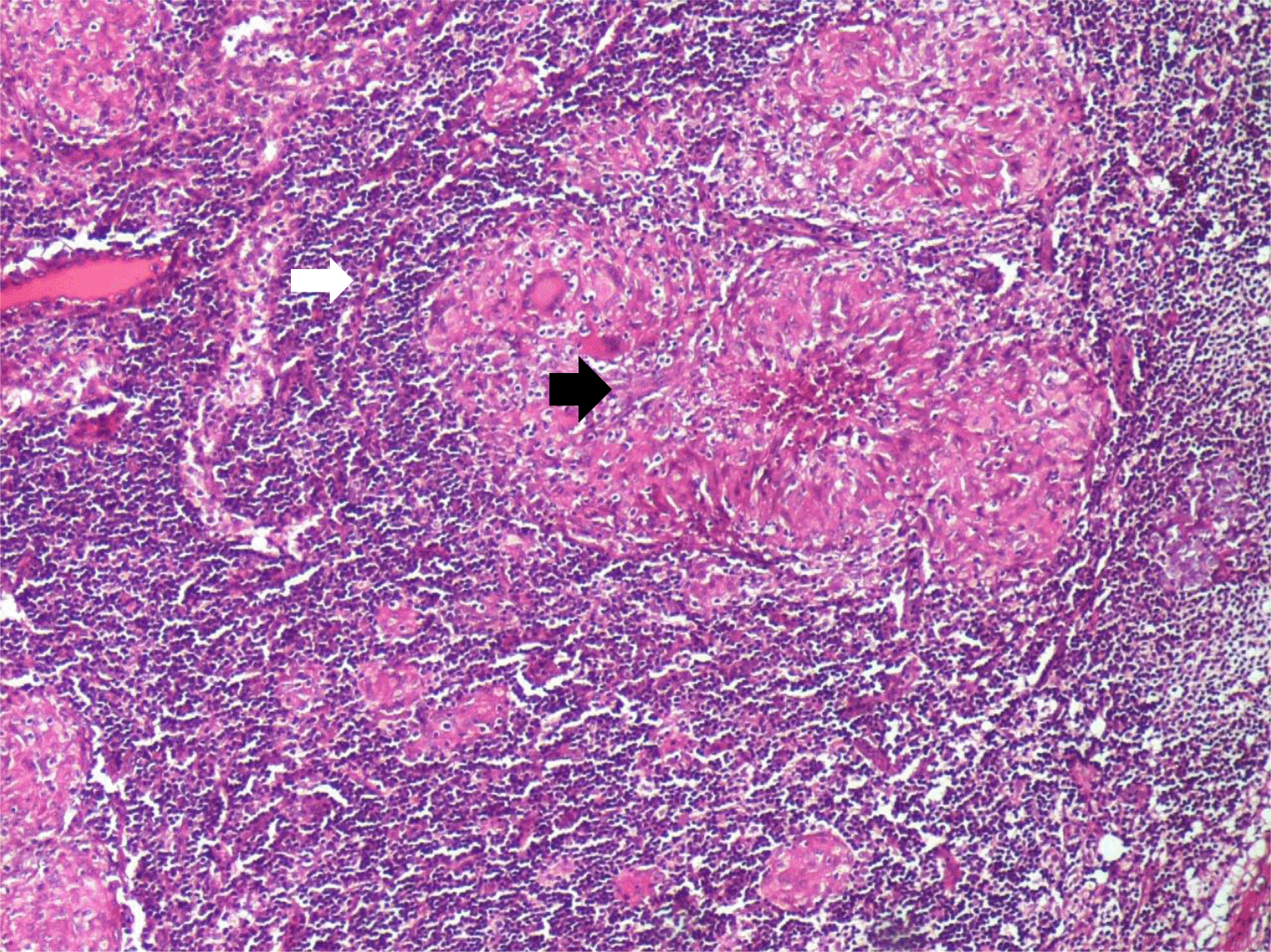
Patient’s lymph node showing numerous necrotizing and non necrotizing epithelioid and giganto-cellular granuloma. Giant cells of Langhan’s type are multinucleated (white arrow). In the center of a granuloma, necrosis is eosinophilc and granular; this is the microscopic appearance of caseous necrosis (black arrow) (HE×10)
Abdominal TBC is always misdiagnosed because of its various clinical manifestations. It is known as a great mimicker, especially when it affects abdominal organs without pulmonary infection, and malignant tumors are the most incriminating as a preoperative diagnoses.2,5 Gastric TBC is an extremely rare form whether it is a primary or secondary infection.1 Debi et al.6 explain the reasons for its rarity such as the bactericidal properties of gastric acid, the scarcity of lymphatic tissue in the gastric wall and the thick gastric mucosa in an intact stomach.
Moreover, mesenteric tuberculose lymphadenitis is an extremely rare cause of intestinal manifestations involving the gastric wall, such as in our case.1 According to the literature, only a few cases have been reported showing tuberculosis lymphadenopathy mimicking submucosal gastric tumors.
Primary gastric TBC, that does not involve any other organ, is generally located on the antrum or prepyloric region involving the duodenum. This location is explained by the presence of lymphoid follicles.1,4 There are six types of gastric tuberculosis in pathological forms: tubercular ulcers; miliary tubercles; hypertrophic tuberculosis; tuberculous pyloric stenosis; solitary tuberculoma; and tubercular lymphadenitis.2
Clinically, patients generally present with nonspecific upper abdominal pain such as epigastric pain, associated with weight loss, anorexia, and weakness.4 The majority of patients with gastric tuberculosis are often diagnosed after surgery because of the lack of specific symptoms.1
An endoscopy is helpful to diagnose this pathology, especially by biopsy results. Endoscopies can show ulcers, masses, or extrinsic compression.7 However, in our case, gastric cancer was suspected and the biopsy did not help to confirm the diagnosis. The poor yield of the biopsy is explained by the submucosal lesion that may not reveal granulomas and is difficult to obtain tissues from.7,8 Endoscopic ultrasonography is also very helpful, especially in the case of submucosal lesions or related lymph node enlargement,7 as it can differentiate between an extrinsic compression and a subepithelial gastric tumor by identifying the relationship between the lesion and the gastric wall.9 Morphologically, no specific imaging findings can help diagnose tuberculosis rather than malignancy, because there are no pathognomonic characteristics that show radiological modalities.5
Using combined radiographic and endoscopic imaging can facilitate an early diagnosis without unnecessary surgical resection. However, it is always difficult to have a final diagnosis by endoscopic biopsy, so it becomes necessary to perform a surgical biopsy using frozen section examination.8
Abdominal lymphadenitis tuberculosis presents a diagnostic challenge and a dilemma for clinicians. It may mimic a long list of differential diagnoses, such as in our case of tuberculosis lymphadenitis eroding the gastric wall. In these cases, endoscopy biopsy is the best modality to identify the pathology. Nevertheless, it could sometimes not be made preoperatively and may require surgery for diagnosis by intraoperative frozen biopsy.
All data underlying the results are available as part of the article and no additional source data are required.
| Views | Downloads | |
|---|---|---|
| F1000Research | - | - |
|
PubMed Central
Data from PMC are received and updated monthly.
|
- | - |
Provide sufficient details of any financial or non-financial competing interests to enable users to assess whether your comments might lead a reasonable person to question your impartiality. Consider the following examples, but note that this is not an exhaustive list:
Sign up for content alerts and receive a weekly or monthly email with all newly published articles
Already registered? Sign in
The email address should be the one you originally registered with F1000.
You registered with F1000 via Google, so we cannot reset your password.
To sign in, please click here.
If you still need help with your Google account password, please click here.
You registered with F1000 via Facebook, so we cannot reset your password.
To sign in, please click here.
If you still need help with your Facebook account password, please click here.
If your email address is registered with us, we will email you instructions to reset your password.
If you think you should have received this email but it has not arrived, please check your spam filters and/or contact for further assistance.
Comments on this article Comments (0)