Keywords
adrenal malignancy, adrenal oncocytoma, adrenal mass, Lin–Weiss–Bisceglia criteria
adrenal malignancy, adrenal oncocytoma, adrenal mass, Lin–Weiss–Bisceglia criteria
With the increasing use of abdominal computed tomography (CT), adrenal masses are common incidental findings, most of which are benign and non-functional (i.e., not metabolically active), hence also known clinically as “incidentalomas”. The estimated overall incidence of adrenal tumors worldwide is 10%.1 However, primary malignant adrenal cancer, namely adrenocortical carcinoma, is rare, with an annual incidence of fewer than two cases per million.2 The oncocytic variant of this malignancy is even rarer, with only a handful of cases documented.3,4
Diagnosing Adrenal Oncocytic Neoplasm is challenging. Adrenal oncocytic neoplasm is extremely rare. A variant of the malignant one is even rarer. Thus, it is really hard to find a reference for diagnosing and treating the case. Radiologically, the adrenal lesion was evaluated from four things: shape, size, density, and heterogeneity. Sometimes, those parameters could show an inconsistent finding which confused both radiologist and surgeon. Histopathological analysis of Adrenal Oncocytic Neoplasm also should be conducted prudently. Since oncocytes have unique characteristics (low proportion of clear cells, diffuse tumor architecture, and high nuclear grade), the application of Weiss criteria would lead to a malignancy diagnosis. Therefore, Adrenal Oncocytic Neoplasm histopathological specimen should be assessed with Lin-Weiss-Bisceglia criteria instead of Weiss criteria. The primary treatment of Adrenal Oncocytic Neoplasm is adrenalectomy. Some references suggested using radiotherapy and mitotane for palliative therapy. Since the rarity of this case, little is known about this tumor behavior in the long term. Therefore, long term follow-up post-surgical procedure is paramount in this patient.
As with other types of cancer, treatment of malignant adrenal lesions necessitates a multidisciplinary approach involving urologic, medical, and radiation oncologists, along with endocrinologists, pathologists, and radiologists. Surgical resection of the primary tumor is the only treatment modality offering a cure, while the roles of chemotherapy and radiotherapy are not well defined in the literature for these variants of a tumor.5–7 In this paper, we present a case report of an adult male with malignant Adrenocortical Oncocytic Neoplasm treated with open right adrenalectomy. This paper is written according to the CARE guideline.8
A 61-year-old male was referred to our department after complaining of progressively worsening right upper quadrant abdominal pain for two years prior to admission. The patient denied any symptoms of palpitations, diaphoresis, fatigue, and headache, although he had been experiencing slight dizziness. He also denied any history of dysuria and hematuria. He had a history of laparoscopic cholecystectomy approximately four years ago. His physical examination was unremarkable; no palpable mass was found at clinical examination. Similarly, his laboratory test results were unremarkable.
About two weeks before he was referred to our center, he had undergone a whole abdominal contrast CT-scan examination, which revealed an inhomogeneous mass with a diameter of 7.695 cm in the right suprarenal region. The pre-contrast image showed that the adrenal lesion has 36 HU. Thus, we conducted a washout evaluation, and the absolute washout result was 40%, and the relative washout was 21%, suggesting a malignant adrenal lesion. Furthermore, the lesion shape and size of the adrenal lesion also leads to malignancy instead of a benign lesion. The author(s) also found that the lesion is heterogeneous since it has a region with lower density, suggesting central necrosis (Figure 1). Additionally, it also showed a small liver cyst measuring 1.16 cm in the right lobe of the liver; the kidneys, bladder, and prostate gland were all normal in appearance. The patient was diagnosed with a right adrenal mass, and he was recommended to undergo surgery to remove the tumor.
Elective open adrenalectomy was performed. The right upper abdomen was opened with a Chevron incision. When the intestines were pushed to the medial, the omentum was adhered to the liver due to the prior cholecystectomy surgery. Adhesiolysis was conducted, and a right retroperitoneal mass was observed. It shoved the right lobe of the liver from the posterior side. There is no infiltration from the mass to the liver or duodenum. The right colon was mobilized, and the Kocher maneuver was performed. After that, the white line was opened. The right kidney and right adrenal gland were observed. The right adrenal gland was then released from the surrounding structure. Surrounding vessels were identified, and the right adrenal artery and vein were ligated and cut. The right adrenal gland can be removed in toto, and the specimen was sent to the Pathological Anatomy department to be analyzed. The bleeding was controlled, and the wound was washed with sterile water. The wound then was closed layer by layer.
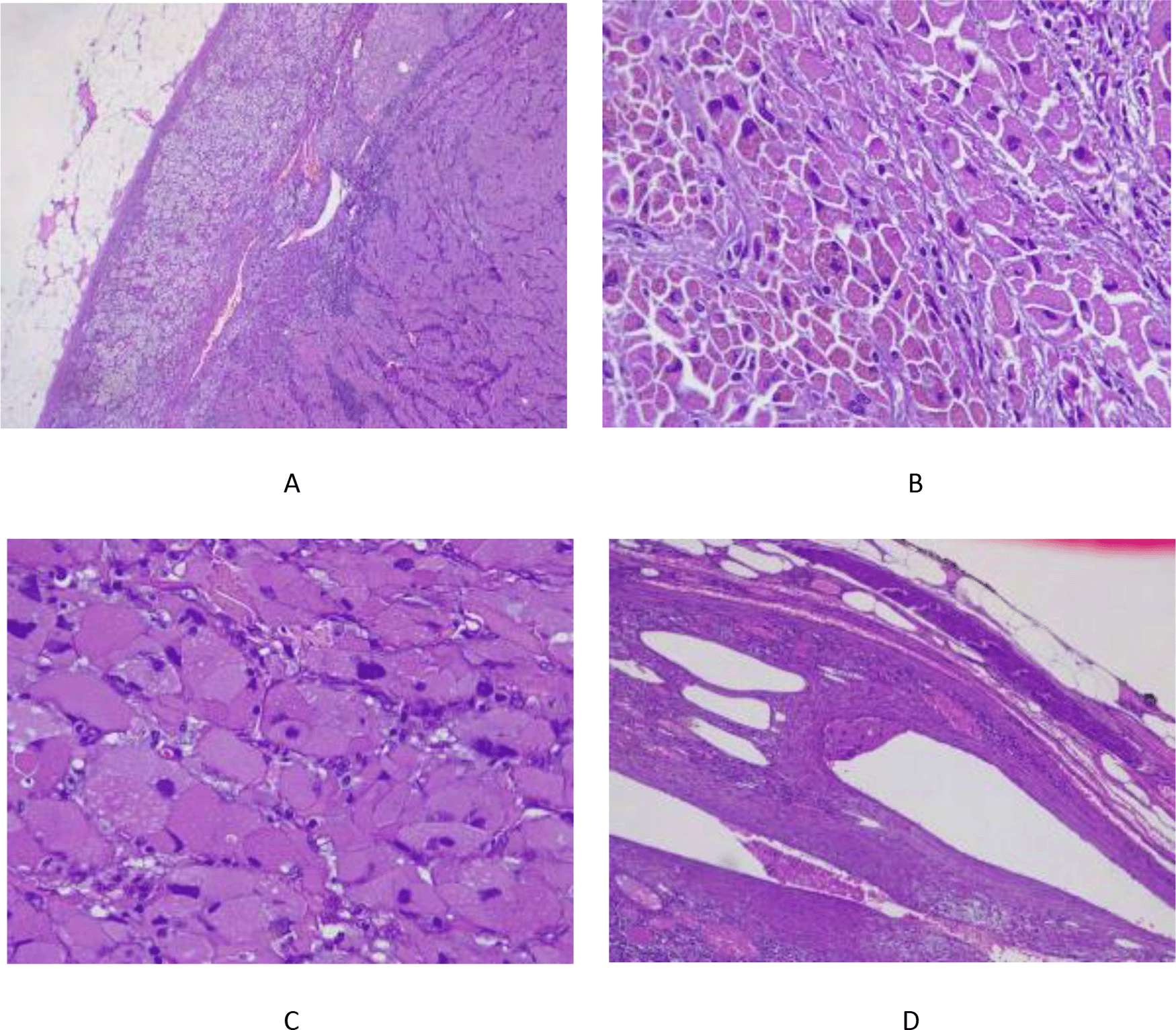
Gross pathologic examination revealed an adrenal tissue measuring 8 cm × 6 cm × 5 cm with a solid, yellowish homogenous, multilobed tumor mass with no apparent infiltration into the adrenal capsule (Figure 2). The histopathologic analysis showed tumor cells with pleomorphic, round/oval shaped, and hyperchromatic, partly vesicular nuclei with clear nucleoli, eosinophilic, granulated, and partly contained brown-pigmented cytoplasm; the mitotic rate was found <5 per 50 high-power fields. As illustrated in Figure 3A, the tumor has lobes separated by septa. It did not breach the adrenal capsule. We can even see a remnant of normal adrenal tissue near the capsule. In 400 times magnification, the author(s) observed tumor cells with pleomorphic nuclei and granulated eosinophilic cytoplasm, a unique feature of oncocytes (Figure 3B and 3C). Figure 3D depicted a tumor invasion into the vascular bed, which highly suggestive of malignant oncocytic lesion according to Lin-Weiss Bisceglia criteria. Based on the Lin-Weiss-Bisceglia (WHO) criteria, it can be concluded that this mass is an Oncocytic Adrenal Cortical Neoplasm, with favor towards malignancy because the tumor has invaded the vein.

(A) Tumor mass and adrenal tissue. (B) Tumor cells with pleomorphic nuclei, round/oval, hyperchromatic with eosinophilic cytoplasm, granular, and brown pigmented. (C) Oncocytic features of the adrenal mass. (D) Tumor invades the vascular bed.
The lesion was subsequently classified as T2, without nodal involvement (N0) nor distant metastasis (M0). Postoperatively, the patient recovered well, and he was discharged within three days of observation. The patient then later had PET-Scan Evaluation six weeks after being discharged from the hospital. The PET-Scan result was remarkable as well. There was no residual mass or pathological metabolic activity on the tumor bed and metastic sign. The patient has given his consent for this publication.
Adrenocortical oncocytic neoplasms, known as adrenocortical oncocytic carcinomas, are rare histologic variants of adrenocortical tumors. As to date, there are less than 200 malignant oncocytic adrenal cases that have been reported.9 Adrenal oncocytomas are usually nonfunctional and benign in nature.3 Some case series have reported that about 20% of these tumors were malignant,3,10 and endocrine abnormalities were found in up to 25% of benign and 30% of borderline/malignant adrenocortical oncocytic neoplasms,10 compared with 15% of conventional adrenocortical adenomas and 60% of conventional adrenocortical carcinomas.11,12
The pathogenesis of adrenocortical oncocytic neoplasms is poorly understood, owing to the rarity of these tumors; hence, the most significant practical concern is related to their poorly defined biologic behavior.3 While the diagnosis is straightforward in obvious malignant lesions, the clinical outcome for benign oncocytomas is more difficult to determine.3 Additionally, as is the case for endocrine neoplasms, the lesion’s size and/or function determines treatment options and prognosis.9
The Weiss criteria were developed to predict adrenal neoplasms' biologic behaviour and distinguish benign from malignant adrenal lesions (i.e., adrenocortical adenomas vs adrenocortical carcinomas). Using several pathologic features, the Weiss system can sensitively and specifically identify malignant adrenal neoplasms.13–15 However, unlike conventional adrenocortical tumors, adrenocortical oncocytic neoplasms are characterized by a low proportion of clear cells, diffuse tumor architecture, and high nuclear grade; thus, the application of Weiss criteria tends to classify these tumors as malignant adrenocortical carcinomas.3,10
For adrenocortical oncocytic neoplasms, the Lin–Weiss–Bisceglia criteria are used instead.3 It discarded three criteria from the Weiss system and rewrote them as “definitional criteria” since these characteristics are universally present in all adrenocortical oncocytic neoplasms. The Lin–Weiss–Bisceglia criteria entails the criteria of mitotic rate, atypical mitoses, and venous invasion, which are considered as “major” criteria, in addition to tumor size, necrosis, capsular invasion, and sinusoidal invasion, which are considered as “minor criteria”, from the original Weiss system. The presence of one of the major criteria is an indication of malignancy, while the presence of one of the minor criteria without any major criteria signifies uncertain malignant potential.3,16 The absence of both major and minor criteria reflects neoplasms of benign behavior. Other than the morphologic criteria, immunohistochemical analysis also aids the recognition of these rare tumors. Based on the landmark paper by Bisceglia and colleagues,3 immunohistochemically, these tumors were reactive for mitochondrial antigen mES-13. In addition, Wong and colleagues10 also found that these tumors were positive for vimentin, synaptophysin, and melan A stainings. In this case, we only found one criterion, invasion into vascular bed (Figure 3D). Thus, a thorough histopathological examination should be evaluated in the future, which might need more sections.
Open adrenalectomy is the treatment of choice for surgically resectable malignant adrenocortical oncocytic neoplasms.9 Laparoscopic adrenalectomy may also be an option, but there are concerns regarding whether this minimally invasive approach can permit satisfactory oncologic resection with enough tumor-free margin.17 Aside from surgery, which is the mainstay of treatment for solid malignant lesions, the treatment of adrenocortical oncocytic tumors is not well described in the literature, perhaps due to the rarity of these tumors.5 However, in practice, the treatment approach for malignant adrenocortical oncocytic neoplasm appears to follow the same general approach as other adrenal cancer. The standard chemotherapeutic regimens should include mitotane and other cytotoxic agents commonly used for adrenocortical carcinomas for advanced disease.9 El-Naggar and colleagues18 reported the use of adjuvant mitotane treatment in one case of adrenocortical oncocytic carcinoma with invasion of the inferior vena cava and the liver after radical right adrenalectomy, en bloc nephrectomy, and partial hepatectomy. Radiotherapy may be utilized for the treatment of malignant adrenocortical oncocytic neoplasm in both adjuvant and palliative settings.19 Juliano and colleagues,20 in their report, suggested that metastasectomy and radiotherapy may be beneficial for treating local recurrence. In this patient, we evaluated the post-operative result with PET-Scan. The result was remarkable, with no residual mass found in the tumor bed. Thus, the patient was not given any chemotherapy or radiotherapy due to this result.
The overall prognosis of adrenocortical oncocytic neoplasms is uncertain but seems to be slightly more favorable than that of conventional adrenocortical carcinomas.21 Wong and colleagues reported a median survival rate of 58 months and a 5-year survival rate of 19.5%; in contrast, the median survival rate for conventional adrenocortical carcinomas (all stages) ranges from 14 to 32 months.22,23 Further molecular biology and clinical studies shall provide a better understanding of the biological behavior of these tumors, as well as the optimal therapeutic approaches, outcomes, and prognosis. In our case, the operation was successful, and the PET-Scan evaluation showed no residual tumor. Therefore, chemotherapy and radiotherapy are not necessary to be given to this patient. There is no adverse or unanticipated event reported. The patient was scheduled for follow-up two months after PET-Scan to monitor his condition and recovery.
We reported a rare malignant oncocytic adrenal case which only based on one histopathology criteria. Therefore, a thorough histopathological examination should be implemented to evaluate the malignant criteria of adrenal case. The oncocytic variants of adrenal tumors are rare, with only a handful of cases reported in the literature. The pathogenesis of adrenocortical oncocytic neoplasms is poorly understood, and these tumors exhibit unique biologic behavior compared with their conventional counterparts, hence the use of different pathological criteria. While most of these tumors are benign and nonfunctional, up to 20% of all adrenocortical oncocytomas are malignant. Surgery is considered the mainstay of treatment for adrenocortical oncocytic neoplasms. The roles of chemotherapy and radiotherapy in the management of malignant adrenocortical oncocytic tumors are less clear, but they appear to be beneficial in both adjuvant and palliative settings. The prognosis of malignant adrenocortical oncocytic neoplasms is likely more favorable than that of conventional adrenocortical carcinomas.
Written informed consent for publication of the patient’s clinical details and clinical images was obtained from the patient. The patient has given his consent for this publication.
All data underlying the results are available as part of the article and no additional source data are required.
The author(s) would like to thank Dr. Edwin and Dr. Jessica for helping the author(s) providing the radiological and histopathological files, which were necessary for writing this manuscript.
| Views | Downloads | |
|---|---|---|
| F1000Research | - | - |
|
PubMed Central
Data from PMC are received and updated monthly.
|
- | - |
Provide sufficient details of any financial or non-financial competing interests to enable users to assess whether your comments might lead a reasonable person to question your impartiality. Consider the following examples, but note that this is not an exhaustive list:
Sign up for content alerts and receive a weekly or monthly email with all newly published articles
Already registered? Sign in
The email address should be the one you originally registered with F1000.
You registered with F1000 via Google, so we cannot reset your password.
To sign in, please click here.
If you still need help with your Google account password, please click here.
You registered with F1000 via Facebook, so we cannot reset your password.
To sign in, please click here.
If you still need help with your Facebook account password, please click here.
If your email address is registered with us, we will email you instructions to reset your password.
If you think you should have received this email but it has not arrived, please check your spam filters and/or contact for further assistance.
Comments on this article Comments (0)