Keywords
atrioventricular block, primary hyperoxaluria, syncope, end-stage renal failure, heart block
atrioventricular block, primary hyperoxaluria, syncope, end-stage renal failure, heart block
Primary hyperoxaluria (PH) type 1 is a scarce hereditary metabolic malady in which an increased production of oxalic acid results in hyperoxalemia and an accumulation of calcium oxalate in different body organs, including the heart.1 Oxalosis involving the myocardium can lead to congestive heart failure, arrhythmias and conduction disturbances.2 We report the case of a 42-year-old-woman followed for PH over 15 years who was admitted to our cardiology department for a paroxysmal syncopal atrioventricular block (AVB). Heart block in patients with primary oxalosis is exceptional, indeed less than eight reports have been previously reported in the medical literature.
A 42-year-old Tunisian housewife was referred eighteen months ago to our cardiology department for repetitive episodes of syncope over two weeks. The patient had PH1 diagnosed at the age of 27 by a genetic study that revealed a c.33-34insC mutation; she had a grandfather with the same disease. Over time, she developed progressive renal failure, and five years earlier, she had undergone a left partial nephrectomy for nephrocalcinosis. Nevertheless, she developed total chronic renal failure and had recently begun hemodialysis via a left humerocephlic fistula.
Upon admission in March 2020, physical examination showed a mucocutaneous pallor. Blood pressure was 100/60mmHg, and heart rate was 70/min. Cardiac auscultation found a 2/6-degree systolic aortic murmur. She had no signs of heart failure. Electrocardiogram (EKG) performed in the emergency room showed a regular sinus rhythm at 75 beats per minute (bpm), a complete left bundle branch block (LBBB), and a first-degree AVB (Figure 1). Laboratory tests showed a normochromic normocytic anemia at 5.9 g/dl, urea level at 37.7 mmol/l, creatinine level at 1771 micromol/l, a normal potassium serum level at 4.3 mEq/l, and a low calcium serum level at 2.08 mmol/l.

The patient’s plasma oxalate level was 98.5 umol/L. Urinary crystallographic exam showed the presence of calcium oxalate crystals in the urine of characteristic icosahedral shape (Figure 2). Plain abdominal X-ray showed images of nephrocalcinosis of the left kidney (Figure 3).


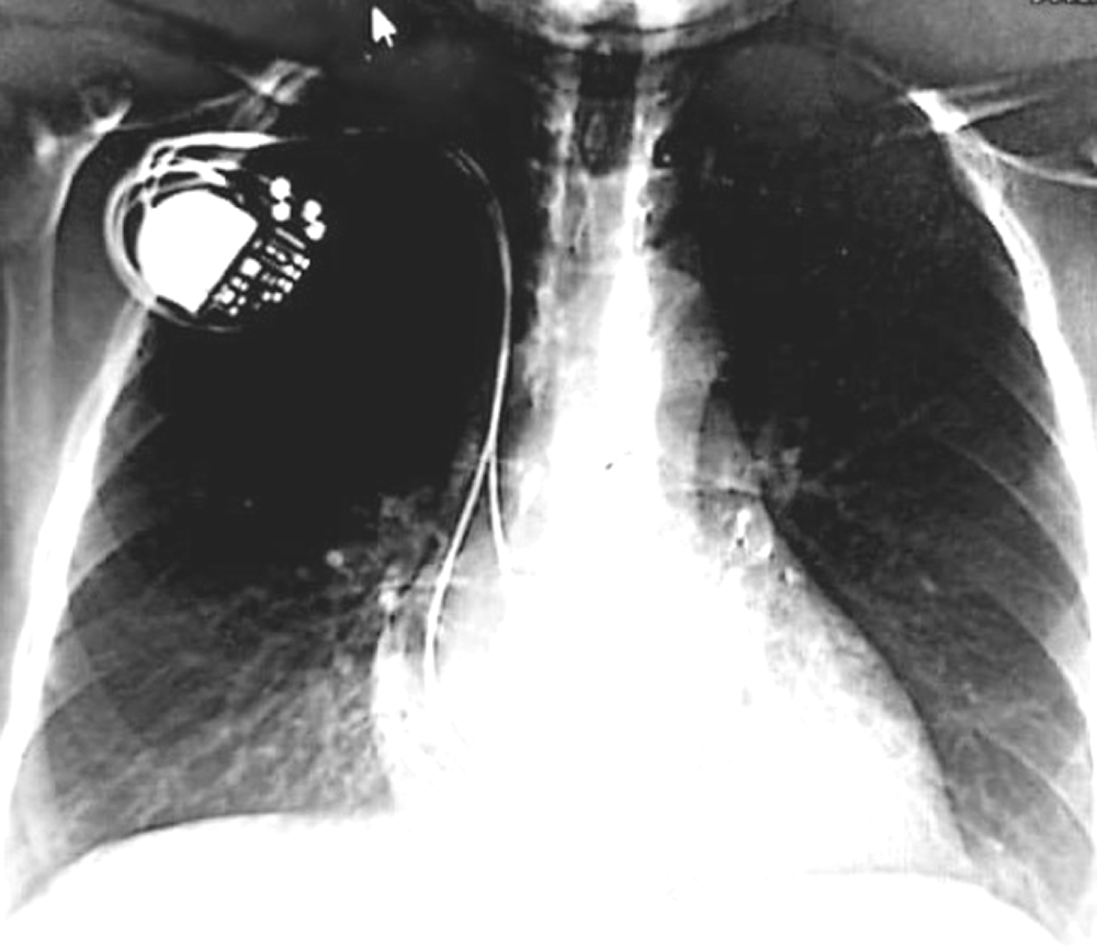
During monitoring in the intensive care unit, the patient presented a further episode of syncope with an EKG (Figure 4) showing a prolonged ventricular asystole (Figure 4A) with second degree AVB 2/1 (Figure 4B) and a ventricular rate of 55 bpm followed by a complete AVB (Figure 4C) with a ventricular rate of 50 bpm. First, the patient received an isoprenaline infusion (1 mL/hour =0.5 microgram/kg/minute) under EKG control, but the heart rate did not accelerate. Within minutes, repeated episodes of syncope imposed the urgent placement of a temporary cardiac pacing lead. Transthoracic echocardiography showed increased left ventricular mass index and diastolic dysfunction. The patient was implanted with a dual-chamber pacemaker (Figure 5) with a good outcome. The patient was discharged three days later.

After one-year follow-up in April 2020, no episodes of syncope were reported. The patient is still waiting for a kidney transplant.
Primary hyperoxaluria type 1 (PH1) is an autosomal recessive inherited metabolic disorder related to the deficiency of alanine glyoxylate aminotransferase, a peroxisomal liver enzyme that allows the transformation of glyoxylate into glycine.1 This deficiency leads to oxalate overproduction by the liver, followed by urine filtration and complexation with calcium to form massive calcium-oxalate nephrolithiasis potentially leading to chronic renal failure (nephrocalcinosis).1 Once end-stage renal failure develops, calcium oxalate is deposited in other organs because of high plasma oxalate concentrations, which exceed the saturation threshold for calcium oxalate.1
Cardiac involvement in PH is rarely described; medical literature is limited to case reports and small case series. Mookadam and colleagues, from the team of the Mayo Clinic Rochester, published in 2010, is the first and only systematic study describing cardiac abnormalities in PH. They reviewed a dataset of 33 patients with primary hyperoxaluria.2 The mean age at the time of cardiac manifestation was 40 years.2 Cardiac anomalies are associated with an alteration in renal function and appear to correlate with the plasma oxalate level.2 Of the patients with cardiac findings, 78% had a history of end-stage renal failure.2 In our case, the oxalate plasma level was much higher. Mookadam, also describes this finding in his study.2 The plasma oxalate level was 79.4 μmol/L in those with cardiac findings, compared to 7.3 μmol/L in those without.2
Conduction deficits and heart block have previously been reported in patients with primary oxalosis.3–7 Massie et al.,4 reported the first case of cardiac electrophysiologic abnormalities due to oxalate infiltration in a 27-year-old man. In that case, oxalate was described to infiltrate the sinoatrial node and to approach the atrioventricular node leading to numerous electrophysiological abnormalities. This finding is also shared by other authors.5–7 Conduction blocks are reported in 13.2% of patients in the Mayo Clinic study.2
Myocardial invasive transjugular biopsy remains the gold standard for the diagnosis of cardiac involvement; but it is not routinely used and does pose a definable risk.2
Two reports in the literature have performed a histologic study of the conduction system, they showed extensive crystal deposits all around the sinoatrial node, atrial myocardium, atrial preferential pathways, common bundle, and both bundle branches associated to secondary degenerative and fibrotic changes.3,4 In addition, the arteries of the atrioventricular and sino-atrial nodes exhibited mural crystals with a narrowing of the lumens.3
Although ventricular pacing may avoid death from complete heart block, it is improbable to prolong life expectancy in oxalosis patients once visceral involvement becomes important.4 Dialysis is supposed to temporarily remove oxalate, but it cannot compensate for the excessive oxalate production rate, thus making it difficult to excrete calcium oxalate in the urine and eventually increasing the blood oxalate level.8
Large left ventricular mass index, impaired left ventricular and right ventricular function, left atrium enlargement, diastolic dysfunction, increased wall thickness suggestive of myocardial dissemination, and rhythm abnormalities are probably due to calcium oxalate deposits in cardiac tissue.2
While kidney transplantation corrects kidney failure, liver transplantation is the only solution for definitive correction of the metabolic defect in PH 1. Combined hepatorenal transplantation is the only therapeutic alternative to dialysis,1 which, unfortunately, is not yet practiced in Tunisia. Cardiac manifestations can be fatal; however, they are reversed after successful liver transplantation.9
The case, which we have presented here, has been limited by the short follow up of the patient. However, it demonstrates that in front of any syncopal signs, particularly in patients with primary hyperoxaluria, monitoring in a cardiological resuscitation unit can be lifesaving.
Oxalate deposits in cardiac tissue compromise prognoses as they can lead to potentially fatal complications. This case demonstrates the need for regular and careful monitoring of cardiac status in patients treated for primary oxalosis, especially when renal function is impaired. Because oxalate cannot be sufficiently removed by hemodialysis, early hepato-renal transplantation is required to improve the chances of survival in these patients.
| Views | Downloads | |
|---|---|---|
| F1000Research | - | - |
|
PubMed Central
Data from PMC are received and updated monthly.
|
- | - |
Provide sufficient details of any financial or non-financial competing interests to enable users to assess whether your comments might lead a reasonable person to question your impartiality. Consider the following examples, but note that this is not an exhaustive list:
Sign up for content alerts and receive a weekly or monthly email with all newly published articles
Already registered? Sign in
The email address should be the one you originally registered with F1000.
You registered with F1000 via Google, so we cannot reset your password.
To sign in, please click here.
If you still need help with your Google account password, please click here.
You registered with F1000 via Facebook, so we cannot reset your password.
To sign in, please click here.
If you still need help with your Facebook account password, please click here.
If your email address is registered with us, we will email you instructions to reset your password.
If you think you should have received this email but it has not arrived, please check your spam filters and/or contact for further assistance.
Comments on this article Comments (0)