Keywords
large gap bone defect, transposition fibular graft reconstruction, inlay and onlay technique, fibula pro tibia, case report
large gap bone defect, transposition fibular graft reconstruction, inlay and onlay technique, fibula pro tibia, case report
Large segmental bone defects of the tibia are challenging for both surgeon and patient.1,2 Segmental bone defects are most commonly caused by fractures from high energy trauma, osteomyelitis, benign or malignant bone tumor surgery, septic nonunion, and congenital abnormality.2–4
A free vascularized fibula graft is commonly used because it is mechanically strong, retains the intrinsic blood supply, osteogenic, and can be used for large bone defects in various places.4,5 However, there are certain drawbacks of free vascularized fibula grafts, such as donor side morbidity (unaffected limb), peroneal nerve injury, and the surgery necessitates a microsurgery technique, thus requiring a long operating time.2,3 In 1884, Hanh described an alternative technique using the ipsilateral vascularized fibula as a graft for a 12 cm segmental tibia defect due to chronic osteomyelitis in an eight-year-old male. Instead of cutting the fibula and performing a reanastomosis at a distance, Hanh simply transposed the fibula with his pedicle into the tibial defect.3,4,6
Ipsilateral vascularized fibula transfer (IVFT) allows the surgeon to transfer the fibula to the tibia as a complete graft without disrupting soft tissue attachment, blood supply and without needing microsurgical techniques, resulting in bone healing at both ends of the defect.1,3,4
In this case, we transposed the ipsilateral fibular with inlay and onlay technique using a locking plate and screw into the tibia gap defect in a one-stage procedure followed with serial radiographic X-ray evaluations and functional outcome evaluation using the Lower Extremity Functional Scale (LEFS) scoring system.7 This report has followed the CARE and Surgical CAse REport (SCARE) checklist and guidelines.8 In addition, written informed consent was obtained from our patient for publication of their data and clinical images.
A 20-year-old male came to our emergency department at RSUD Dr. Saiful Anwar Malang Indonesia with an open fracture grade III B of his left lower leg (Figure 1). The cause of the injury was a high-speed motorcycle collision. The patient did not consume any routine medication and did not have any other illness. We performed debridement, serial irrigation and applied an external fixation device on the affected leg (Figure 2). After regular wound care for ten months, the soft tissue condition improved, but we found a nonunion of the tibia with a large defect of approximately 7.5 cm (Figures 3 and 4). We planned to perform external fixation removal with a reconstruction of the bone defect using an ipsilateral transposition fibular graft with a combined inlay and onlay technique using a locking plate and screw fixation (fibula pro tibia technique).

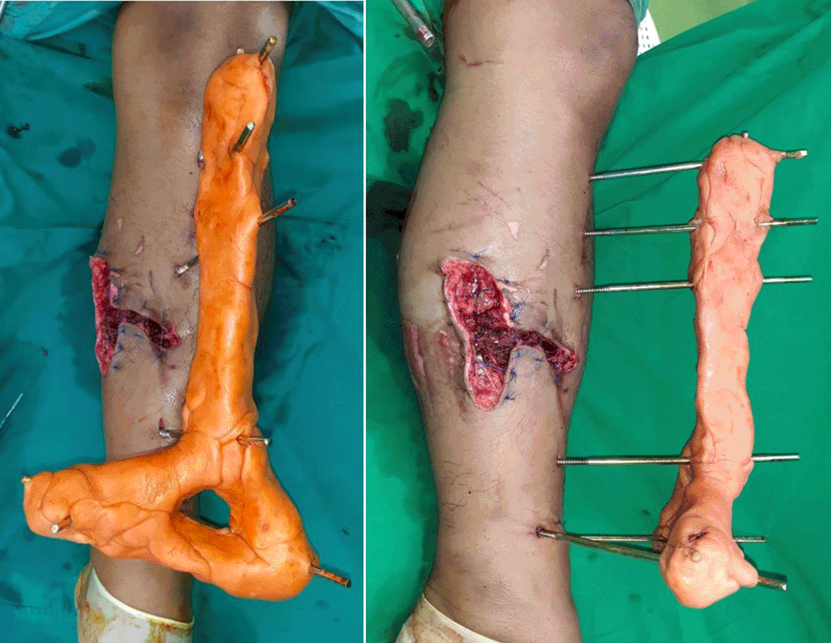
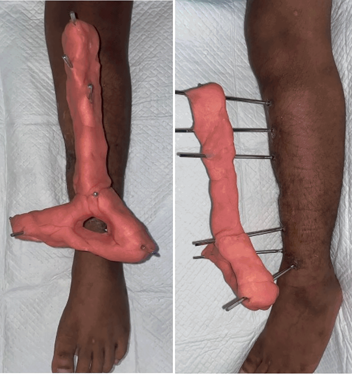

Before the operation, we did a thorough preoperative evaluation that included measuring the extent of the bone defect and assessing the quality of the adjacent soft tissue and joints. A thorough excision of all avascular bone back to bleeding tissue was performed through an anterolateral approach. The peroneal nerve and its branches were identified and protected. The fibula is dissected from its surrounding soft tissue, keeping muscles and periosteum to protect the periosteal vascularization. The dissected fibula was moved to the posterior interosseus membrane of the tibial defect with the preserved vascularization without tension on the soft tissues. Then, the graft was fixed proximally and distally with a locking plate to improve stability. Meticulous care was taken during the transpose to avoid kinking or stretching of the vascularization. In addition, we performed an onlay technique using an ipsilateral avascular fibular graft fixated with screws to provide more mechanical strength and minimize the gap defect between fracture fragments, therefore enhancing the union rate of the bone (Figures 5 and 6). To ensure ankle stability, at least 10 cm of the distal fibula must be preserved. After receiving intravenous antibiotics for one week, the patient was allowed to go home. His left leg was braced and weight-bearing was prohibited until the vascularized bone graft healed and, periodically, the patient was followed up clinically and radiographically (Figure 7). No infections occurred in the four-month postoperative evaluation, and progressive callus formation with extending ossification along the periosteal tissue was seen in serial radiographic X-ray evaluations. Clinically, the LEFS score was 62 (good), there was no leg length discrepancy, and the union sign showed as the patient could achieve full range of movement (ROM) and walk with crutches without pain.


(A, B) One month. (C, D) Three months. (E, F) Four months.
Large segmental tibial defects have been treated successfully using a variety of procedures, including autogenous corticocancellous bone grafting, tibiofibular synostosis, ipsilateral vascularized fibula transfer (IVFT), allograft tibial reconstruction, free vascularized fibula transfer, and bone transport accompanied with Ilizarov technique.1–3 Proximal transtibial amputation is one of the options of treatment, but amputation should be avoided if the foot and ankle vascularization is normal.1 In addition, for most patients, shortening of limbs or amputation is an unacceptable condition.3,9
Bone transfer or corticocancellous bone grafting can be used to treat shorter defects of up to 5 cm. In contrast, larger tibial defects usually need a more complicated repair procedure, such as bone transfer or a free vascularized fibula transfer.3,9 The optimal treatment would provide appropriate vascularization as well as the availability of the essential osteoinductive, osteoconductive, and osteoprogenitor components. It should also allow for early mobilization while reducing the possibility of leg length discrepancy or axial deformity.3
A variety of vascularized grafts are available for treatment. Vascularized bone grafts from the fibula or iliac crest have been applied for large lesions with satisfactory functional results. However, the iliac crest can only be used to treat defects 10–15 cm long, and the anatomical aspects of the iliac bone pose a congruency issue when replacing tubular bone such as the tibia and pose a high rate of donor morbidity, primarily pain and incisional hernias.2,3
The fibula is a suitable graft material because of its long, straight cortical bone that can bridge most defects, good structural strength, osteogenic potential, does not cause distant donor site morbidity and, unlike allografts, it has no immunogenicity, thus making the fibula a popular donor site for long bone defects.1,5,10 The fibula also bears just 6–15% of the weight transmitted via the leg, and it is considered expendable.4 The size of the fibular graft and its straight configuration allows it to fit into the femur or tibia medullary canal, allowing restoration of significant defects up to 26 cm in length.3,5
Fibula grafts can be harvested from the ipsilateral or contralateral limb. However, contralateral vascularized or non-vascularized fibula transfer carries a high risk of endangering the unaffected limb. The risk of donor-site morbidity and microvascular thrombosis should always be taken into account. Deep infection, peroneal nerve damage, long operation time, and contralateral unaffected limb ankle instability are all detrimental complications.2 Infection, rejection, fracture, and nonunion have also been reported with these procedures.1,9
The fibula possesses dual vascularity, with endosteal and periosteal vessels, and this is preserved in fibula pro tibia and provide a firm mechanical and biological framework for union.1,5 Experiments on Macaca monkeys showed that a vascularized pedicle graft of the ipsilateral fibula's shaft could be placed across a tibia defect and remained alive even when separated from the surrounding tissue.1,4
The main benefit of a perfused transplant is that the biological potential of living bone is preserved. In vascularized grafts, the osteocytes and other osteoprogenitor cells are maintained.11 Therefore, the grafts take less time to consolidate, have more remodeling potential, are more resistant to infection, and have better long-term mechanical characteristics. In addition, unlike an allograft, it has no immunogenicity.1,4,5,10 The vascularized bone graft maintains its mass and architecture better than an avascular fibular graft, is biomechanically stronger and has better healing potential and hypertrophy. Furthermore, in scarred and avascular recipient locations, the vascularized bone transplant provides a significant source of vascularity.5
More benefits of a perfused transplant are that primary or secondary bone healing is used to integrate viable grafts rather than creeping substitution because the vascularized bone graft skips the creeping substitution process. Creeping substitution process characterized by graft necrosis, resorption, and new bone growth in avascular transplants.5,10,11
In a study by Föhn et al., the ipsilateral fibula was used as a bone graft and positioned into the proximal and distal medullary canal of the fractured site with its peroneal and periosteal vascularization.9 The technique used in this case was the same, but we performed an additional onlay technique using an ipsilateral avascular fibular graft to provide more mechanical strength to the injury site. Moreover, the purpose of combining ipsilateral fibular transport with inlay and onlay technique is to minimize the gap defect between fracture fragments, therefore enhancing the union rate of the bone. Meanwhile, the downside of this technique is the need for a large incision to do the operation.
We use a locking plate to do the internal fixation because a locking plate is a type of internal fixator that combines the benefits of external fixation techniques with biological plating technique into one unit. Therefore, the lesion is stabilized, reducing interfragmentary motion and inflammation and providing a better environment for graft incorporation and bone union.5
Mechanical stress or stress loading on bone is widely acknowledged as an essential aspect in maintaining a proper balance between bone formation and resorption. Adaptive response in which bone formation outpaces resorption can occur when mechanical stress on long bones is increased. However, if the external mechanical loading is greater than the bone's strength, a stress fracture will occur.10 We planned gradual weight bearing with serial radiography follow-up to avoid stress fractures until the bone graft had hypertrophied sufficiently before total weight-bearing.
Föhn et al. accomplished a single-step fibula pro tibia procedure with no contralateral limb morbidity because they used ipsilateral fibula and less operation time because the graft vascularization was intact, thus micro anastomosis was not required.9
In this patient, an infection on the surgery site was not seen. This outcome was also found in the study of Koulouvaris et al.3 He reported that the average infection rate was 2.1 percent in ipsilateral vascularized fibula transfer papers, albeit this varied depending on the number of osteomyelitis patients treated. In an average of 8.5 percent of cases, the fibula graft fractured, but a sound union was achieved within six months, and patient mobilization and the outcomes were described as good in the majority of cases.3
The fibula originally takes only one-sixth of the leg's static load. However, the fibula will grow if it is subjected to higher loading forces.1 In the fibula pro tibia, the fibula undergoes hypertrophy and becomes an integral part of the static supporting architecture of the leg when it is subjected to more than usual weight-bearing loads.2 Föhn et al., also described that the periosteum of the remaining fibula stumps also played a significant role in neo-ossification in fibula remodeling into a tibia-shaped dimension.9 Gayito et al., in their study, reported that the bone remodeling process was observed with the gradual growth of the transferred fibula in the fibula pro tibia postoperatively. In their study, compared to the unaffected fibula, the diameter of the transferred fibula increased substantially by at least twice its initial size in eight years of observation.2
The weaknesses of the fibula pro tibia technique are that this technique cannot be done for tibia defects that are very proximal or distal. The fibular graft can only be moved a certain distance without disrupting its vascularization and the disrupted vascularization can make the graft avascular.1,4 Moreover, when implanted on an avascular and scarred bed, these avascular transplants are doomed to fail because if the union of the allograft is not achieved, a vascularization is impossible, and healing will never occur.4 It is also important to ensure the preservation of 8–10 cm or the distal fibular length to maintain ankle stability and cause no substantial ankle morbidity.12
The ‘fibula pro tibia’ technique is an inexpensive, simple, and efficient method compared to allografts. The advantages of ‘fibula pro tibia’ include the transfer of a living autograft with remodeling capability, infection resistance, and better long-term mechanical qualities.2
Ipsilateral transposition fibular graft reconstruction with a combined inlay and onlay technique using plate and screw fixation (fibula pro tibia technique) can be used as an alternative treatment option for large gap bone defects in lower extremities with minimal complication.
| Views | Downloads | |
|---|---|---|
| F1000Research | - | - |
|
PubMed Central
Data from PMC are received and updated monthly.
|
- | - |
Provide sufficient details of any financial or non-financial competing interests to enable users to assess whether your comments might lead a reasonable person to question your impartiality. Consider the following examples, but note that this is not an exhaustive list:
Sign up for content alerts and receive a weekly or monthly email with all newly published articles
Already registered? Sign in
The email address should be the one you originally registered with F1000.
You registered with F1000 via Google, so we cannot reset your password.
To sign in, please click here.
If you still need help with your Google account password, please click here.
You registered with F1000 via Facebook, so we cannot reset your password.
To sign in, please click here.
If you still need help with your Facebook account password, please click here.
If your email address is registered with us, we will email you instructions to reset your password.
If you think you should have received this email but it has not arrived, please check your spam filters and/or contact for further assistance.
Comments on this article Comments (0)