Keywords
Sarcoidosis, lymph node, late onset sarcoidosis, granuloma, corticosteroids
Sarcoidosis, lymph node, late onset sarcoidosis, granuloma, corticosteroids
Sarcoidosis is a chronic granulomatous condition with an unclear etiology that can affect people of various ethnicities, ages, and genders.1 The annual incidence of sarcoidosis in Asian and Hispanic people is 1-2 per 100,000 population.2 Almost half of all sarcoidosis patients are asymptomatic. Patients with symptoms present with respiratory issues (shortness of breath, dry cough) and constitutional symptoms.1 Elderly onset sarcoidosis (EOS), defined as sarcoidosis identified in adults above the age of 65 years, is uncommon and frequently has a poor prognosis at the time of diagnosis.3 More than 90% of patients have abnormal chest radiography.4 Computed tomography (CT) is also used since it is more sensitive for detecting parenchymal abnormalities.5 Most treatment guidelines for pulmonary sarcoidosis recommend dosages ranging from 0.5 to 1 mg/kg per day.6 The dose is titrated as per response and the patient is followed up every 2-4 months.4 We present a case of a 69-year-old woman who presented with nonspecific symptoms initially and was later diagnosed with sarcoidosis and treated.
A 69-year-old Asian woman, housewife by occupation, presented to the clinic with complaints of generalized body ache, fatigue, fever, joint pain, and mild joint swelling for one month. She also complained of chest discomfort, shortness of breath, and dry cough during the next clinic visit. Sweating during fever was prominent. The patient noticed multiple nodular lesions, about the size of a pea, in her hands and legs for 2 weeks. However, there was no weight loss, productive cough, visual changes, dry eyes or mouth, palpitations, or syncope.
On examination, her general appearance was ill-looking. On gross examination of the extremities, there were multiple mildly tender firm, oval, flesh-colored skin nodules over the dorsum of the bilateral forearm and hands about 1-2 cm in diameter. In addition, she had violaceous tender nodules of about 2-3 cm on the anterior surface of both legs. Multiple palpable, non-tender, firm, and mobile lymph nodes on the right side of the neck were palpable. The largest node measured about 2.0 × 1.5 cm in the anterior triangle of the right neck. None of the parotid glands were enlarged. On auscultation of the lungs, bilateral basal crackles were audible. However, there were no significant cardiovascular, neurological or abdominal examination findings including hepatomegaly and splenomegaly. She was hypertensive and was under treatment. She had no prior history of lung disease including pulmonary tuberculosis or any other connective tissue disease. There was no significant family history.
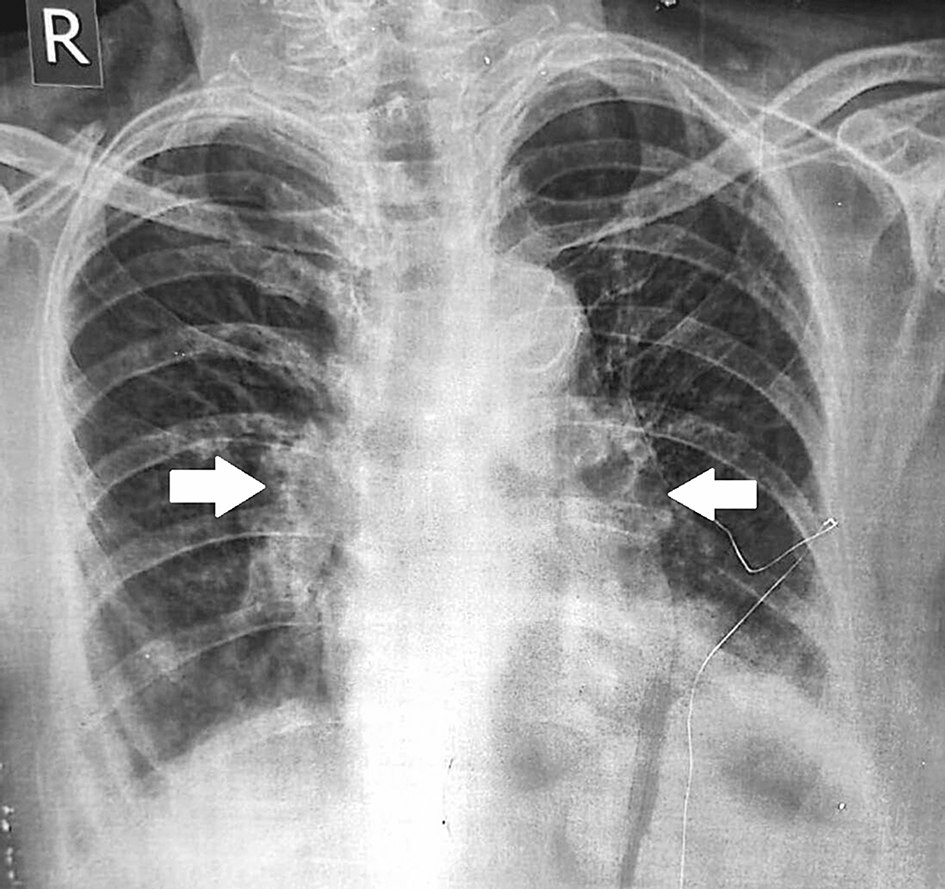
On investigation, her hemoglobin was low (10.3 gm/dl), with leukocytosis (11,800 per cumm) and eosinophilia (10%). Erythrocyte sedimentation rate (ESR) and C-Reactive protein (CRP) were elevated to 60 mm/hr and 14.7 mg/L, respectively. Sputum examination for gram stain and acid-fast bacilli stain were negative. The tuberculin skin testing was negative (4 mm) and her rheumatoid factor levels were normal (7.19 IU/ml). The antistreptolysin O titer was negative (57.6 IU/ml). Angiotensin-converting enzyme (ACE) level was raised to 98.78 U/L. Vitamin D level was slightly low at 18.63 ng/mL and serum calcium level was normal. Alkaline phosphatase level and other liver and renal function test parameters were normal. Further investigation revealed bilateral hilar lymphadenopathy on her chest X-ray (Figure 1).
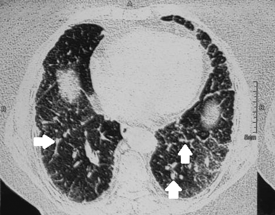
CT scans revealed bilateral pulmonary perilymphatic micronodules, mediastinal and bilateral hilar lymphadenopathy, and interlobar and interlobular septal thickening. The imaging findings were suggestive of sarcoidosis. Further, fine needle aspiration cytology of the right superior supraclavicular lymph node was done and it showed noncaseating granulomatous inflammation (Figure 2).
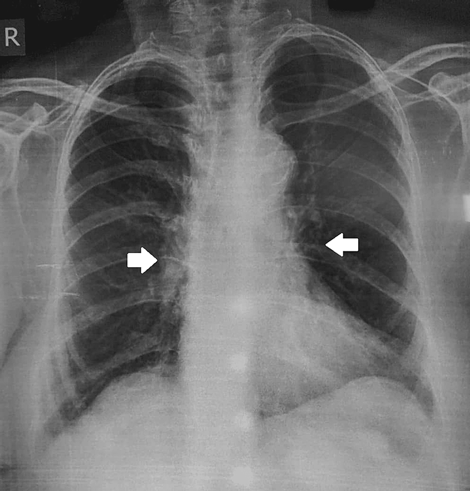
The patient was started on prednisolone 40 mg daily for 3 months and she was regularly followed up for the resolution of symptoms and radiographic improvement. She was also given calcium and vitamin D supplementation with regular monitoring of the serum and urine calcium and serum vitamin D levels. At the end of three months, the patient had significant relief of symptoms with marked radiological improvement as evidenced by serial chest X-rays. However, during the course of prednisolone therapy, the patient developed hyperglycemia and she was then managed with metformin 500 mg once daily (Figure 3).
Sarcoidosis is a chronic granulomatous condition with a complex etiology caused by a combination of host factors and environmental stimuli. The annual incidence of sarcoidosis in Asians and Hispanic people is 1-2 per 100,000 population. A retrospective cohort study conducted by Tripipitsiriwat et al. found female predominance (80.9%) with a mean age of 46.8 years at diagnosis.2 In line with this study, the patient in our case was an Asian woman, however, the age at presentation and diagnosis was at 69 years, which contrasts with this study’s finding.
Sarcoidosis has a wide range of clinical symptoms, owing to its multisystem involvement. The lungs (90%), liver (60%-80%), peripheral lymphatics (50%-75%), heart (30%), skin (10%-30%), eye (17 %), spleen (15%), joints (10%-15%), salivary glands (5%), and bone (1%-35%) are the most commonly involved organ systems pathologically. A study done in Nepal showed that the most frequent respiratory problems are cough and dyspnea. Fever affects between 15% and 20% of patients. Erythema nodosum occurs seldom. Weight loss, fatigue, weakness, and malaise are frequent constitutional complaints.7 Our patient had generalized body weakness, fatigue, and joint pain as the initial presenting symptoms. She later developed a cough, dyspnea, skin nodules, and erythema nodosum. These features were consistent with the findings of the preceding study. Cervical lymphadenopathy is the most prevalent head and neck symptom of sarcoidosis as shown in a study done by Chen et al.4 The patient in our case also had right cervical adenopathy on physical examination which is similar to the results of this study.
Sarcoidosis is diagnosed using three key criteria: consistent and appropriate clinical presentation, the presence of non-caseating granulomas in one or more tissue samples, and the exclusion of alternative causes of granulomatous diseases.8 The patient in our case had pulmonary symptoms and the biopsy of the cervical lymph node revealed noncaseating granulomatous inflammation. Other granulomatous diseases including tuberculosis were ruled out with negative tuberculin skin testing results and sputum acid-fast bacilli stain. More than 90% of patients have abnormal chest radiography.4 Although chest X-ray is the most often used imaging modality for pulmonary sarcoidosis and may be enough to rule out the disease, CT is commonly used for additional examination since it is more sensitive for detecting parenchymal abnormalities and thoracic lymphadenopathy.5 Although other illnesses such as lymphoma, tuberculosis, and metastases can also produce hilar and mediastinal adenopathy, sarcoidosis is characterized by symmetric involvement, which can assist distinguish it from other entities.5 CT findings in our patient also had symmetric involvement suggesting sarcoidosis.
The diagnostic workup of a patient with hilar adenopathy involving many systems or distant extrapulmonary regions and a probable diagnosis of sarcoidosis is dependent on the accurate exclusion of other prevalent chronic illnesses and a high level of suspicion.7 Because of the ambiguity of the symptom presentation as a result of multisystem involvement, most patients with sarcoidosis are diagnosed late, with only 15.3 percent of cases detected at the initial clinic visit.7 Similarly, in our situation, the patient was initially discharged with only analgesic and symptomatic therapy without a pulmonary radiograph as her initial symptoms were nonspecific. Only after a thorough workup, a definite diagnosis of sarcoidosis was obtained on the further clinic appointment. One aspect might be the patient's age upon presentation, which is rare after the age of 40, compared to our patient's age at diagnosis, which was 69 years.2 Another cause could be the possibility of other provisional diagnoses like bronchitis, asthma, or other lung diseases in such patients.9 As a result, while dealing with patients who exhibit these symptoms, a high level of suspicion should be maintained.
Increased ACE levels, hypercalcemia, hypercalciuria, hypergammaglobulinemia, increased erythrocyte sedimentation rate, and a positive Kveim test are all laboratory results.4 Serum ACE levels are elevated in 30 to 80 percent of sarcoidosis patients.1 The ACE level in our patient was raised along with elevated ESR and CRP.
The majority of sarcoidosis patients have spontaneous remission. Around 10-20% of patients have a chronic illness, with a death rate of 1-5%.4 Most treatment guidelines for pulmonary sarcoidosis recommend starting with dosages ranging from 20 to 40 mg daily. In practice, the dose is often titrated based on the response.6 The optimal duration of therapy for pulmonary sarcoidosis is uncertain, however, it is usually at least 6 months for sarcoidosis that has recently developed (i.e., 5 years).6
In our case, treatment with corticosteroids at a dose of 40mg/day was carried out for 3 months, with clinical improvement. Subsequently, slow and progressive corticosteroid weaning over the next 12 months was done, with a resolution of symptoms. Clinicians should examine the involvement of vital organs such as the lungs, heart, brain, liver, and kidneys throughout follow-up and provide routine examinations every 2-4 months based on clinical requirements. Chest X-rays, pulmonary function tests, electrocardiograms (ECG), blood counts, and urinalysis, as well as testing for liver function, renal function, and calcemia, should all be performed.4 Our patient was kept under routine follow-up every 3 months. Radiographic improvement assessed with serial chest X-rays was obtained. Other tests including ECG, urinalysis, liver and renal function test, and calcium levels were normal. At the end of 12 months, pulmonary changes in chest X-ray resolved and the skin lesions disappeared.
Other corticosteroid-sparing agents used for treatment are methotrexate, hydroxychloroquine, azathioprine, and other immunosuppressive agents.10 Patients with pulmonary sarcoidosis frequently experience recurrence and relapse. Those using corticosteroids and immunosuppressive drugs are at risk of side effects such as diabetes, hypertension, and opportunistic infections.10 Even though the patient has not developed any features of relapse following completion of therapy, she developed hyperglycemia during the course of treatment with steroids which was managed with metformin. The patient is doing well within a follow-up duration of 4 years.
Although sarcoidosis in elderly has worse prognosis, the patient in our case had a good recovery and prognosis.3 The patient was clinically investigated in detail, diagnosed, followed up for about three months, and managed as per the recent guidelines available in literature, thus increasing the strength of the study.
Sarcoidosis is an uncommon etiology of granulomatous illness in the Asian population. This case report adds to the existing literature about the clinical presentation, workup, and treatment of a patient with sarcoidosis. Physicians should be aware of sarcoidosis in elderly patients since it is associated with a poor overall prognosis. A high degree of suspicion, familiarity with imaging features, and prompt treatment in selected cases may help in diagnosing, evaluating the extent of disease, and guiding optimal health care.
| Views | Downloads | |
|---|---|---|
| F1000Research | - | - |
|
PubMed Central
Data from PMC are received and updated monthly.
|
- | - |
Provide sufficient details of any financial or non-financial competing interests to enable users to assess whether your comments might lead a reasonable person to question your impartiality. Consider the following examples, but note that this is not an exhaustive list:
Sign up for content alerts and receive a weekly or monthly email with all newly published articles
Already registered? Sign in
The email address should be the one you originally registered with F1000.
You registered with F1000 via Google, so we cannot reset your password.
To sign in, please click here.
If you still need help with your Google account password, please click here.
You registered with F1000 via Facebook, so we cannot reset your password.
To sign in, please click here.
If you still need help with your Facebook account password, please click here.
If your email address is registered with us, we will email you instructions to reset your password.
If you think you should have received this email but it has not arrived, please check your spam filters and/or contact for further assistance.
Comments on this article Comments (0)