Keywords
Polysomnographic signals, OSA, CSA, Mixed Apnea and Discrete Wavelet Transform
This article is included in the Computational Modelling and Numerical Aspects in Engineering collection.
Polysomnographic signals, OSA, CSA, Mixed Apnea and Discrete Wavelet Transform
Sleep disorders are most commonly seen in humans due to life style changes. Sleep is interrupted based on the person’s behaviour during sleep and due to changes in physiological signals, i.e., electrical rhythms in human brain or scalp. These electrical rhythms are recorded as electroencephalogram (EEG) signals. Sleep is defined as quiescent sleep or non-rapid-eye-movement (NREM) sleep or rapid-eye-movement (REM) sleep based on the subject’s physiological behaviour and the EEG.1,2 The study of normal sleep and sleep disorders is critical because these are the most frequently encountered difficulties in people. Sleep apnea (SA) is a common sleep disorder defined by intermittent breathing during sleep phases. It is a partial lack of airflow for more than 10 seconds in patients.3,4
If the amplitude of the breathing signal falls below 75% of the normal respirative signal level for a period 10 seconds or more, then SA is considered to be significant. SA is of three types: obstructive sleep apnea (OSA), central sleep apnea (CSA) and mixed sleep apnea (MSA). OSA is caused due to blockage of airway flow, this event occurs during sleep where the soft tissue in throat may be collapsed which then closes. In CSA, the blockage of airflow does not occur but the brain stops giving signals to the muscles to breathe.5,6
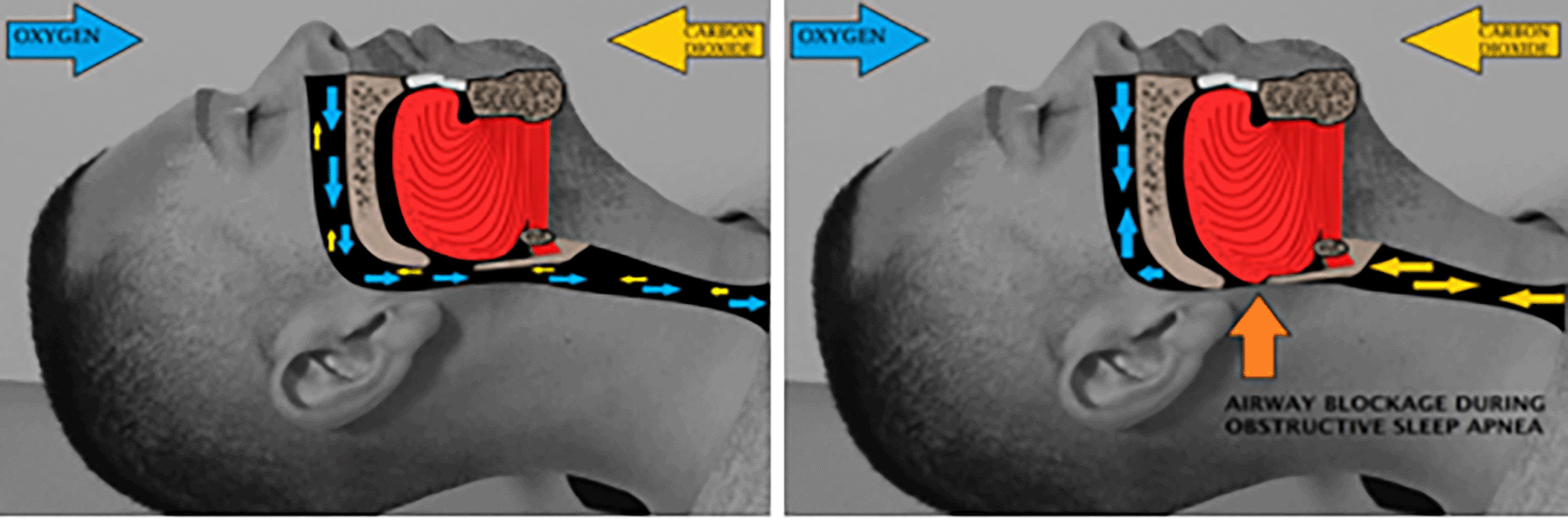
OSA is the most common type of SA, accounting for approximately 84% of all patients diagnosed. As illustrated in Figure 1, this syndrome is defined by complete or partial obstruction of the airways, which prevents oxygen from reaching the lungs. OSA is seen in loud snoring people because of blockage of upper airways in the respiratory system. A blockage can be resulted due to various physiological behaviours, that is genetically and progresses over time. The blockage is more common in obese individuals or those with a bigger neck with more surrounding fat and tissue. SA can be difficult to identify since the signs and symptoms are sometimes generalised and misconstrued as signs and symptoms of less serious conditions. OSA patients are frequently unaware they have the illness; rather, it is diagnosed by a partner or spouse due to the loud snoring and repetitive gasping while sleeping.7,8
CSA is generalized by continuous cessation of oxygen intake throughout the duration of sleep, whereas OSA is the cessation event occuring due to reduced ventilatory drive for obese subjects.9,10 CSA-related disorder is seen when cessation in breath occurs for a period greater than 10 seconds and less than 30 seconds.11,12 When a patient has more than five central apneas per hour of sleep, as well as the concomitant symptoms of frequent awakenings and/or daytime sleepiness, CSA syndrome is diagnosed. Because central apneas can occur in people who have obstructive apneas, 50 to 80% of apneic events must be central rather than obstructive in order to determine CSA. Cheyne–Stokes respiration (CSR), which is common in patients with heart failure, neurovascular disorders, and dementia, is strongly linked to CSA. Doctors struggle to make a diagnosis of CSA syndrome, and OSA requires full-night polysomnography to determine the frequency and pattern of central apnea.13
Complex or mixed sleep apnea syndrome (CompSAS) illustrates the characteristics of sleep-disordered breathing. In this, repetitive central apneas (≥5 per hour) can continue or appear when such obstructive events are eliminated through positive airway pressure (PAP). CompSAS is very dangerous combination consists of both OSA and CSA.14 The subjects suffering from both experience blockage of uppper airway and brain stops giving signals to muscles to breathe. This is inability of feedback mechanism which controls the brain. When the central-apnea-index (CAI) exceeds 5 events per hour, it becomes a major and disruptive event.15
Currently, various methods have been published in establishing sleep apnea from biomedical signals. With reference to,16 OSA along with CSA events can be separated with wavelet packet analysis of electrocardiogram signals. With reference to,17 SVM classifier gained a precision of 91.08%. A new approach was developed to categorize and evaluate the electroencephalogram (EEG) and identify the EEG features for detection of SA stages with the DWT techniques and ANN classifier.18
The feature extraction and optimization techniques for OSA, CSA and MSA is investigated. Based on decomposition stages along with bivariate methods was used to examine the features of EEG signals via a Hilbert–Huang transform. Once an apnea has happened then there will be an energy waveform in low frequencies, which is related to the delta power and it represents the body autonomous system and homeostasis regulation. Another sleep apnea review, the result of transient, apnea-induced hypoxemia on electrocortical activity, five patients having adverse obstructive sleep apnea syndrome (OSAS) were analysed during nocturnal sleep. While non rapid eye movement (NREM) apneas, amplitude of the delta band rises, the abnormality of sleep in adverse OSAS may form such a tendency for slow-wave sleep that stages pass too much rapid than in normal persons.
The co-efficients are used as inputs to the classifiers. The novel classfication method uses features as inputs for apnea stages identification. The primary goal of sleep apnea study is to develop an algorithm for sleep stage recognition, which is done by analysing the electrooculography (EOG) signals. The secondary goal is to use sleep quality features to categorize and screen sleep apnea–hypopnoea syndrome (SAHS) patients and periodic limb movements of sleep (PLMS) patients, as different from healthy control subjects. Brain condition can be measured by EEG, which records the electrical state of the scalp. Electroencephalogram (EEG is more convenient tool for knowing the typical actions of the scalp. The goal of this paper is to visualize the abnormalities in the EEG signal.
The proposed work describes the raw electroencephalogram (EEG) data obtained from the MIT-BIH database (physionet data base). The obtained EEG dataset is in the form of .mat and it is loaded in to the matrix laboratory (MATLAB) workspace and generates the EEG signal. The resulting EEG signal is segmented using the Daubechies wavelet transform, which yields all of the electroencephalogram signal’s fundamental frequency components. The segmented bars correspond to various brain states. Energy, standard deviation, mean, maximum, minimum, and variance are all computed (extracted) from EEG bands and can be used to detect sleep apnea. The flowchart depicting the diagnosis and classification of sleep apnea is illustrated in Figure 2. Five steps comprise the process flow. The first stage involves acquiring the signal from the MIT-BIH data base, the second stage involves segmenting the EEG signal, the third stage involves extracting features from the segmented signal, the fourth stage involves detecting the severity of apnea, and the fifth stage involves classifying the abnormality as OSA, CSA, or CompSAS.

The signals are extracted from polysomnographic data taken from the MIT-BIH Polysomnographic Database. The monitoring of patient overnight in a sleep laboratory is called polysomnography (PSG). In general, PSG is the common test for identification of OSAS. PSG is also used to evaluate irregularities of sleep and different physiologic disorders which influence the health. One-night PSG is enough to determine if OSA is present.9,10 A PSG test constitutes concurrent recording of several physiologic parameters which belong to sleep and wakefulness. Electrical signals are transmitted by using specialized sensors or electrodes which are applied to the various body parts. The translation of these signals into records is done by specialized amplifiers and filters which are present in recording instrument.13,14
There are many signals that are acquired. These include:
1. Electroencephalogram (EEG) signal
2. Electrocardiogram (ECG) signal
3. Airflow signal
4. Thoracic+abdomen positional sum signal
5. Electroculogram (EOG) signal
6. Electromyogram (EMG) signal
7. Oxyhemoglobin saturation signal (SpO2)
Electrical activity of the brain can be recorded from the scalp and which is quite small, measured in microvolts. The EEG waveform can be decomposed into five frequency bands, namely delta, theta, alpha, beta and gamma. These bands can give important information for observing, diagnosing and managing neurological features and disorders11,12
Feature extraction is the technique of extracting specific information from the electroencephalogram (EEG) as recorded by neuronal activity in the brain. A feature extraction methodology is an ideal way for dissecting the input EEG signal into a set of characteristics and differentiating samples with distinct patterns. With the purpose of minimising the loss of vital information, patterns are represented by features. Feature extraction is the process of identifying a characteristic or characteristic vector from a pattern vector.
The discrete wavelet transform method is used to extract features in this article. Wavelet transform (WT) provides the general approaches, which can be used to a wide variety of signal processing jobs. Wavelets can be used to analyse abrupt, short-duration signal changes. The primary application is to calculate and regulate data that has been reduced to a few parameters, referred to as features. As a result, the time-varying biological signal contains a large number of data points that can be condensed via WT to a few parameters. These parameters describe the behaviour of a time-varying biological signal. The time-varying biomedical signal is represented by a smaller number of parameters, which are particularly important for recognition and diagnostic purposes. Statistical features are computed for each sub bands which are mean, energy, standard deviation, maximum, minimum and variance. Signal energy characterizes the power at all instances of time interval. Standard deviation (SD) is easy to calculate the changeability of a data set. Variance parameter gives the distance between values in the data set from mean.
The analysis was performed on EEG signal and features are extracted from the each frequency band of the EEG signal by using wavelet transform.
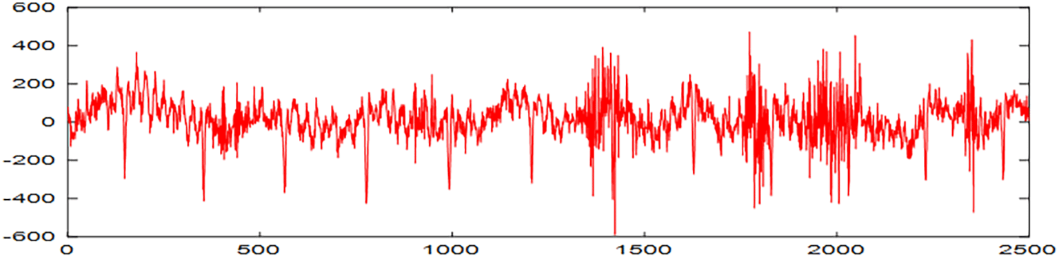
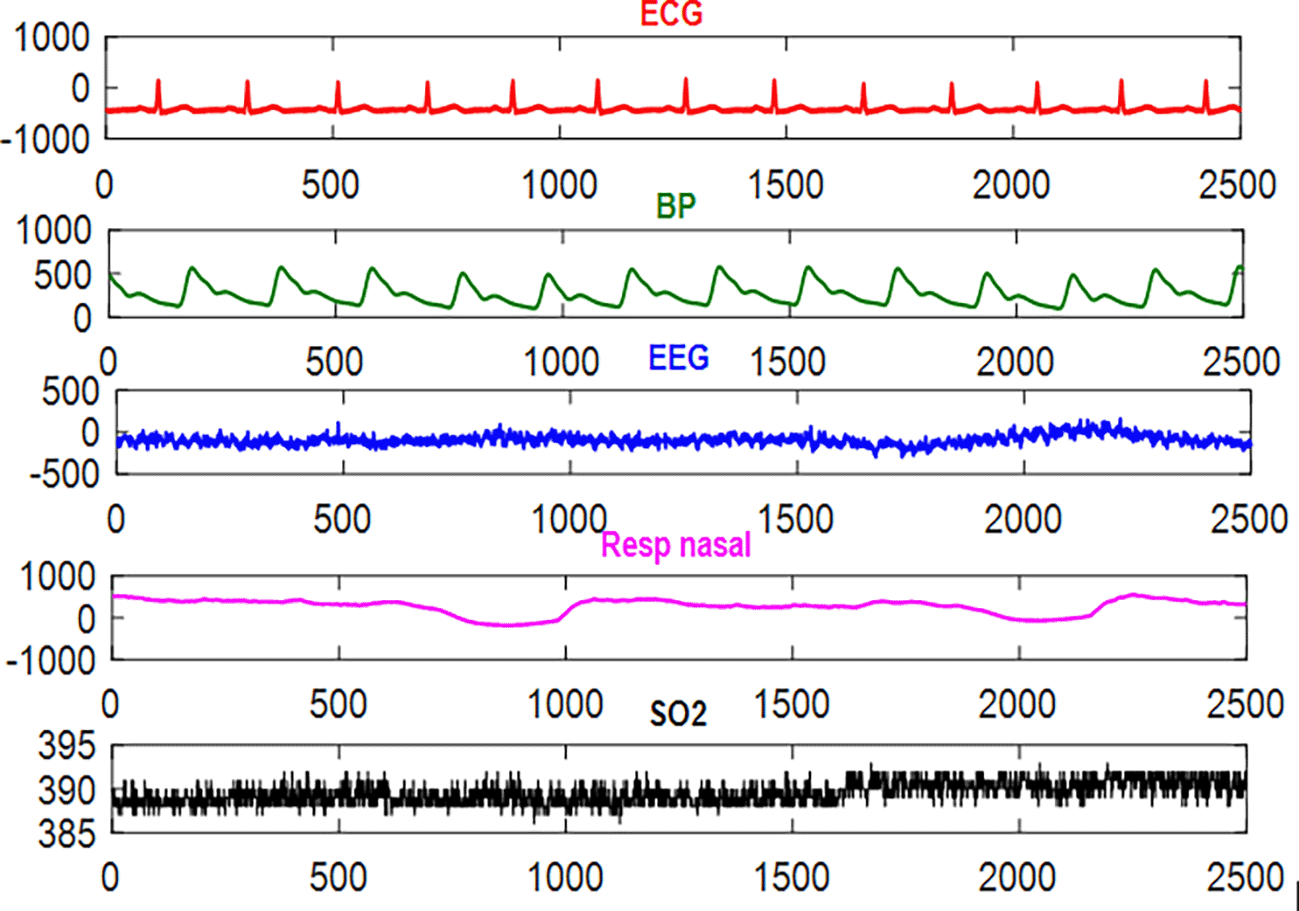
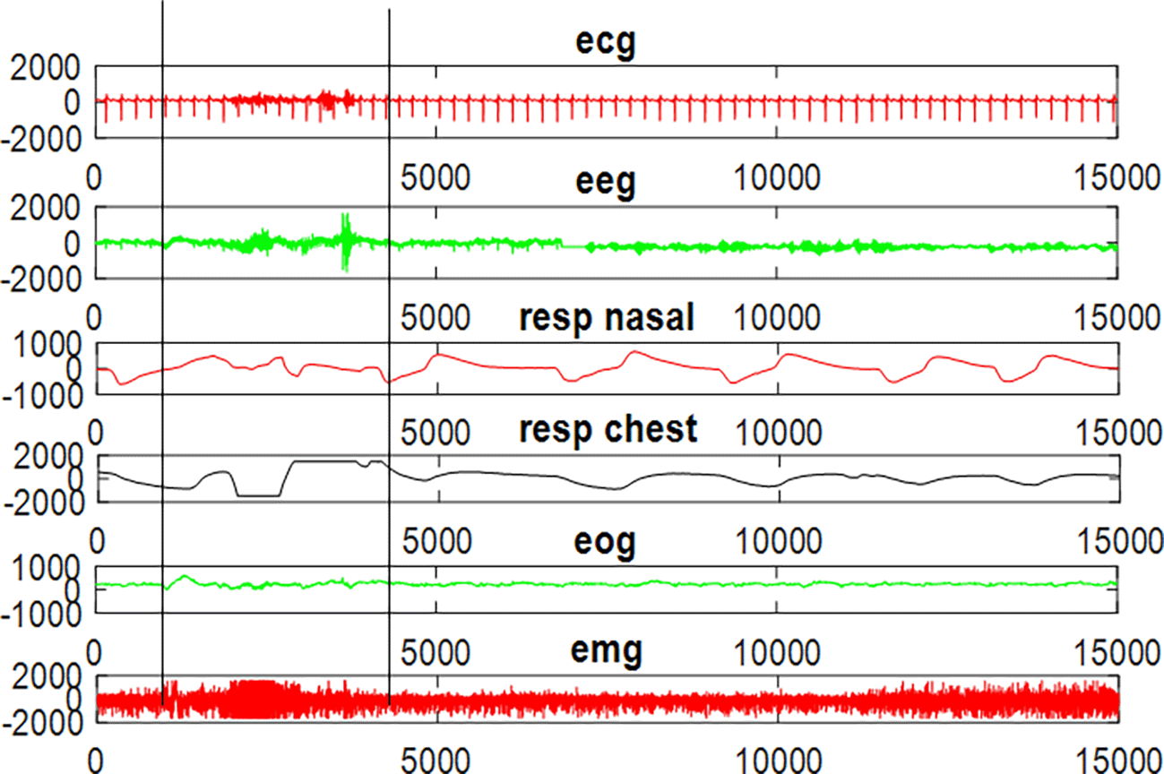
The EEG signal acquisition for one subject is shown in Figure 3; it is the normal EEG signal without any abnormality. Various physiologic signals sampled at 250 Hz frequency and plotted for 60 seconds for the subject slp32m is shown in Figure 4. These physiologic signals are ECG, BP (blood pressure), Resp nasal (respiration at nasal) and SpO2 (oxygen saturation). In Figure 5 the area between two lines indicated the sleep apnea event has occured. This clearly shows the abnormality in the ECG, EEG, Nasal, EOG and EMG. It is characterized by reduction in breathing. These changes in signals is compared to the normal PSG.
The EEG signal is divided into subbands such as gamma, beta, theta, alpha, and delta using the wavelet technique as illustrated in Figure 6 DWT is used to extract features from the EEG signals during the feature extraction stage. Table 1 summarises the computed properties, such as standard deviation, variance, and energy, for each frequency band that can be utilised to depict the EEG signals’ characteristics.

In this paper the physiologic signals ECG, BP, EEG, respiratory signal at nasal, oxygen saturation (SpO2) for normal subject and abnormal subject is analyzed. The EEG signal is segmented into delta, theta, alpha, beta, gamma for both normal and abnormal subjects. Various features like standard deviation, variance and energy, mean, maximum and minimum are extracted from both normal and abnormal subjects. Using DWT Daubechies order 2, the detailed and approximate coefficients are extracted. These inputs are given to the classifier for detection of sleep disorder stages.
Ch. Usha Kumari, K. Swaraja: Conception and design of study. Ch. Usha Kumari, K. Meenakshi: Acquisition, analysis and/or interpretation of data. Ch. Usha Kumari, T Padma: Drafting the manuscript and revising the manuscript critically for important intellectual content.
Underlying data Raw polysomnographic data were from taken from the MIT-BIH database (https://physionet.org/content/slpdb/1.0.0/).
Figshare: Underlying data for “Detection of sleep apnea using polysomnographic signals” https://figshare.com/s/08009ff4301370a5f58d
Data are available under the terms of the Creative Commons Attribution 4.0 International license (CC-BY 4.0).
| Views | Downloads | |
|---|---|---|
| F1000Research | - | - |
|
PubMed Central
Data from PMC are received and updated monthly.
|
- | - |
Provide sufficient details of any financial or non-financial competing interests to enable users to assess whether your comments might lead a reasonable person to question your impartiality. Consider the following examples, but note that this is not an exhaustive list:
Sign up for content alerts and receive a weekly or monthly email with all newly published articles
Already registered? Sign in
The email address should be the one you originally registered with F1000.
You registered with F1000 via Google, so we cannot reset your password.
To sign in, please click here.
If you still need help with your Google account password, please click here.
You registered with F1000 via Facebook, so we cannot reset your password.
To sign in, please click here.
If you still need help with your Facebook account password, please click here.
If your email address is registered with us, we will email you instructions to reset your password.
If you think you should have received this email but it has not arrived, please check your spam filters and/or contact for further assistance.
Comments on this article Comments (0)