Keywords
PEEK, laser surface texturing, fractional CO2 laser, roughness, wettability
PEEK, laser surface texturing, fractional CO2 laser, roughness, wettability
Polyetheretherketone (PEEK) is a high-performance polymer that has been recently investigated as an alternative biomaterial for metallic materials in dentistry.1 The increased interest in PEEK as an implant material is due to its biocompatibility and its superior physical and mechanical properties over titanium and other metallic materials.2,3
Studies have shown that PEEK causes fewer hypersensitive and allergic reactions than titanium.4,5 Radiolucency is one of the benefits of using PEEK as an implant material since it can be imaged by X-ray without any distortion or artifacts.6,7 Besides, it has more aesthetic appeal than titanium due to its beige color.8 Most importantly, the elastic modulus value of neat PEEK is about 3.6 GPa, which lies between the values of trabecular (1.4 GPa) and cortical bone (14 GPa).9,10 The close proximity in stiffness between PEEK and human bone causes less stress shielding and bone resorption around the implant in comparison with stiffer titanium (elastic modulus = 110 GPa).11
This polymer is not bioactive due to its hydrophobicity and poor interfacial interaction, meaning it has a limited ability to bind with the surrounding bone tissue. Wettability is a crucial property of an implant material since the first event of implantation is the adsorption of water and protein from the blood on the implant surface, which signals the process of new bone formation.12,13 However, it is well-established that wettability is affected by the biomaterials' surface roughness and texture, which play important roles in establishing a functional interface with the surrounding tissue in the body.14,15
The topography of a surface has a direct impact on biological reactions at the cellular level, including cell orientation and migration as well as the development of ordered cytoskeletal structures. Successful osseointegration of implants has been shown to be associated with surface roughness in the nano- and micro-scales.16,17 Previous studies18,19 demonstrated that a moderately rough implant surface with an average surface roughness of more than 1 μm allowed for bone ingrowth and provided mechanical interlocking with bone tissue. High bone-to-implant contact was seen with rough surfaces due to the increase in surface area.18,19
Several methods have been experimented to alter the surface topography of PEEK polymer, including acid etching, grit blasting, plasma treatment, and laser texturing.20 Laser surface texturing (LST) is one of the simplest methods to modify the surface of polymers. It can simultaneously alter the surface roughness at the macro-, micro-, and nano-sized scales without using different tools.21 A laser can be used to create surface features like pits and grooves with an equidistance ranging from several nanometers to even micrometers between the pits or grooves.22 These topographic features influence cellular adhesion, migration, proliferation, and differentiation behavior.23,24
A fractionated laser is a mode of laser delivery in which energy is conveyed in an array of parallel vertical columns of multiple microscopical thermal spots termed microscopic treatment zones (MTZs) with a constant distance between spots.25 Fractional CO2 was introduced in 2004 in dermatology for skin resurfacing and it may have several advantages in dentistry, especially in the surface treatment of biomaterials.26–28 The device allows for a precise irradiation area with a more homogenous texturing pattern to be set without the need for manual movement of the device handpiece. Unlike with traditional CO2 lasers, there is less overall material bulk heating and thermal degradation with fractional lasers.25 Additionally, with the correct selection of laser parameters (power, pulse duration, distance between spots, and spot pattern), the desired melting, ablation, and penetration depth can be achieved.29,30
The aim of this research was to improve the surface roughness and wettability of a PEEK substrate by fractional CO2 laser texturing without affecting the material’s microstructure. The null hypothesis is that laser surface texturing by fractional CO2 laser does not improve the surface roughness nor the wettability of PEEK.
PEEK samples were supplied by (Energetic Industry Co., Ltd., China) by means of cutting continuous extruded rods into discs with dimensions of 10 mm × 2 mm (diameter × thickness). The samples were gradually ground with 500, 800, 1200, 2000, and 2400 grit silicon carbide abrasive papers, and they were polished to obtain a mirror finish. After that, all samples were cleaned with ethanol and water, successively.
Laser surface texturing of PEEK samples was carried out using a fractional CO2 laser system (CICU-f, Ilooda Co., Ltd., South Korea). This device has a fixed output power of 11.25 W and a laser wavelength of 10600 nm. Different pulse durations (PD) were tried (100, 200, 300, 400, and 500 μs) and two patterns (straight dots and 60° staggered dots) were performed for each pulse duration at a dot separation distance of 0.2 mm. The handpiece of the device was affixed by a clamp to prevent any movement during the procedure and to ensure the incident laser beam was perpendicular to the sample (0° incidence angle). The texturing procedure was done with 1 scan (pass). After laser treatment, the samples were cleaned with ethanol in an ultrasonic cleaning device.
Initial examination of laser texturing was done using an optical microscope (Olympus BH-2, Japan) at 10× magnification. The observations included the effect of laser heating on PEEK material and any signs of burning or carbonization. The samples that were treated with 100 μs PD did not show any observable laser effect on the PEEK surface, and those treated with 500 μs PD resulted in the burning of the material. For those reasons, these parameters were not considered for further surface characterizations.
The surface morphology of untreated (control) and laser treated PEEK samples was examined by field emission scanning electron microscopy (FESEM) (MIRA3, TESCAN, Czech Republic) to observe the texturing pattern, spot shape, melting pattern around the spot, ablation at the heat-affected zone, and presence of cracks. Energy dispersive X-ray spectroscopy (EDX) was performed to determine the effect of laser on the microstructure of the material. Elemental weight percentages and atomic percentages were compared between control samples and laser treated samples. For laser treated samples, elemental analysis was done in three areas: at the center of the dot, at the edge of the dot, and outside the dot (within the heat-affected zone).
Surface microroughness was measured by a digital profilometer (TR220, Beijing Time High Technology Ltd., China). The device has a sharp stylus with an 8 mm travelling distance across the surface. Three readings of the average surface roughness (Ra) in micrometers were obtained for each sample, and the average of these readings was recorded for each sample.
A contacting AFM device (BenYuan CSPM-5500, Being Nano-Instruments Ltd., China) was used to obtain 3D topographical images of PEEK samples. The AFM was operated in tapping mode with a 299.6 kHz tapping frequency. The scanning area was a square with a size of about 4550 × 4550 nm. The average surface roughness (Sa) and the surface area ratio (Sdr) were obtained for each sample.
The wettability of PEEK samples was assessed by a contact angle goniometer (Cam110 contact angle goniometer, Creating Nano Technologies Inc., Taiwan) with a sessile drop of deionized water. The device consists of a table for holding the sample, a syringe with a revolving knob on top to release the water drop, and a camera that is connected to a computer to analyze the image with an included imaging software (Touchmate, Cam110). The sample was placed on the table and a 7 μm drop of deionized water was released from the syringe. The water drop was left to spread on the sample for 30 seconds, and then an image was captured. The computer software was used to calculate the contact angles.
Prism 8 (GraphPad Software, USA) (RRID:SCR_002798) was used for statistical analysis of the data (an open-source alternative able to perform similar analysis is JASP). The results were depicted as bar charts, with the mean values (n = 10) placed inside the bars and the standard deviation written above the bars. To assess for statistical significance among groups, one-way analysis of variance (ANOVA) was used, and for multiple comparisons, Tukey's HSD (honestly significant difference) post-hoc test was performed. A P-value > 0.05 is statistically non-significant (NS), < 0.05 is significant (S), and < 0.01 is highly significant (HS).
Optical microscope images of control and laser textured samples36 are shown in Figure 1. Samples treated with 100 μs PD did not show any noticeable laser effect for both patterns. With increasing the pulse duration over 200 μs, the dots pattern became more prominent with larger heat-affected zones. The dots of samples with 400 μs PD had more depth than other samples with less PD due to the melting and evaporation of the material as the absorbed heat increased. Carbonization can be seen over the edges of the dots with samples treated with 500 μs PD. The staggered dots pattern seemed to cover more surface area than the straight dots pattern with fewer unaffected zones between the dots due to dots staggering or zig-zag dots formation.
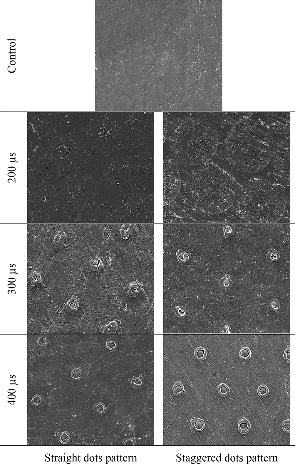
The surface morphologies of PEEK samples at 200× and 2000× magnifications are shown in Figure 2 and Figure 3, respectively. The FESEM images were consistent with the optical microscope images in terms of the effect of heat on the material. The laser effect became more prominent as the pulse duration was increased. With PD of 200 μs, there was some uniform ablation at the center of the dot and in the heat-affected zone with no signs of melting. There was little melting at the center of the dot with 300 μs PD, but with 400 μs PD complete melting occurred at the center of the dot with formation of uniform edges. There were no cracks caused by laser in all samples.

Elemental analysis with an EDX spectrum of untreated PEEK sample is presented in Figure 4. An area analysis was done for the whole image shown in the figure. For laser treated sample with 400 μs PD, EDX spectra were obtained at three points, as shown in Figure 5 to assess the effect of laser on the microstructure of the material. The elemental analysis of the laser treated samples showed very comparable weight percentages of carbon and oxygen to that of unmodified samples. There was no increase in the percentage of carbon at the center or at the margins of the spot, indicating no presence of carbonization or burning.
Mean values of surface roughness (Ra) in micrometers are presented in Figure 6. One-way ANOVA showed that there was a high significance among groups (p-value < 0.01). All laser treated samples showed a highly significant increase in surface roughness compared to the control sample. The highest microroughness values belonged to samples treated with 400 μs pulse duration and a staggered dots pattern.
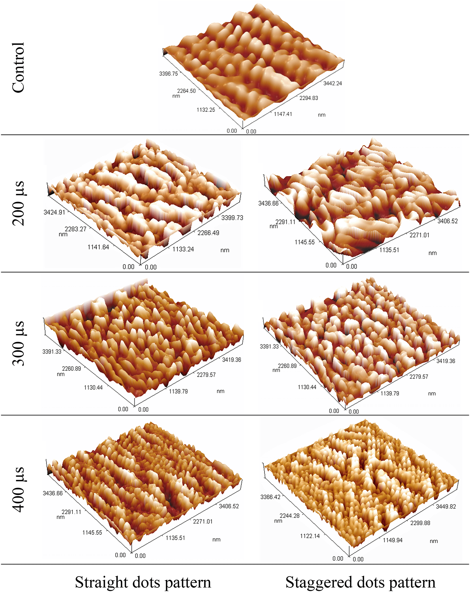
The 3-dimensional topographical images of control and laser textured samples are presented in Figure 7. There was an increase in summits and valleys density in all laser treated samples compared to the control group.

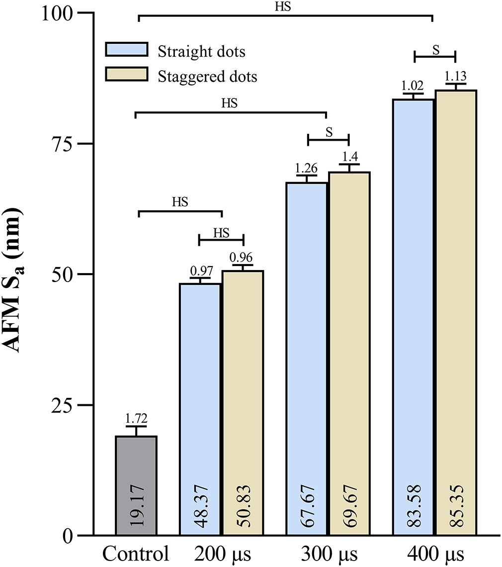
Mean values of AFM average surface roughness (Sa) and surface area ratio (Sdr) are presented in Figure 8 and Figure 9, respectively. There were highly significant increases in Sa and Sdr values for all laser samples in comparison to control samples. The samples of 400 μs PD treatment with a staggered dots pattern showed the highest mean values of both Sa and Sdr.

Images of water contact angle measurements are shown in Figure 10, while the mean values are presented in Figure 11. One-way ANOVA resulted in a high significance among groups. All laser textured samples showed a highly significant decrease in the water contact angle. There were highly significant improvements in wettability with laser PD of 400 μs especially with a staggered dots pattern, which showed the most improvement in this property.
This study was conducted to assess the efficiency of a fractional CO2 laser system used for skin resurfacing in surface texturing and enhancement of surface properties of PEEK implant substrate. PEEK has proven itself to be a valuable material in the field of implantology due to its biocompatibility, high mechanical strength, inherent radiolucency, and exceptional chemical resistance. Unfortunately, PEEK's bioinert surface does not support cellular adhesion and bone tissue bonding, limiting its use in the medical field.12
Laser texturing with a fractional laser is easy, fast, and applicable since the whole surface can be treated with a single run without the need for a moving device. When a laser beam of high intensity comes in contact with the surface of a material, some of the beam is absorbed by the material, and the remaining intensity is lost to the surrounding environment through the processes of reflection and scattering. The effect of laser on a polymeric material can be either photothermal or photochemical. A photothermal effect occurs when the absorbed laser energy converts to heat that raises the temperature of the material. When the temperature of the substance exceeds its boiling point, it turns into vapor and is generally ejected from the surface as it boils. This effect induces diverse phenomena, including melting or vaporization (ablation), which modify the topography.31
The photochemical effect occurs when the laser used has a wavelength in the UV range (like excimer lasers) in which the photon energy is enough to break molecular bonds at the polymer surface and induce a change in the surface chemistry.
Since the wavelength of CO2 laser is within the infrared range (10600 nm), the effect induced on the PEEK surface was photothermal by a process called photothermolysis without any chemical change in the microstructure of the material as confirmed by EDX in Figure 4 and Figure 5. The fractional laser causes selective photothermolysis by splitting the laser beam into an array of microbeams with a fixed distance between them. The ablation and texturing pattern of PEEK material became clearer and more distinct as the pulse duration was increased, as can be seen in Figure 1. By increasing the pulse duration, the spot energy increases and causes more heat to be absorbed by the material. With a short pulse duration of 100 μs, no effect was seen because the heat was below the melting threshold of PEEK, while increasing the duration to 500 μs caused burning of the material due to intense heating.
Surface topography, roughness, and wettability have been shown to significantly affect cellular responses to biomaterials in a number of studies.32–34 The behavior of cells on biomaterial surfaces is determined by the interaction between implant and cells, which is related to surface roughness. Naturally, bone cells encounter and interact with nanostructures in their environment, such as extracellular matrix (ECM) proteins. To improve bone cell adhesion and proliferation, it is thoughtful to mimic the cellular environment by creating materials with nanotopography. While nanotopographic features are important for the recruitment and migration of bone cells to the implant surface, microtopographically complex surfaces are equally vital in increasing the degree of bone-to-implant contact. Microcraters created on PEEK’s surface by the fractional laser could encourage bone ingrowth and enhance the anchorage of implants with bone tissue. The results of this study confirmed that laser texturing with fractional CO2 laser significantly increase surface roughness on both nano- and micro-level as demonstrated by AFM and microroughness tests. The samples treated with 400 μs pulse duration showed surface roughness values of about 1.3 μm. This value is well within the optimum roughness range (1–2 μm) to enhance bone integration reported by other studies.32,35
In regards to wettability, it is an essential property for cell spreading, which in turn is an important stage in cell adhesion prior to proliferation and differentiation. The wettability was significantly enhanced after laser texturing, particularly with samples treated with 400 μs PD which showed the least water contact angles among other samples. These results are consistent with Wenzel’s theory, which stated that adding surface roughness and increasing the surface area leads to an enhancement in the wettability.
Since the fractional CO2 laser surface texturing improved the surface properties of PEEK material, the null hypothesis was rejected.
Although this study has some limitations, it can be concluded that laser texturing of PEEK biomaterial by fractional CO2 laser significantly enhanced its surface properties in terms of surface topography, roughness, and wettability. The best results were achieved with pulse duration = 400 μs, dot separation distance = 0.2 mm, and a 60° staggered dots pattern. With further in vitro (like cell culture) and in vivo (like animal study) investigations, this method of PEEK modification might have the potential to be used in the implant field.
Figshare: Enhancement of surface properties of polyetheretherketone implant material by fractional laser texturing. https://doi.org/10.6084/m9.figshare.21518136.v5. 36
This project contains the following underlying data:
- Surface microroughness.csv (Raw data of surface microroughness test)
- AFM Sa (nm).csv (Raw data of AFM average surface roughness)
- AFM Sdr.csv (Raw data of AFM surface area ratio)
- Wettability.csv (Raw data of water contact angle measurements)
- Microscope images.rar (All raw images taken with microscopes)
Data are available under the terms of the Creative Commons Attribution 4.0 International license (CC-BY 4.0).
| Views | Downloads | |
|---|---|---|
| F1000Research | - | - |
|
PubMed Central
Data from PMC are received and updated monthly.
|
- | - |
Is the work clearly and accurately presented and does it cite the current literature?
Yes
Is the study design appropriate and is the work technically sound?
Yes
Are sufficient details of methods and analysis provided to allow replication by others?
Yes
If applicable, is the statistical analysis and its interpretation appropriate?
Not applicable
Are all the source data underlying the results available to ensure full reproducibility?
No
Are the conclusions drawn adequately supported by the results?
Partly
Competing Interests: No competing interests were disclosed.
Reviewer Expertise: Surface Egnieering
Is the work clearly and accurately presented and does it cite the current literature?
Yes
Is the study design appropriate and is the work technically sound?
Yes
Are sufficient details of methods and analysis provided to allow replication by others?
Yes
If applicable, is the statistical analysis and its interpretation appropriate?
Yes
Are all the source data underlying the results available to ensure full reproducibility?
Yes
Are the conclusions drawn adequately supported by the results?
Yes
Competing Interests: No competing interests were disclosed.
Reviewer Expertise: Laser Material interactions applications in medical and scientific applications
Alongside their report, reviewers assign a status to the article:
| Invited Reviewers | ||
|---|---|---|
| 1 | 2 | |
|
Version 1 05 Dec 22 |
read | read |
Provide sufficient details of any financial or non-financial competing interests to enable users to assess whether your comments might lead a reasonable person to question your impartiality. Consider the following examples, but note that this is not an exhaustive list:
Sign up for content alerts and receive a weekly or monthly email with all newly published articles
Already registered? Sign in
The email address should be the one you originally registered with F1000.
You registered with F1000 via Google, so we cannot reset your password.
To sign in, please click here.
If you still need help with your Google account password, please click here.
You registered with F1000 via Facebook, so we cannot reset your password.
To sign in, please click here.
If you still need help with your Facebook account password, please click here.
If your email address is registered with us, we will email you instructions to reset your password.
If you think you should have received this email but it has not arrived, please check your spam filters and/or contact for further assistance.
Comments on this article Comments (0)