Keywords
African Human Iris images, Age Prediction, Ethnicity Prediction, Gender Prediction, Biometrics, Personal Recognition
This article is included in the Artificial Intelligence and Machine Learning gateway.
This article is included in the Data: Use and Reuse collection.
African Human Iris images, Age Prediction, Ethnicity Prediction, Gender Prediction, Biometrics, Personal Recognition
The iris recognition system is one of the most widely used and acceptable means of personal recognition and authentication. It has recently become an official means of national identification in India. The Unique Identification Authority of India (UIDAI) has successfully captured 1.5 billion irises from Indian citizens for identification and recognition purposes.1 Many countries have done the same and this increasing popularity and acceptability of human iris as a means of national identification and recognition has called for additional benchmarks, and therefore, new publicly available databases of human iris images.2 Though several human iris datasets exist,3–6 iris datasets of African descents are presently not publicly available. This research effort aimed at bridging this gap in the African continent by embarking on the capture and subsequent creation of human iris images of people of African descent to make them publicly available for research purposes. In the words of Prof. John Daugman:
“There is a more urgent need for an African FACE image database because researchers into face recognition have famously (or infamously) used primarily non-African face images, leading to high levels of bias in algorithms, and disastrous classification performance when they are tested on African face images”.
Therefore, the authors believe the human iris dataset presented in this data article7 will be of great value to researchers willing to advance iris-related research across the African continent. The following are some of the uniqueness of the iris dataset described in this article:
• The dataset presented in this Data Note is the first publicly available human iris dataset of African descent.
• In addition to the iris images, the dataset provides soft biometric features about each volunteer. This additional information will open up new multi-modal biometric research.
• The dataset can serve as a benchmark for evaluating iris recognition methods and other human iris-related research.
• The dataset can be used to validate results obtained from existing iris-related research that used non-African iris images for their research validation.
• The dataset can be used to enhance studies such as personal recognition, age, gender, or ethnicity prediction as well as iris color pigment research on the African continent.
Approvals were obtained from the Ethical and Review Committee of participating Universities before the commencement of the data collection exercise. This was done to ensure and guarantee that the data collection task would not hurt the health of the volunteers and that the publication of the data collected would not infringe on their privacy in any way. Also, all volunteers (willingly without any form of cohesion or pressure) agreed to participate in the iris data collection task with the awareness that the collected data would be made publicly available for research purposes. The privacy of the volunteers was respected as personal details that could make the data traceable to them were not collected.
A Vista EY2H dual iris camera was employed for the iris image capture. The iris camera is a RoHS compliant device that uses a cutting-edge, high-resolution CMOS sensor to produce ISO/IEC 19794-6-compliant images of both irises simultaneously. The camera uses a multi-wavelength near-infrared band of light (NIR: 700nm - 900nm) illumination for superior iris images in all environments. The capturing process meets international eye safety requirements and also has a live eye anti-Spoof detection feature that can be used to reliably detect when a subject is alive. At a click of the capture button, a large 2560 by 720-pixel iris image was produced from the camera; this image is automatically separated into four images with a dimension of 640 by 480 pixels. These are the left and right iris images with the iris region localized, and another set of left and right iris images without the iris section localized. An overview of the camera is provided in Figures 1a-c:
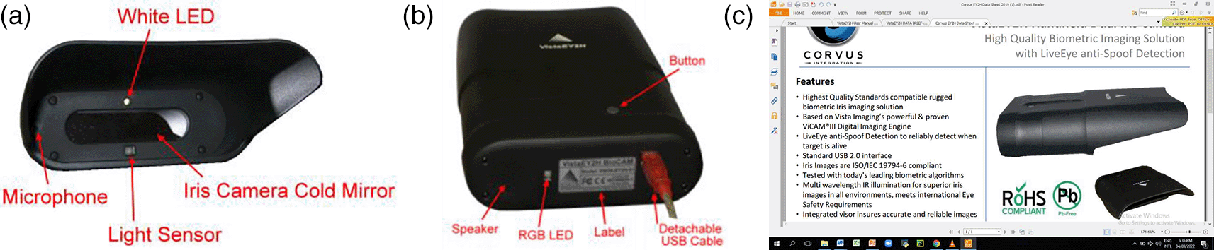
Images have been reproduced from Vista Imaging8 with the appropriate permissions.
Digital Weighing scale: a digital weighing scale as shown in Fig. 2 was employed to measure the weight (soft biometric data) of volunteers.

Image reproduced from www.jumia.com.ng with the appropriate permissions.
Digital Height Measurement Scale: a height measurement scale as shown in Fig. 3 was employed to measure the height of the volunteers.
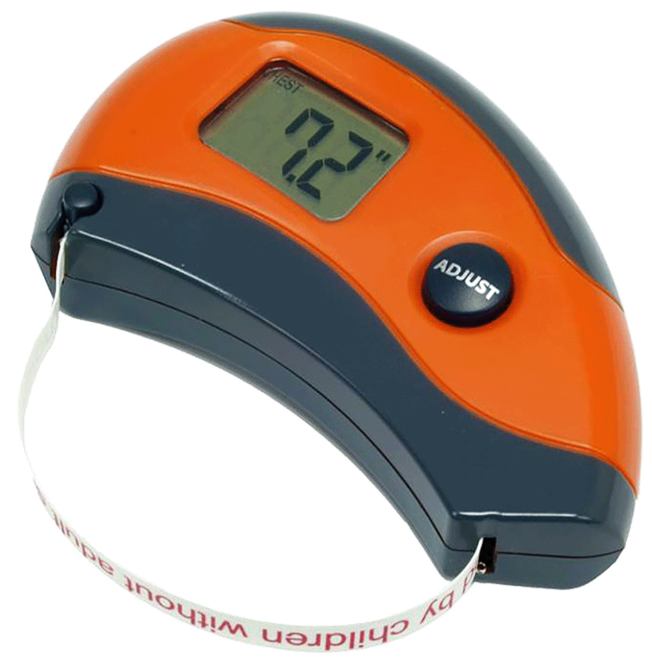
Image reproduced from www.jumia.com.ng with the appropriate permissions.
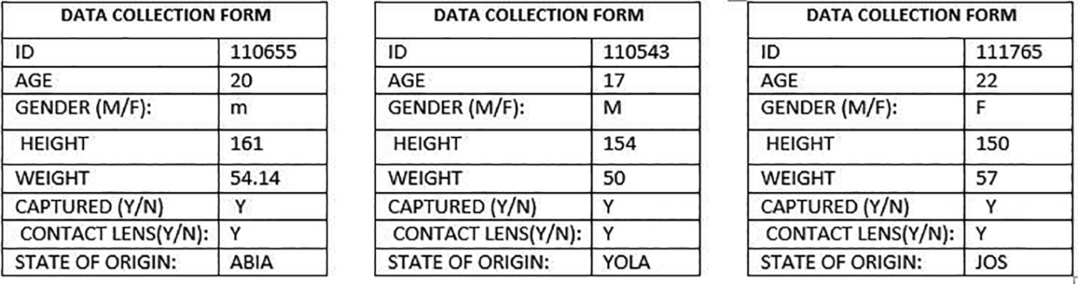
Three standard approaches are generally employed for iris image capturing. They are self-enrollment; handheld, self-enrollment (fixed on a tripod); and operator-assisted enrolment. For fast and accurate data capture, the operator-assisted enrolment method was used. The capture device was fixed on a tripod and volunteers were only asked to place their forehead horizontally with their eyes gazing at the lens of the camera. When a perfect range of the irises had been set, the operator clicked the capture button on the camera to initiate the capture process. The captured images were automatically saved on the investigator’s personal computer connected to the scanning device. Afterward, soft biometric features such as: volunteer’s height, weight, age, gender, and state of origin were collected with the respective measuring devices. The data collection sheet shown in Figure 4 was used to initially document the data collected
Detailed information about the iris data collected is presented in this section.
The human iris images presented in this data article are publicly available on Mendeley Data. The dataset contains 8192 human iris images obtained from 1028 volunteers who were students and members of staff in two Nigerian Universities. The human iris images were captured using a handheld VistaEY2H dual iris camera. The first category of images was captured when the volunteers wore spectacles while the second category of images was captured when the volunteers wore no spectacles. The third category contains iris images captured from volunteers that used print-patterned contact lenses. Moreover, the capture device automatically took four images for each category. All images were saved in .bmp image format. In addition, soft biometrics such as height, weight, age, gender, and state of origin were also collected.
(a) First category of images collected
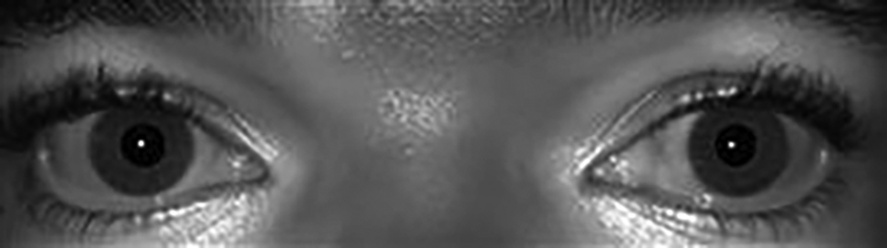
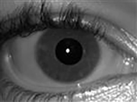
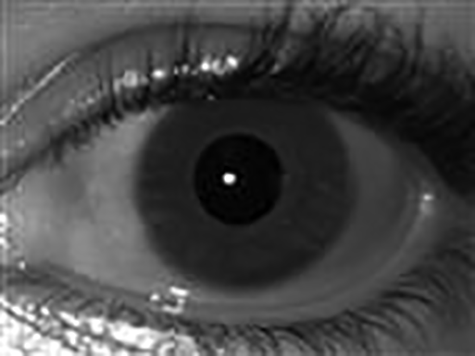
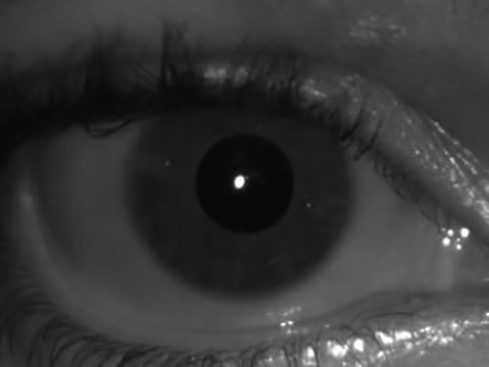
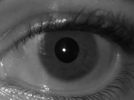
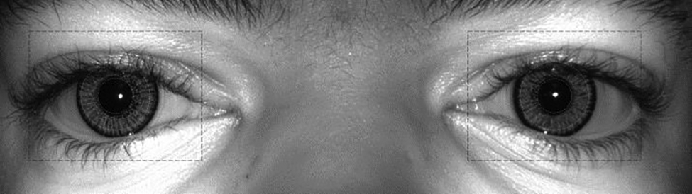
The VistaEY2H dual iris camera used for the automatic capture produced four images per volunteer at each capturing instance. These are the right and left iris images of each volunteer as shown in Figure 5, the right and left iris images of each volunteer with the iris region automatically localized; this is shown in Figure 6, the right iris image of the volunteer as shown in Figure 7 and left iris image of the individual as shown in Figure 8.

(b) Second category of images collected
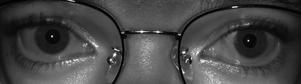
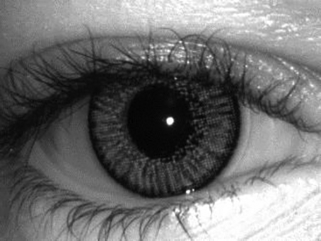
The second category of images was collected from volunteers that wore spectacles. The images generated were the right and left iris images of each volunteer (as shown in Figure 9), the right and left iris images of each volunteer with the iris region automatically localized (this is shown in Figure 10), right iris image of the volunteer as shown in Figure 11 and left iris image of the volunteer as shown in Figure 12.

(c) Third category of images collected
The third category was captured from volunteers that used print-patterned contact lenses. Only four images were captured from volunteers in this category as they were not asked to put on spectacles. However, the use of print-patterned contact lenses was not popular among the population considered, therefore, only eight volunteers used lenses. Examples of these images are provided in Figures 13-16.


(d) Soft biometric features
Soft biometric features of each volunteer were also recorded. These are the age, gender, height, weight, and State of origin of volunteers. For instance, the soft biometric features for volunteer B are shown in Table 1:
To easily distinguish the images, the capturing device automatically generated a unique identification number for each image. For instance, the corresponding unique identification number for each image captured from volunteers A and B are presented in Table 2:
| Sample number | Figures | Unique identification number |
|---|---|---|
| 1. | Figure 1: Right and left irises of volunteer A | Iris_20210429_110713_Dual |
| 2. | Figure 2: Localized right and left irises of volunteer A | Iris_20210429_110713_Diag |
| 3. | Figure 3: Right iris image of volunteer A | Iris_20210429_110713_Right |
| 4. | Figure 4: Left iris image of volunteer A | Iris_20210429_110713_Left |
| 5. | Figure 5: Right and left irises of volunteer A with spectacles | Iris_20210429_110730_Dual |
| 6. | Figure 6: Right and Left Irises of Volunteer A with Spectacles and Localized iris region | Iris_20210429_110730_Diag |
| 7. | Figure 7: Right iris image of volunteer A | Iris_20210429_110730_Right |
| 8. | Figure 8: Left iris image of volunteer A | Iris_20210429_110730_Left |
The supplementary file in Excel format contains the detailed unique identification number for each iris image as well as the biometric features for each volunteer. cell A of the supplementary file is the serial number of the image, cell B contains the last six digits of the unique identification number of a volunteer without spectacles, and cell C contains the last six digits of the unique identification number of the same volunteer with spectacles while cells D to H contain the soft biometric traits of the same volunteer.
This data article has extensively described the experimental setup behind 1056 human iris datasets of African descent collected to enhance iris-related research in the African continent and beyond. It is believed that the collection could serve as a benchmark for evaluating existing and new iris recognition techniques. Most importantly, the dataset could be used to validate results obtained from existing iris-related research that used non-African iris images for their research validation. The authors look forward to creating more iris datasets captured under different circumstances.
| Views | Downloads | |
|---|---|---|
| F1000Research | - | - |
|
PubMed Central
Data from PMC are received and updated monthly.
|
- | - |
Provide sufficient details of any financial or non-financial competing interests to enable users to assess whether your comments might lead a reasonable person to question your impartiality. Consider the following examples, but note that this is not an exhaustive list:
Sign up for content alerts and receive a weekly or monthly email with all newly published articles
Already registered? Sign in
The email address should be the one you originally registered with F1000.
You registered with F1000 via Google, so we cannot reset your password.
To sign in, please click here.
If you still need help with your Google account password, please click here.
You registered with F1000 via Facebook, so we cannot reset your password.
To sign in, please click here.
If you still need help with your Facebook account password, please click here.
If your email address is registered with us, we will email you instructions to reset your password.
If you think you should have received this email but it has not arrived, please check your spam filters and/or contact for further assistance.


Comments on this article Comments (0)