Keywords
Transfersomes, Nanoemulsion, Centella asiatica, Rosemary, Skin aging, UVB radiation
RETRACTION NOTICE (6th October 2023): The article by Khotimah H, Dewi Lestari Ismail D, Widasmara D et al. ‘Ameliorative effect of gel combination of Centella
... a extract transfersomes and rosemary essential oil nanoemulsion against UVB-induced skin aging in Balb/c mice’ [version 1; peer review: 3 approved with reservations]. F1000Research 2022, 11:288. (https://doi.org/10.12688/f1000research.109318.1) has been retracted from F1000Research by the F1000 Editorial Team.During prepublication checks of a new article by these authors, the F1000 Editorial Team noticed that the dataset and methodology of the new article were very similar to this published article. Investigation into the content of these two articles was performed by the F1000 Editorial Team, which revealed discrepancies in the ethical approval (approval document mentioned rats when the study was in mice; new article mentioned consent when study in animals) and methodology (wrong description of mouse model used); and concerns over the treatment of the mice (wellbeing concerns during shaving procedure and euthanasia) and authenticity of the data. The new article was rejected.
For this published article, the F1000 Editorial Team requested an explanation from the authors regarding the above issues, including a request for a copy of the ethical approval document, on 22nd March and 29th March 2023. The authors' institution was contacted on 24th April and 21st June 2023. The authors were unable to respond adequately to our queries; the institution has not responded to our requests.
The decision was made by the F1000 Editorial Team to retract this article, due to the inability to verify the ethical approval for this study, and concerns over data integrity and the study design, including the treatment of the mice.
The authors have seen a copy of this retraction notice. Husnul Khotimah and Dina Dewi Lestari Ismail agree with the retraction of the article. No responses were received from the other authors.
This article is included in the Nanoscience & Nanotechnology gateway.
RETRACTION NOTICE (6th October 2023): The article by Khotimah H, Dewi Lestari Ismail D, Widasmara D et al. ‘Ameliorative effect of gel combination of Centella asiatica extract transfersomes and rosemary essential oil nanoemulsion against UVB-induced skin aging in Balb/c mice’ [version 1; peer review: 3 approved with reservations]. F1000Research 2022, 11:288. (https://doi.org/10.12688/f1000research.109318.1) has been retracted from F1000Research by the F1000 Editorial Team.
During prepublication checks of a new article by these authors, the F1000 Editorial Team noticed that the dataset and methodology of the new article were very similar to this published article. Investigation into the content of these two articles was performed by the F1000 Editorial Team, which revealed discrepancies in the ethical approval (approval document mentioned rats when the study was in mice; new article mentioned consent when study in animals) and methodology (wrong description of mouse model used); and concerns over the treatment of the mice (wellbeing concerns during shaving procedure and euthanasia) and authenticity of the data. The new article was rejected.
For this published article, the F1000 Editorial Team requested an explanation from the authors regarding the above issues, including a request for a copy of the ethical approval document, on 22nd March and 29th March 2023. The authors' institution was contacted on 24th April and 21st June 2023. The authors were unable to respond adequately to our queries; the institution has not responded to our requests.
The decision was made by the F1000 Editorial Team to retract this article, due to the inability to verify the ethical approval for this study, and concerns over data integrity and the study design, including the treatment of the mice.
The authors have seen a copy of this retraction notice. Husnul Khotimah and Dina Dewi Lestari Ismail agree with the retraction of the article. No responses were received from the other authors.
Transfersomes, Nanoemulsion, Centella asiatica, Rosemary, Skin aging, UVB radiation
Editorial note (24th April 2023): Since publication, the Editorial team have been made aware of multiple concerns with this article regarding the ethical approval, treatment of mice, description of the mouse model, and authenticity of the data. F1000 have contacted the authors’ institution to gain clarification about these concerns. This Editorial Note has been posted to inform readers of a potential, not yet resolved, issue with this article, and will remain in place until clarifications are gained from the institution. Peer review activity has been suspended for this article until this time.
People's life expectancy is increasing with age and economic growth, leading them to care about health and skincare because having smooth skin will increase self-confidence1. Many studies have been carried out and led to for the development of skincare products, especially for inhibiting and delaying skin aging2. Skin aging is promoted by intrinsic and extrinsic factors that cause changes in skin function and esthetics3. Intrinsic aging occurs naturally with age, whereas extrinsic aging is caused by external factors, such as solar ultraviolet B (UVB) radiation4. Premature aging known as photoaging due to UVB radiation causes wrinkling of the skin through decreased collagen biosynthesis and direct degradation of collagen in the extracellular matrix (ECM). Degradation of the protein components of the ECM occurs due to the production of intracellular reactive oxygen species (ROS) generated by UVB radiation, which stimulates the production of mitogen-activated protein kinase (MAPK), and activates activator heterodimer 1 (AP-1) protein consisting of c-Fos and c- Jun 3; these further induce the synthesis of matrix metalloproteinases (MMPs)5. It is known that matrix metalloproteinase-9 (MMP-9) can act as collagenase and gelatinase, which can further degrade and decompose collagen fragments into peptides6. Furthermore, decreased collagen synthesis occurs due to ROS generated from UVB radiation activating the MAPK/NF-κB signaling pathway, and can inhibit the TGF-β/Smad signaling pathway and ultimately reduce collagen biosynthesis7. TGF-β is a specific receptor complex binder on the cell surface, whereas Smad7 is a negative factor in the TGF-β/Smad signaling pathway, Smad7 which interacts with TβRI to prevent Smad2/3 activation8. In addition, to inhibiting collagen biosynthesis and promoting collagen degradation, UVB radiation can stimulate the production of ROS in skin cells thereby increasing oxidative stress levels in the skin9. Therefore, the use of natural ingredients that have antioxidant activity and collagen synthesis is considered as a way to develop anti-aging skin products10.
Centella asiatica L. Urban (CA), commonly known in Indonesia as “Pegagan” has a wide range of uses in Indonesian traditional medicines. CA is widely found in humid regions of topical countries, and has long been known in ethnopharmacology to cure various diseases11. Triterpenoids are the main chemical components of CA and asiaticoside, asiatic acid, madecassoside, and madecassic acid are responsible for its pharmacological activity12. Asiaticoside is a compound of CA, proven to increase the synthesis of type 1 collagen in dermal fibroblast cells through activation of TGF-β, and can be used as a functional ingredient in anti-aging cosmetics13–16. Besides CA, several aromatherapy plant product such as rosemary essential oil (REO) can also prevent aging of the skin due to its biological activity17. The main chemical components of REO including -pinene, camphor, 1-8-cineole, and verbenone, have been reported to exhibit antioxidant, antimicrobial, and anti-aging activities18,19. Many properties make CA and REO potential candidates in preventing skin aging20,21. However, their use in preclinical and clinical pharmacokinetics is limited due to physicochemical characteristics such as a high molecular weight and instability, that impede skin absorption and low bioavailability after topical applications12,22. Therefore, in this study, we developed a lipid-based nanocarrier as a CA and REO delivery system, encapsulated in a topical application to enhance effective and efficient permeation into the skin.
Lipid-based nanocarriers such as transfersomes and nanoemulsions can facilitate the passage of drugs or active compounds through the skin, increase penetration, protect against physicochemical instability and increase their effectiveness23. Transfersomes are also known as liposomes in relation to their preparation method and structural features; however, the difference relates to their deformability and penetration mechanism in the skin. Transfersomes are added with edge activators in the form of surfactants together with phospholipids, so that they are highly deformable, elastic, or highly flexible, which allows passage via intercellular routes to penetrate the stratum corneum24. In a study by Avadhani et al. (2017), an optimized transfersomes synergizing epigallocatechin-3-gallate (EGCG) and hyaluronic acid played an important role in reducing free radicals and lipid peroxidation, as well as inducing MMP in HaCaT cells25. In addition, mangiferin-loaded transfersomes exhibited wound healing activity by increasing mangiferin deposition in the epidermis and dermis, inhibiting oxidative stress, as well as stimulating proliferation26.
Nanoemulsion is a transparent emulsion of oil in water or water in oil with a droplet diameter of 20-200 nm; it has thermodynamically stable properties and can be stabilized by an interface layer27. The physicochemical properties of nanoemulsions can be easily adjusted and modified depending on specific target organs, thereby increasing the bioavailability, stability, and solubility of highly lipophilic drugs and active compounds28. Various studies have been carried out on the potential of nanoemulsions in anti-aging, such as the topical application of coenzyme Q10 (CoQ10) in nanoemulsions that can increase the solubility and permeability of CoQ10 thereby reducing wrinkles and making skin appear smooth29. A recent study by Asasutjarit et al. (2021) reported that andrographolide-loaded nanoemulsion has benefits for transdermal applications, especially for the treatment of skin disorders, by reducing pigmentation, damage, melanin index value, and was shown to heal rat skin after exposure to UVB radiation30. A combination of transfersomes and nanoemulsion can be applied into a semisolid dosage form; in our case, a gel was chosen because it has a high water content to hydrate the skin and quickly spreads when applied31.
In this study, we hypothesized that nanoencapsulation of a CA transfersomes and REO nanoemulsion with a lipid-based nanocarrier as topical delivery systems may act synergistically for the prevention of UVB radiation as well as providing anti-aging effects. To test this hypothesis, we investigated the effects a combined of topical application of gel combination of CA transfersomes and REO nanoemulsion in wrinkle formation, epidermal hyperplasia, and degradation of collagen fibers, the mechanism underlying its ameliorative effects, were studied as well by measuring the expressions of MDA, MMP-9, TGF-β, and type I collagen in the skin by immunohistochemical staining on UVB-induced skin aging in hairless mice.
The CA plants were obtained and certified from UPT Material Medica Batu City, East Java, Indonesia, with an asiaticoside content of 0.29%. Fresh leaves were washed, cut, dried, crushed into a fine powder, and subjected to extraction. Briefly, the CA powder was macerated for 14 hours in distilled ethanol 96% v/v. The solvent in the extract was evaporated at 45°C and low pressure with a rotary evaporator (Büchi Rotavapor R-300 System B-301, Switzerland). After that, the filtrate was evaporated and maintained at -20°C. The CA extract was stored in a dark and dry place until required.
Transfersomes of CA leaf extract was prepared using a thin-film hydration method described by El-Gizawy et al. and Surini et al.32,33. Briefly, CA leaves extract, soya phosphatidylcholine (SPC), tween 80, and tween 80 were dissolved in a flask with methanol and placed in a round-bottom flask. The solution mixture was evaporated using a rotary vacuum evaporator (Buchi V-850 Vacuum Controller, Switzerland) at 37°C, at a maximum speed of 150 rpm, for 70 minutes. After all the solvent evaporated and formed a thin layer on the walls of the flask, the hydration process was continued. Hydration was carried out with phosphate buffer saline (PBS, pH 7,4) containing CA in a rotary vacuum evaporator at 37°C, at a maximum speed of 100 rpm, for 60 minutes. The dispersion was then sonicated with a sonicator for 10 minutes to reduce particle size and then stored in a refrigerator at 4°C for further characterization.
The formulation of the REO nanoemulsion consisted of tween 80, aquades, propylene glycol, and REO. The gel composition formulation contained the CA extract transfersomes, REO nanoemulsion, Carbopol 940, triethanolamine (TEA), and distilled water. Gel preparation was started by dispersing Carbopol 940 into distilled water then keeping it for 24 h at room temperature. Then, the Carbopol gel base was homogenized using a homogenizer at 1.500 rpm until the Carbopol base gel formed. Then, TEA was added to the gel base. Once the gel was made, CA extract transfersomes and REO nanoemulsion were suspended. The gel formulation was homogenized with a stirring speed of 500 rpm for 15 minutes.
The particle size distribution (PS), polydispersity index (PDI), and zeta potential (ZP) of the CA-loaded transfersomes and REO-loaded nanoemulsion were analyzed by dynamic light scattering technique, using Malvern Zetasizer (Zetasizer Nano ZS-90, Version 7.01 Malvern Instruments Ltd, UK). Before analysis, dry CA transfersomes and REO nanoemulsion were resuspended in the distilled water and then vortexed. This dispersion was further diluted with deionized water, and 1 ml of this sample was taken on a disposable plastic cuvette and the reading was taken at 25°C. Size and PDI values were obtained from the correlogram by the ZetaSizer Nano ZS software (Malvern Panalytical, Malvern, UK). Zeta potential was measured by electrophoretic light scattering. All analyses were performed in triplicate at 25°C, and the mean value ± standard deviation (SD) was reported.
The eight week-old male Balb/c hairless mice (n = 24, average weight of 20 ± 22 g) used in this study were obtained from the Laboratory of Pharmacology, Universitas Brawijaya. All mice were maintained in a standard environment condition, consisting of a temperature of 25 ± 2°C, with a relative humidity of 50 ± 5%, and a 12 h light/dark cycle, and received standard food and water ad libitum. Mice were acclimatized for one week before treatment. At the beginning of the experiment, mice were randomly divided into six groups, with four mice in each group (Table 1). In this study, the dorsal skin surface of mice was shaved with a razor on an area of 3 × 3.5 cm2 and kept hairless during the experimental period. The animal experimental protocol was approved by the Ethical Committee, Faculty of Medicine, Universitas Brawijaya, Indonesia (Number 82/EC/KPEK-2/03/2020), and performed in accordance with seven WHO 2011 Standards: 1) Social values, 2) Scientific values, 3) Equitable assessment and benefits, 4) Risks, 5) Persuasion/exploitation, 6) Confidentiality and privacy, and 7) Informed consent, referring to the 2016 CIOMS Guidelines. In addition, the animal study was conducted according to the ARRIVE guidelines 2.0 using the ARRIVE Essential 10 checklist for pre-clinical animal research. All efforts were made to ameliorate any suffering of animals.
CA: Centella asiatica REO: rosemary essential oil.
The frequency of exposure was set to three times a week (Monday, Wednesday, and Friday) for two weeks. UVB radiation exposure was done using an array of two Exo Terra Reptile UVB100 25 watt (Rolf C. Hagen Inc., CA, USA) UVB lamps, which emitted radiation of wavelength 290-320 nm, thrice a week for two weeks. Details of exposure were 125 mJ/cm2 in the first week, 155 mJ/cm2 in the second week so that the total dose reached 840 mJ/cm2. Five minutes before UVB radiation, the shaved dorsal skin of mice a gel combination of CA transfersomes and REO nanoemulsion was applied topically (2 g/mice), spread evenly thrice a week for two weeks. The application schedule is shown in Table 1. Throughout the experimental period, the dorsal skins of mice were photographed on days 1, 3, 7, 9, and 14, using a Panasonic LUMIX DMC-GF8 KIT 12-32mm Camera (Panasonic Corp, Osaka, Japan). The wrinkle formation was evaluated and quantified through the ImageJ 1.53e software (National Institutes of Health, Maryland, USA.). The mice were sacrificed using Ketamine HCl after two weeks, and the skin tissues were collected for further analysis.
The dorsal skin tissue of hairless mice was collected, fixed with 10% formalin neutral buffered solution (Sigma-Aldrich, Chemicals. St. Louis, MO, USA), embedded in paraffin, and cut into parcels of 4 μm thickness, deparaffinized with xylene, and rehydrated through graded alcohol. Hematoxylin and eosin (HE) stains was used for histological observations of skin structure and epidermal thickness. Masson's trichrome stain were used to evaluate the density of collagen fibers in the dermis. HE staining was carried out in several stages starting with deparaffinization, hydration, hematoxylin staining, eosin staining, and finally dehydration. Masson's trichrome staining was also carried out in several stages starting from staining with hematoxylin solution, staining with bietrich scarlet, and fuchsin acid solution. Then, the slides were put in phosphomolybdic acid-phosphotungstic acid, finally, aniline blue was used to stain the collagen epidermal thickness, and collagen fiber density was analyzed at 10 random locations per slide using an Olympus Cx21 light microscope (Olympus Optical Co. Ltd., Tokyo, Japan) with 1000× magnification. Each specimen was photographed under a Sony Alpha A6000 Mirrorless Camera (Sony Corp, Minato-Ku, Tokyo, Japan). Histological alterations and collagen fiber density were evaluated and quantified using the ImageJ 1.53e software (National Institutes of Health, Maryland, USA).
Immunohistochemical staining was used to assess the expression of MDA, TGF-β, MMP-9, and type I collagen in skin tissue. After dehydration, the antigens on the slides were recovered with citrate buffer in a water bath for three minutes, then endogenous peroxidase in the tissue was inactivated with 0.3% hydrogen peroxide for 20 minutes. After that, the slides were incubated with primary antibodies (anti-MDA, anti-TGF-β, anti-MMP-9, or anti-type I collagen) at 1:200 at 4°C overnight. A secondary antibody, Biotin Conjugate (1:200) was added to slide sections and incubated for 30 min at room temperature. After PBS washing, the slides were incubated with Strep Avidin Horseradish Peroxidase (SAHRP) for 30 minutes and washed with PBS-Tween 20 buffer; then, the slides were stained with a 3,30-diaminobenzidine (DAB) solution and counter-stained with hematoxylin. Finally, the slides were dehydrated, mounted, and observed under an Olympus Cx21 light microscope (Olympus Optical Co. Ltd., Tokyo, Japan). The images were photographed using a Sony Alpha A6000 mirrorless camera (Sony Corp, Minato-Ku, Tokyo, Japan). The quantitative analysis of the expression of each photo was analyzed using ImageJ 1.53e software (developed by the National Institutes of Health, Maryland, USA).
Results are presented as mean ± standard deviation. Differences between groups was analyzed by one-way analysis of variance (ANOVA) followed by post hoc Tukey test analysis using SPSS software (SPSS, Version 21.0, IBM Inc. USA). The difference was considered statistically significant when the p-value < 0.05.
The physicochemical properties of lipid-based nanocarriers affected drug delivery through the skin; in addition, vesicle size lower than 200 nm increased transdermal and topical delivery. Characterization results of CA-loaded transfersomes and REO-loaded nanoemulsion in terms of particle size, polydispersity index, and zeta potential are illustrated in Table 2. The average particle size of the CA-loaded transfersomes and REO-loaded nanoemulsion were less than 200 nm (43.97 ± 5.6 nm, and 7.03 ± 0.95 nm respectively). In addition, the CA-loaded transfersomes and REO-loaded nanoemulsion had average PDI values of 0.64 ± 0.01, and 0.72 ± 0.10 respectively, PDI value between 0.08 and 0.70 is an indication of acceptable size distribution34. The zeta potential values negatively charged for the tested CA transfersomes (-10.91 ± 1.99 mV), and REO nanoemulsion (-4.73 ± 0.82 mV), with values between +30 and −30 mV, indicating good stability.
The process of wrinkle formation may indicate the extent of skin damage35. The macroscopic appearance of skin wrinkle formation in mice in the last week of the experimental period for each group was photographed, recorded, and are shown in (Figure 1A). During the experimental period, the control group (normal) showed mild wrinkles, related to age5. After two weeks of UVB exposure, in the UVB group, there was a dry, coarse wrinkle, rough wrinkles, and slight damage to the skin surface. However, the UVB-induced thick macroscopic skin wrinkle changes were significantly ameliorated in the group treated with a gel combination of non-transfersomes or CA extract and REO nanoemulsion (UVB + NT 0.3%) and groups treated with the gel combination of CA transfersomes and REO nanoemulsion (UVB + T 0.15%, UVB + T 0.3%, and UVB + T 0.6% group) in a concentration-dependent manner (Figure 1B). Wrinkle formation was quantified using the ImageJ 1.53e analysis software. These results indicate that topical application of a gel combination of CA transfersomes and REO nanoemulsion could act synergistically and effectively prevent UVB-induced skin damages compared to non-transfersomes or CA extract only.
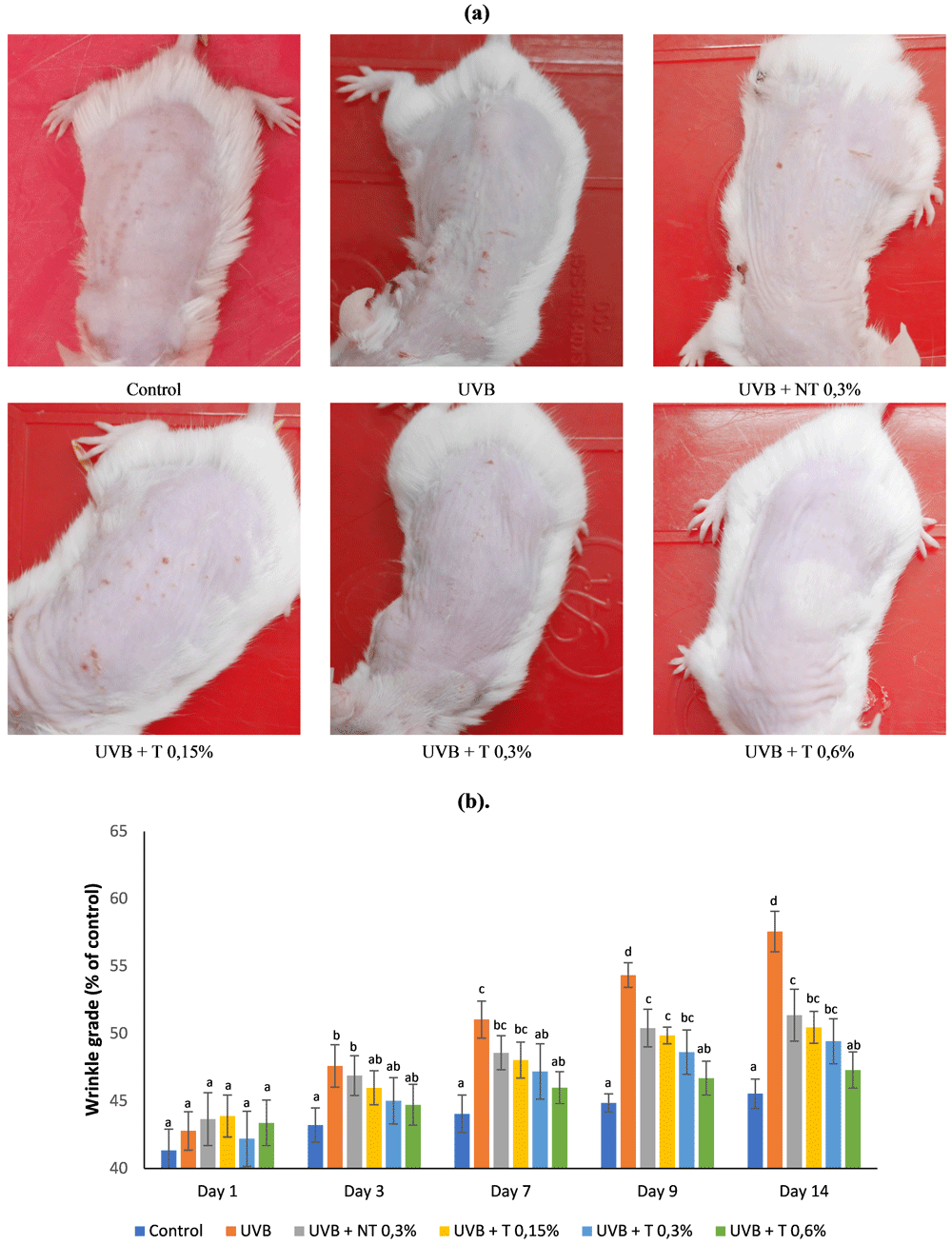
Mice were radiated with UVB (840 mJ/cm2) three times a week for two weeks. Control group: normal; UVB group: UVB exposure + topically applied with Carbopol base gel; UVB + NT 0.3% group: UVB exposure + topically applied with gel combination 0.3% CA non-transfersomes (CA extract) and REO 1% nanoemulsion; UVB + T 0.15% group: UVB exposure + topical application of gel combination of 0.15% CA transfersomes and 1% REO nanoemulsion; UVB + T 0.3% group: UVB exposure + topically applied with gel combination 0.3% CA transfersomes and 1% REO nanoemulsion; UVB + T 0.6% group: UVB exposure + topical application of gel combination 0.6% CA transfersomes and 1% REO nanoemulsion. (a). Skin macroscopic appearances at the end of the experiment period. (b). The quantitative analysis of wrinkle. Quantitative analysis of wrinkles was performed by ImageJ 1.53e software. Data are presented as mean ± SD (n=4). Statistical analysis was done by one-way ANOVA followed by post hoc test analysis using the SPSS software. Data with different notation in the same chart implied a significant difference (p < 0.05).
Epidermal thickness is a histological parameter that reflects UVB-induced skin damage. HE staining was used to analyze the histological effects of gel combination of CA transfersomes and REO nanoemulsion (Figure 2A). The mean values of different skin thicknesses were calculated by measuring the 10 chosen locations for each section. HE staining showed that the skin epidermis appeared thin and homogeneous with a thin layer of stratum corneum for the control group; the average thickness of the epidermis was 48.8 μm. In comparison, the UVB-treated group showed that the epidermis on the dorsal skin of mice was significantly thicker at 83.8 μm (Figure 2B). However, UVB-induced epidermal thickness on the dorsal skin of mice was significantly decreased upon topical application with a gel combination of non-transfersomes or CA extract and REO nanoemulsion (UVB + NT 0.3% group) by 68.57 μm compared to the UVB group. When the gel combination of CA transfersomes and REO nanoemulsion (UVB + T 0.15%, UVB + T 0.3%, and UVB + T 0.6% groups) was applied topically, the epidermal thickness was significantly decreased to 57.8 μm, 54.1 μm, and 51.95 μm respectively, in a concentration-dependent manner (all p < 0.05 versus UVB group). These results indicated that topical application of gel combination of CA transfersomes and REO nanoemulsion could synergistically have an anti-aging effect and significantly reduce the increased thickness of the stratum corneum and epidermis induced by UVB radiation compared to non-transfersomes or CA extract only.
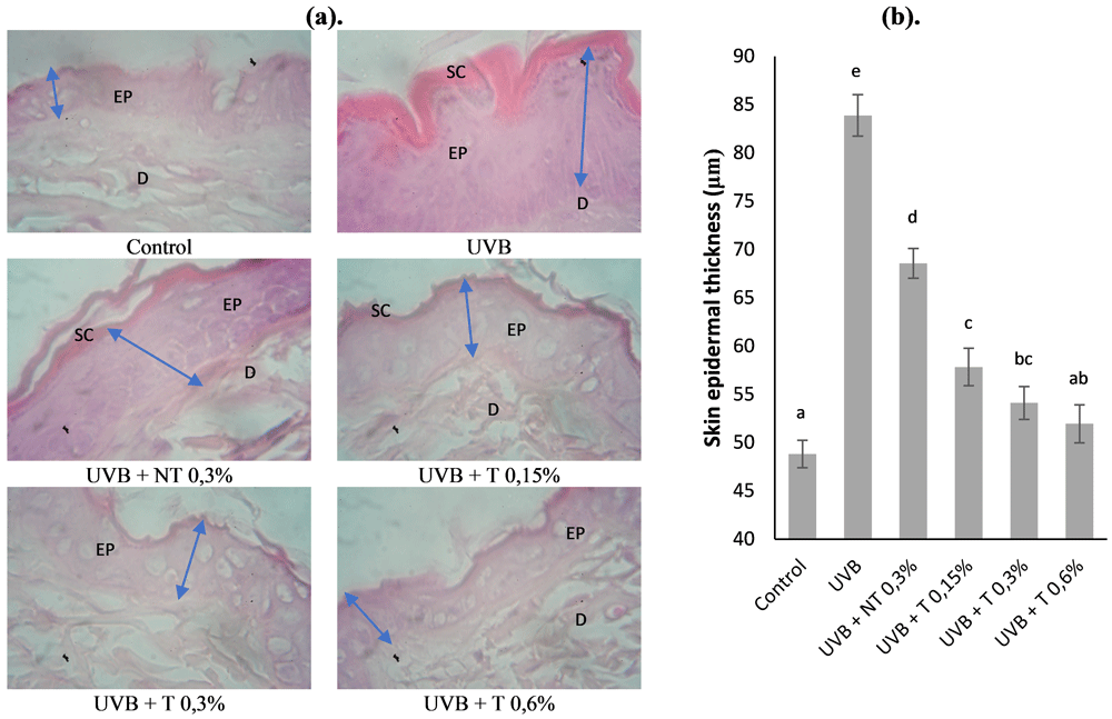
Mice were radiated with UVB (840 mJ/cm2) three times a week for two weeks. Control group: normal; UVB group: UVB exposure + topical application of base gel Carbopol; UVB + NT 0.3% group: UVB exposure + topical application of gel combination 0.3% CA non-transfersomes (CA extract) and REO 1% nanoemulsion; UVB + T 0.15% group: UVB exposure + topical application of gel combination 0.15% CA transfersomes and 1% REO nanoemulsion; UVB + T 0.3% group: UVB exposure + topical application of gel combination of 0.3% CA transfersomes and 1% REO nanoemulsion; UVB + T 0.6% group: UVB exposure + topical application of gel combination of 0.6% CA transfersomes and 1% REO nanoemulsion. (a). Dorsal skin sections were stained with hematoxylin and eosin (H&E) (magnification 1000×); scale bar = 50 μm. (b). The quantitative analysis of epidermal thicknesses. Quantitative analysis of epidermal thicknesses was performed using ImageJ 1.53e software. Data are presented as mean ± SD (n=4). Statistical analysis was done by one-way ANOVA followed by post hoc test analysis using SPSS software. Data with different notation in the same chart implied a significant difference (p < 0.05). SC: stratum corneum; EP: epidermis; D: dermis.
Collagen fiber density and irregular arrangement of collagen fibers are manifestations of UVB-induced skin damage36, so Masson's trichrome staining was used to determine the effect of the gel combination of CA transfersomes and REO nanoemulsion on collagen fiber damage due to UVB exposure (Figure 3A). In the control group, the density of collagen fibers (blue) was mostly found in the dermis of the dorsal skin of mice (Figure 3A). However, in the UVB group, the collagen fiber density significantly decreased by 4.75% in the dermis compared to the control group (Figure 3B). In contrast, in the UVB + NT 0.3% group, the reduction in collagen fiber density appeared to be greater in the dermis compared to the UVB group Figure 3B. While topical application with the gel combination of CA transfersomes and REO nanoemulsion significantly reversed the reduction of collagen fibers triggered by UVB by 11.7%, 15.5%, and 17.2% in the UVB + T 0.15%, UVB + T 0.3%, and UVB + T 0.6% groups respectively, in a concentration-dependent manner (all p < 0.05 versus UVB group). These results demonstrate that UVB radiation contributes to a reduction in collagen fibers. In contrast, the reduction of collagen fibers density was markedly reversed by topical application of gel combination of CA transfersomes and REO nanoemulsion, which could have better effects than non-transfersomes or CA extract only through synergistic activity.
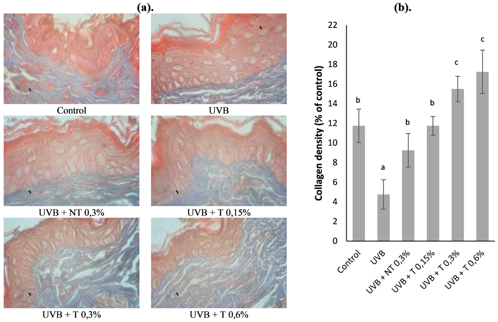
Mice were radiated with UVB (840 mJ/cm2) three times a week for two weeks. Control group: normal; UVB group: UVB exposure + topical application of base gel Carbopol; UVB + NT 0.3% group: UVB exposure + topical application of gel combination of 0.3% CA non-transfersomes (CA extract) and REO 1% nanoemulsion; UVB + T 0.15% group: UVB exposure + topical application of gel combination of 0.15% CA transfersomes and 1% REO nanoemulsion; UVB + T 0.3% group: UVB exposure + topical application of gel combination of 0.3% CA transfersomes and 1% REO nanoemulsion; UVB + T 0.6% group: UVB exposure + topical application of gel combination of 0.6% CA transfersomes and 1% REO nanoemulsion. (a) Masson’s trichrome staining for the visualization of collagen fibers (magnification 1000×); scale bar = 50 μm; slices stained with dark blue indicate collagen fiber, and red indicate cytoplasm and muscle fiber. (b) Quantitative analysis of collagen fiber density. Quantitative analysis of collagen fiber density was performed using ImageJ 1.53e software. Data are presented as mean ± SD (n=4). Statistical analysis was done by one-way ANOVA followed by post hoc test analysis using SPSS software. Data with different notation in the same chart implied a significant difference (p < 0.05).
MDA is one of the main parameters of oxidative stress and a by-product of lipid peroxidation produced by ROS37. As shown in Figure 4A, MDA expression in the UVB group was significantly increased, compared with the control group. The UVB + NT 0.3% group’s topical application caused a reduction in MDA expression compared to the UVB group, but there was no significant difference in these groups. In the three groups treated with topical gel combination of CA transfersomes and REO nanoemulsion the skin MDA expression induced by UVB was significantly suppressed in the UVB + T 0.15%, UVB + T 0.3%, and UVB + T 0.6% groups respectively (all p < 0.05 versus UVB group) in a concentration-dependent manner. Interestingly, UVB + T 0.6 % individuals showed no significant difference compared to the control group (non-UVB exposure). The gel combination of CA transfersomes and REO nanoemulsion had a greater effect than non-transfersomes or CA extract on the reduction of oxidative stress in the skin induced by UVB radiation through the improvement of lipid peroxidation38.
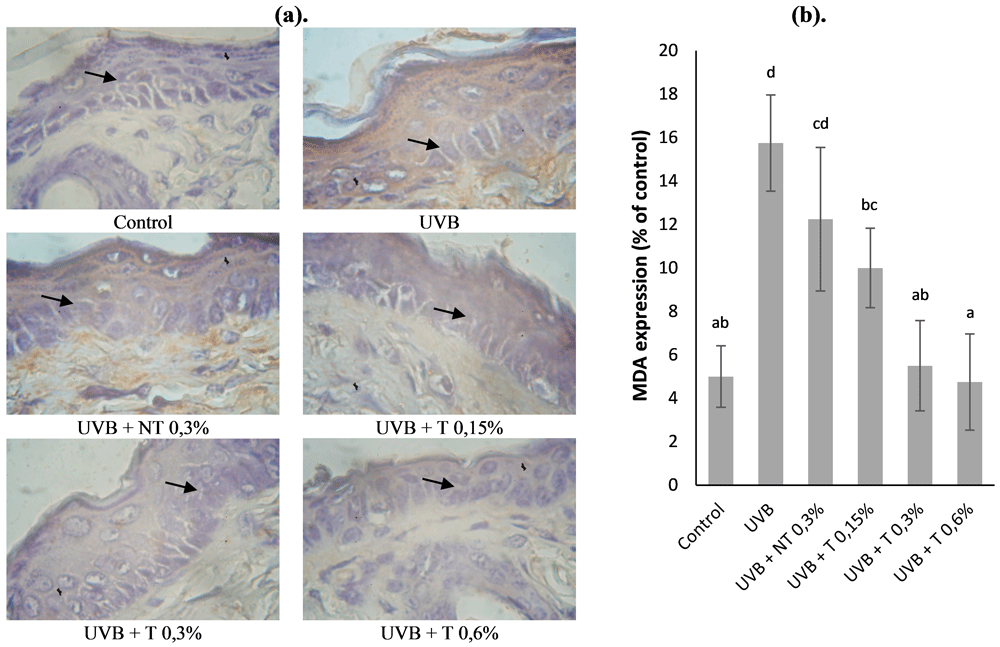
Mice were radiated with UVB (840 mJ/cm2) three times a week for two weeks. Control group: normal; UVB group: UVB exposure + topical application of base gel Carbopol; UVB + NT 0.3% group: UVB exposure + topical application of gel combination of 0.3% CA non-transfersomes (CA extract) and REO 1% nanoemulsion; UVB + T 0.15% group: UVB exposure + topical application of gel combination of 0.15% CA transfersomes and 1% REO nanoemulsion; UVB + T 0.3% group: UVB exposure + topical application of gel combination of 0.3% CA transfersomes and 1% REO nanoemulsion; UVB + T 0.6% group: UVB exposure + topical application of gel combination of 0.6% CA transfersomes and 1% REO nanoemulsion. (a) Immunohistochemistry sections of MDA (magnification 1000×); scale bar = 50 μm; brown staining indicates positive cells. (b) Quantitative analysis of MDA expression. Quantitative analysis of expression intensity was performed using ImageJ 1.53e software. Data are presented as mean ± SD (n=4). Statistical analysis was done by one-way ANOVA followed by post hoc test analysis using the SPSS software. Data with different notation in the same chart implied a significant difference (p < 0.05).
The TGF-β expression in mice skin was significantly suppressed by 9.25% after UVB radiation (UVB group versus control group) (Figure 5B). In contrast, TGF-β expression was increased by topical application of gel combination of CA non-transfersomes (CA extract) and REO nanoemulsion (UVB + NT 0.3% group) and gel combination of CA transfersomes and REO nanoemulsion (UVB + T 0.15%). There was no significant difference between the UVB + NT 0.3% and UVB + T 0.15% groups regarding the expression of TGF-β compared to that in the UVB group. However, the expression of TGF-β was much higher by 13.25% and 16.5% in the UVB + NT 0.3% and UVB + T 0.15% groups respectively, than in the UVB group. When the gel combination of CA transfersomes and REO nanoemulsion was topically applied (UVB + T 0.3% and UVB + T 0.6% groups), the expression was significantly increased by 18.25% and 22.25%, respectively that in the UVB group (Figure 5B). These results suggest that the antioxidant effect of the gel combination of CA transfersomes and REO nanoemulsion could act synergistically and increase collagen density. However, whether the gel combination of CA transfersomes and REO nanoemulsion directly mediates the interaction of the TGF-β/Smad signaling pathway and collagen synthesis gene expression remains to be elucidated.
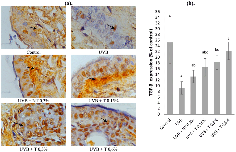
Mice were radiated with UVB (840 mJ/cm2) three times a week for two weeks. Control group: normal; UVB group: UVB exposure + topical application of base gel Carbopol; UVB + NT 0.3% group: UVB exposure + topical application of gel combination of 0.3% CA non-transfersomes (CA extract) and REO 1% nanoemulsion; UVB + T 0.15% group: UVB exposure + topical application of gel combination of 0.15% CA transfersomes and 1% REO nanoemulsion; UVB + T 0.3% group: UVB exposure + topical application of gel combination of 0.3% CA transfersomes and 1% REO nanoemulsion; UVB + T 0.6% group: UVB exposure + topical application of gel combination of 0.6% CA transfersomes and 1% REO nanoemulsion. (a) Immunohistochemistry sections of TGF-β (magnification 1000×); scale bar = 50 μm; brown staining indicates positive cells. (b) Quantitative analysis of TGF-β expression. Quantitative analysis of expression intensity was performed by ImageJ 1.53e software. Data are presented as mean ± SD (n=4). Statistical analysis was done by one-way ANOVA followed by post hoc test analysis using the SPSS software. Data with different notation in the same chart implied a significant difference (p < 0.05).
In this study, we analyzed the expression of MMP-9 to determine the underlying molecular mechanism of the gel combination of CA transfersomes and REO nanoemulsion as a protective effect against UVB-induced collagen degradation (Figure 6A). As expected, UVB exposure increased MMP-9 expression in the skin tissues of mice. The UVB group had a significantly increased MMP-9 expression by 14.25% of compared to the control group. As shown in Figure 6B, the MMP-9 expression was lowered by 10 %, but no significant difference was found between the UVB + NT 0.3% and UVB groups. MMP-9 expression was significantly decreased upon topical application of gel combination of CA transfersomes and REO nanoemulsion in UVB + T 0.15%, UVB + T 0.3%, and UVB + T 0.6% groups, showing an approximate 6.75%, 6.25%, and 4.5% reduction respectively compared to the control UVB group (Figure 6B). Therefore, topical application of gel combination of CA transfersomes and REO nanoemulsion could significantly suppress the UVB-induced increase in MMP-9 expression through synergistic action.
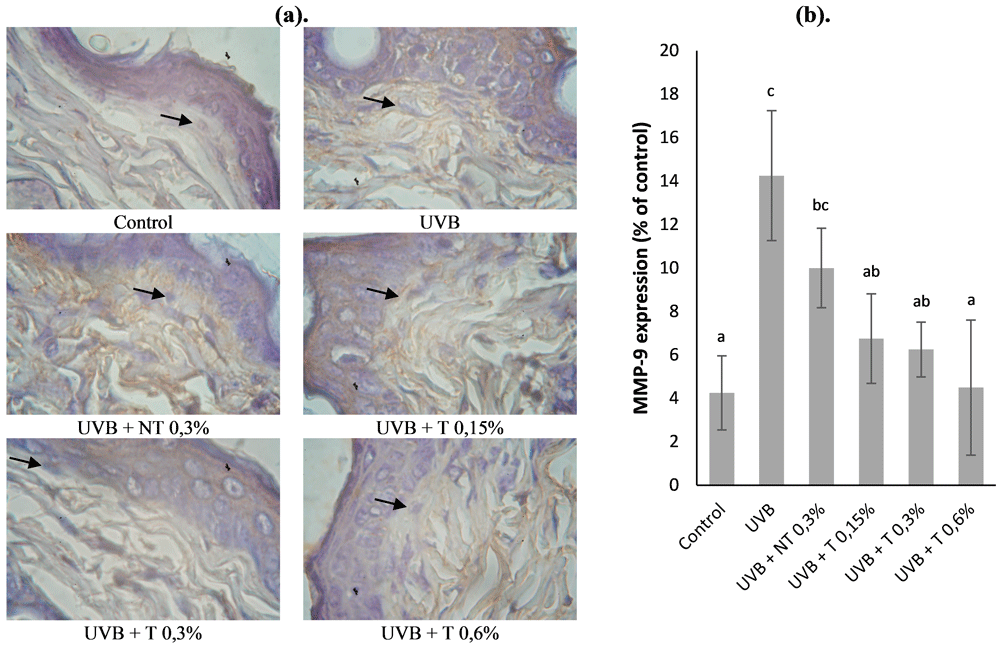
Mice were radiated with UVB (840 mJ/cm2) three times a week for two weeks. Control group: normal; UVB group: UVB exposure + topical application of base gel Carbopol; UVB + NT 0.3% group: UVB exposure + topical application of gel combination of 0.3% CA non-transfersomes (CA extract) and REO 1% nanoemulsion; UVB + T 0.15% group: UVB exposure + topical application of gel combination of 0.15% CA transfersomes and 1% REO nanoemulsion; UVB + T 0.3% group: UVB exposure + topical application of gel combination of 0.3% CA transfersomes and 1% REO nanoemulsion; UVB + T 0.6% group: UVB exposure + topical application of gel combination of 0.6% CA transfersomes and 1% REO nanoemulsion. (a) Immunohistochemistry sections of MMP-9 (magnification 1000×); scale bar = 50 μm; brown staining indicates positive cells. (b) Quantitative analysis of MMP-9 expression. Quantitative analysis of expression intensity was performed using ImageJ 1.53e software. Data are presented as mean ± SD (n=4). Statistical analysis was done by one-way ANOVA followed by post hoc test analysis using SPSS software. Data with different notation in the same chart implied a significant difference (p < 0.05).
Type I collagen expression was analyzed using immunohistochemistry to determine the effect of the gel combination of CA transfersomes and REO nanoemulsion; the results are shown in Figure 7A. Type I collagen gene expression in the UVB group was significantly decreased by 9% in the dorsal skin compared to those in the control group (Figure 7B). The expression of type I collagen was not significantly different between the UVB + NT 0.3% and UVB and the UVB + T 0.15% groups. However, the expression of type I collagen was much higher by 11.75%, and 12.75%, respectively, in the UVB + NT 0.3%, and UVB + T 0.15% groups than in the UVB group. When the gel combination of CA transfersomes and REO nanoemulsion was topically applied (UVB + T 0.3% and UVB + T 0.6% groups), the expression was significantly increased by 16.5% and 22% respectively compared to that in the UVB group (Figure 7B). The results showed that UVB exposure contributed to a reduction in type I collagen expression. In contrast, the reduction of type I collagen expression was markedly prevented by topical application of the gel combination of CA transfersomes and REO nanoemulsion (especially the UVB + T 0.6% group).
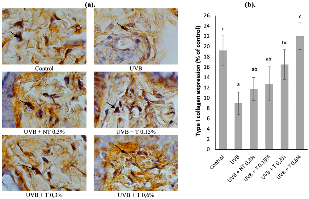
Mice were radiated with UVB (840 mJ/cm2) three times a week for two weeks. Control group: normal; UVB group: UVB exposure + topical application of base gel Carbopol; UVB + NT 0.3% group: UVB exposure + topical application of gel combination of 0.3% CA non-transfersomes (CA extract) and REO 1% nanoemulsion; UVB + T 0.15% group: UVB exposure + topical application of gel combination of 0.15% CA transfersomes and 1% REO nanoemulsion; UVB + T 0.3% group: UVB exposure + topical application of gel combination of 0.3% CA transfersomes and 1% REO nanoemulsion; UVB + T 0.6% group: UVB exposure + topical application of gel combination of 0.6% CA transfersomes and 1% REO nanoemulsion. (a) Immunohistochemistry sections of type I collagen (magnification 1000×); scale bar = 50 μm; brown staining indicates positive cells. (b) Quantitative analysis of type I collagen expression. Quantitative analysis of expression intensity was performed using ImageJ 1.53e software. Data are presented as mean ± SD (n=4). Statistical analysis was done by one-way ANOVA followed by post hoc test analysis using SPSS software. Data with different notation in the same chart implied a significant difference (p < 0.05).
Interest in anti-aging has grown with the increase in people's life expectancy; in particular various efforts are made to prevent skin aging, and many studies have been carried out and dedicated to skin health and beauty39. Skin aging factors can be classified as intrinsic or extrinsic. Extrinsic aging or premature skin aging is caused by external factors, such as exposure to UVB radiation, thereby increasing skin damage such as sagging, wrinkle formation, and skin roughness40. This study was designed to investigate the preventive effect of a gel combination of the CA transfersomes and REO nanoemulsion on skin aging, using a UVB-exposed BALB/c mice skin model to explore the underlying anti-aging mechanism. The delivery method becomes important if CA and REO are anti-aging because the drug must pass through complex skin structures such as the stratum corneum. Many studies of lipid-based nanocarriers have been carried out to transfer large proteins and peptides to the skin via transfersomes and nanoemulsions41,42. For ease of use in topical application to the skin, a combination of transfersomes and nanoemulsions can be formulated into semisolids. In a recent study by Surini et al. (2020), recombinant human epidermal growth factor (rhEGF) encapsulated into transfersomal emulgels showed increased skin penetration33. Osmotic force theory is the main mechanism associated with skin penetration of transfersomes43. Lipid-based nanocarriers systems are encapsulated into transfersomes and nanoemulsions for topical and transdermal drug delivery based on skin penetration, generally having a average particle size distribution between 1-100 nm44. In the present study, the particle size distributions of CA transfersomes and REO nanoemulsion were less than 70 nm. Additionally, the polydispersity indices of the CA transfersomes and REO nanoemulsion were appropriately less than 0.8. In topical drug delivery using lipid-based nanocarriers, PDI values greater than 0.7 indicate that the sample has a very broad particle size distribution and is probably not suitable to be analysed by dynamic light scattering (DLS) technique, while a PDI value below 0.7 indicates a very uniform and acceptable vesicle sample population45. This study showed the average zeta potential values negatively charged of the CA transfersomes and REO nanoemulsion encapsulated with lipid-based nanocarriers (Table 2), the zeta potential values between +30 and −30 mV indicating good stability. The negative surface charge has the potential to enhance skin penetration46. These findings suggest that transfersomes and nanoemulsions can be an effective approach to enhancing skin delivery via topical and transdermal applications.
The appearance of skin wrinkles, roughness, sagging and other aging processes due to direct UVB exposure is also associated with epidermal thickness, disorganization of connective tissue, and collagen fibers47. In this study, we demonstrated that topical application of a gel combination of CA transfersomes and REO nanoemulsion significantly alleviated wrinkle formation, epidermal hyperplasia, and density of collagen fibers caused by UVB exposure. Epidermal hyperplasia is one of the histological parameters used to determine the level of skin damage due to UVB exposure48. In addition, epidermal hyperplasia also contributes to wrinkle formation49. As expected, the topical application of a gel combination of CA transfersomes and REO nanoemulsion reduced these effects and was shown to minimize skin damage and the appearance of wrinkles. Skin aging caused by UVB exposure increases collagen breakdown and decreases collagen synthesis, resulting in an overall reduction in collagen levels50. As expected, the gel combination of CA transfersomes and REO nanoemulsion had a protective effect against UVB radiation-induced pathological changes of collagen fibers in skin aging, as observed by Masson's trichrome staining (Figure 3). Collagen is the main component of ECM which is responsible for maintaining tensile strength, wrinkle formation, skin resistance and can be directly degraded by UVB exposure8. Moreover, the abnormal arrangement of collagen fiber density in the dermis due to UVB radiation could be inhibited by the topical application of gel combination of CA transfersomes and REO nanoemulsion.
Generally, all cells possess three natural antioxidant enzymes: catalase (CAT), glutathione peroxidase (GSH-Px), and superoxide dismutase (SOD), which act as a defense system to scavenge free radicals and repair oxidative damage, and play an important role in oxidative balance51. The pathogenesis of UVB-induced skin aging is associated with an imbalance between antioxidants and free radicals, resulting in elevated intracellular ROS levels and oxidative stress, which severely destroys the skin's antioxidant defense system52. A study has previously demonstrated that UVB exposure in hairless mice caused a significant decrease in SOD, GSH-Px, and CAT activity, but increased MDA activity37. As expected, UVB exposure in hairless mice led to an increase in MDA activity. However, comparisons between antioxidant activities were not performed in this study, as the focus was on oxidative stress activity. MDA is the product of lipid peroxidation degradation which is an important biological marker of oxidative stress and was significantly increased in UVB-irradiated mice skin36. It is well known that UVB exposure can result in the production of ROS in skin cells, which further promotes lipid peroxidation by reacting with unsaturated fatty acids in cell membranes, which in turn enhances collagen degradation53. In this study, topical application of a gel combination of CA transfersomes and REO nanoemulsion on the skin (especially with the concentration of 0.6% CA transfersomes and 1% REO nanoemulsion) significantly decreased the upregulated MDA expression induced by UVB exposure. The accumulation of MDA lipid peroxidation products increase oxidative stress in skin tissues38, which we verified by macroscopic and histological studies. The results showed that the topical application of a gel combination of CA transfersomes and REO nanoemulsion improved skin structure and epidermal thickness, which further affects the macroscopic appearance of the skin and prevents the occurrence of inflammation and skin damage due to UVB exposure.
Oxidative stress due to UVB exposure stimulates MAPK and activates AP-1, which further induces the synthesis of MMPs54. In addition, MMPs play an important role in the physiological mechanisms of skin aging, including through the degradation of collagen and elastin in the ECM55. Several types of MMPs are elevated in response to UVB exposure and indicate skin aging, such as MMP-1, MMP-8, MMP-13, and MMP-18 (collagenase), MMP-2 and MMP-9 (gelatinase), MMP-3, MMP-10, MMP-11, and MMP-19 (stromelysin), and MMP-14, MMP-15, MMP-16, MMP-17, MMP-24, and MMP-25 (membrane-associated)54. MMP-9 (gelatinase) plays a major role in the degradation of type IV collagen and gelatin, degrades basement membrane collagen and denatures structural collagen, and is also responsible for wrinkle formation56. This study shows that topical application of a gel combination of CA transfersomes and REO nanoemulsion on the skin (especially with the concentration of 0.6% CA transfersomes and 1% REO nanoemulsion) significantly suppressed the UVB-induced increase of MMP-9 expression (Figure 6). In addition, these results also correlated with macroscopic and histological studies, where the topical application of a gel combination of CA transfersomes and REO nanoemulsion significantly increased the density of collagen fibers, inhibited epidermal thickness, which in turn affected the macroscopic appearance of the skin, such as reducing the formation of wrinkles and erythema. To investigate this mechanism further, we verified the expression of TGF-β, since the TGF-β/Smad pathway is the main pathway that controls procollagen production35,57.
The dynamic balance between the degradation and synthesis of collagen in the ECM is critical for combating skin aging, whereby collagen synthesis increases via activation of the TGF-β/Smad pathway, while the opposite collagen degradation pathway is associated with the MAPK signaling pathway58. A novel approach in the prevention of photoaging is investigating the molecular mechanism of photoaging, whereby the TGF-β/Smad pathway is the main activator of procollagen production in human skin. It is well known that TGF-β acts through its cell surface receptor to activate the transcription factor Smad 2/3, and acts as an antagonist to Smad759. UVB exposure can impair TGF-β and Smad2/3 signaling, thereby decreasing type 1 procollagen synthesis, and causing collagen loss in the dermis60. Park et al. (2018), reported that UVB exposure downregulates the expression of TGF-β/Smad signaling, but that topical application of Eucalyptus globulus restored this effect35. Similarly, our data showed that UVB exposure also reduced the expression of TGF-β, whereas topical application of a gel combination of CA transfersomes and REO nanoemulsion reversed the UVB-induced expression of TGF-β. The present data indicates that the potential anti-photoaging mechanism of a gel combination of CA transfersomes and REO nanoemulsion may be related to the up-regulation of TGF-β expression, which stimulates the phosphorylation of the Smad-related pathway, followed by suppression of the reduction of type I collagen expression caused by UVB exposure35.
Collagen is a major protein in the ECM and an important indicator of skin aging because it is closely related to the mechanical properties and structure of the skin, and a stable collagen structure is the basis of skin elasticity61. Collagen type I and collagen type III are the main parameters in anti-aging effects and are associated with elasticity properties, where the expression of type I collagen decreases, while type III collagen increases8. In addition, Young et al. (2019), reported that UVB exposure inhibited collagen synthesis, mainly through decreased expression of type I and III collagen62. In the present study, UVB exposure contributed to a reduction in type I collagen expression. In contrast, the decrease of type I collagen expression was significantly prevented by topical application of a gel combination of CA transfersomes and REO nanoemulsion (especially with the concentration of 0.6% CA transfersomes and 1% REO nanoemulsion), which could effectively act synergestically.
This study showed that lipid-based nanocarriers containing two bioactive molecules of CA transfersomes and REO nanoemulsion synergistically acted as a topical drug delivery systems with anti-aging benefits. The topical application of a gel combination CA transfersomes and REO nanoemulsion can effectively ameliorate UVB-induced macroscopic aspect (wrinkle formation) and histological appearance of the skin by downregulating the lipid peroxidation and MMPs’ expression while increasing TGF-β/Smad pathway and synthesis of type I collagen in the skin.
Figshare: Supplementary Data – Ameliorative effect of gel combination of Centella asiatica extract transfersomes and rosemary essential oil nanoemulsion against UVB-induced skin aging in balb/c mice, https://doi.org/10.6084/m9.figshare.17399351.v363
This project contains the following underlying data:
Raw data ameliorative effect of gel combination of CA transfersomes and REO nanoemulsion.xlsx
Supporting information ameliorative effect of gel combination of CA transfersomes and REO nanoemulsion.docx
Statistic result effect of gel combination of CA transfersomes and REO nanoemulsion.docx
Images showing skin macroscopic appearances at the end of the experiment period
skin macroscopic appearances control.jpg
skin macroscopic appearances UVB.jpg
skin macroscopic appearances UVB + NT 0,3%.jpg
skin macroscopic appearances UVB + T 0,15%.jpg
skin macroscopic appearances UVB + T 0,3%.jpg
skin macroscopic appearances UVB + T 0,6%.jpg
Hematoxylin and eosin (HE) stains images for histological observations of skin structure and epidermal thickness
epidermal thicknesses control.jpg
epidermal thicknesses UVB.jpg
epidermal thicknesses UVB + NT 0,3%.jpg
epidermal thicknesses UVB + T 0,15%.jpg
epidermal thicknesses UVB + T 0,3%.jpg
epidermal thicknesses UVB + T 0,6%.jpg
collagen fiber density control.jpg
collagen fiber density UVB.jpg
collagen fiber density UVB + NT 0,3%.jpg
collagen fiber density UVB + T 0,15%.jpg
collagen fiber density UVB + T 0,3%.jpg
collagen fiber density UVB + T 0,6%.jpg
MDA expressions control.jpg
MDA expressions UVB.jpg
MDA expressions UVB + NT 0,3%.jpg
MDA expressions UVB + T 0,15%.jpg
MDA expressions UVB + T 0,3%.jpg
MDA expressions UVB + T 0,6%.jpg
TGF-β expressions control.jpg
TGF-β expressions UVB.jpg
TGF-β expressions UVB + NT 0,3%.jpg
TGF-β expressions UVB + T 0,15%.jpg
TGF-β expressions UVB + T 0,3%.jpg
TGF-β expressions UVB + T 0,6%.jpg
Figshare: The ARRIVE Essential 10: author checklist – Ameliorative effect of gel combination of Centella asiatica extract transfersomes and rosemary essential oil nanoemulsion against UVB-induced skin aging in balb/c mice, https://doi.org/10.6084/m9.figshare.19222071.v164
Data are available under the terms of the Creative Commons Attribution 4.0 International license (CC-BY 4.0).
| Views | Downloads | |
|---|---|---|
| F1000Research | - | - |
|
PubMed Central
Data from PMC are received and updated monthly.
|
- | - |
Is the work clearly and accurately presented and does it cite the current literature?
Yes
Is the study design appropriate and is the work technically sound?
Yes
Are sufficient details of methods and analysis provided to allow replication by others?
Yes
If applicable, is the statistical analysis and its interpretation appropriate?
Partly
Are all the source data underlying the results available to ensure full reproducibility?
Yes
Are the conclusions drawn adequately supported by the results?
Yes
Competing Interests: No competing interests were disclosed.
Reviewer Expertise: Photoprotection, dermatology, Skin drug delivery, cosmeceuticals, and pharmaceutics.
Is the work clearly and accurately presented and does it cite the current literature?
Yes
Is the study design appropriate and is the work technically sound?
Yes
Are sufficient details of methods and analysis provided to allow replication by others?
Partly
If applicable, is the statistical analysis and its interpretation appropriate?
Yes
Are all the source data underlying the results available to ensure full reproducibility?
Partly
Are the conclusions drawn adequately supported by the results?
Partly
Competing Interests: No competing interests were disclosed.
Reviewer Expertise: Extraction, Isolation, Identification & Characterization of Bioactive compounds.Purification by Column Chromatography, Thin layer Chromatography & HPLC. Atomic Absorption Spectroscopy (AAS), Gas Chromatography (GC), High Performance Thin Layer Chromatography (HPTLC). Data interpretation using IR, UV, LC-MS, NMR. Evaluation of biological activity in-vitro (Angiotensin Converting Enzyme inhibition, Anti-cholinesterase activity, Anti-tyrosinase activity, α-glucosidase inhibition).
Is the work clearly and accurately presented and does it cite the current literature?
Yes
Is the study design appropriate and is the work technically sound?
Partly
Are sufficient details of methods and analysis provided to allow replication by others?
Yes
If applicable, is the statistical analysis and its interpretation appropriate?
Yes
Are all the source data underlying the results available to ensure full reproducibility?
Yes
Are the conclusions drawn adequately supported by the results?
Partly
Competing Interests: No competing interests were disclosed.
Reviewer Expertise: Pharmaceutics, Cosmetic Sciences, Topical and transdermal delivery system
Alongside their report, reviewers assign a status to the article:
| Invited Reviewers | |||
|---|---|---|---|
| 1 | 2 | 3 | |
|
Version 1 08 Mar 22 |
read | read | read |
Provide sufficient details of any financial or non-financial competing interests to enable users to assess whether your comments might lead a reasonable person to question your impartiality. Consider the following examples, but note that this is not an exhaustive list:
Sign up for content alerts and receive a weekly or monthly email with all newly published articles
Already registered? Sign in
The email address should be the one you originally registered with F1000.
You registered with F1000 via Google, so we cannot reset your password.
To sign in, please click here.
If you still need help with your Google account password, please click here.
You registered with F1000 via Facebook, so we cannot reset your password.
To sign in, please click here.
If you still need help with your Facebook account password, please click here.
If your email address is registered with us, we will email you instructions to reset your password.
If you think you should have received this email but it has not arrived, please check your spam filters and/or contact for further assistance.
Comments on this article Comments (0)