Keywords
bilateral peritonsillar abscess, upper airway obstruction, peritonsillar abscess puncture, incision and drainage, infectious diseases, health risk
bilateral peritonsillar abscess, upper airway obstruction, peritonsillar abscess puncture, incision and drainage, infectious diseases, health risk
There are some updates to the text, which are the precision size of the abscesses, additional information on the HIV rapid antibody test, additional information of what classification of tonsil enlargement was used in this case, and minor correction of misplacement of symbol in Figure 2.
To read any peer review reports and author responses for this article, follow the "read" links in the Open Peer Review table.
Unilateral peritonsillar abscess is a common complication of acute tonsillitis, whereas bilateral peritonsillar abscess is rare. The incidents of bilateral peritonsillar abscess are unknown, the rough estimate is 4.9% of all peritonsillar abscess cases.1,2 The prevalence of peritonsillar abscess is approximately 1.9% to 24% of tonsillectomy cases since abscess is also exists on the contralateral side.2,3 The comprehensive report of bilateral peritonsillar abscess incidents reaches 4.9%.1,2,4 Peritonsillar abscess patient age ranges from 10 to 40 years old, with an average of 27 to 28 years old. It is mainly found in young adult males on a ratio of 2:1 compared to females.2,4,5 Diagnosis is performed through puncture of the abscess in the most bomban area with CT scan examination to determine the extent of the disease spread. The treatment includes incision and drainage of bilateral peritonsillar abscess and also appropriate medications.2,3,5
This paper reports the clinical experiences and management of a bilateral peritonsillar abscess patient.
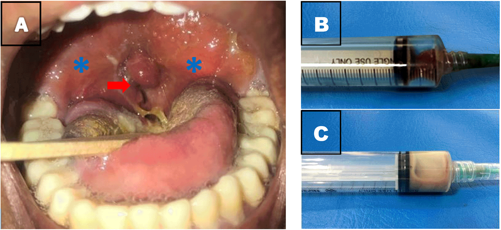
A 20-year-old Javanese Indonesian non-diabetic male came to the emergency room at Dr. Soetomo General Academic Hospital on October 22, 2017, with a chief complaint of shortness of breath going for two weeks that had worsened in the last two days. The shortness of breath came and went and even worsened when the patient laid down. Additional complaints included pain and swallowing difficulty, throat felt lumpy, fever, and cough with a thick sputum consistency. The patient had a history of sore throats approximately two to three times per year that got better after oral amoxicillin 500 mg every 8 hours and normal saline gurgle, drank alcohol frequently, and smoked. He also reported having snoring issues for several years. Physical examination showed muffled voice, an axillary temperature of 37.2 °C, a pain scale of 7, 82% oxygen saturation (normal > 95%), and inspiratory stridor. Throat examination showed smooth-surfaced T4-T4 tonsils by using Brodsky tonsil classification, palate, right and left peritonsillar areas of the bombans, and hyperemia. The isthmus faucium was narrow, with about 30% remaining (Figure 1A).

Results of laboratory blood tests showed an increase in leukocytes at 34,03 × 10³/uL (normal 3,80-10,60 × 10³/uL), C-reactive protein (CRP) at 86.54 mg/L (normal 0,00-10,00 mg/L) glutamic oxaloacetic transaminase serum (SGOT) and glutamic pyruvic transaminase serum (SGPT) at 51 and 123 U/L (normal SGOT <41 U/L, SGPT 0-35 U/L), and hepatitis B surface antigen (HBsAg) came out reactive. Human Immunodeficiency Virus (HIV) Rapid examination with qualitative methods obtained negative results. Therefore, based on the history, physical and laboratory examination, a bilateral peritonsillar abscess was suspected with obstruction of the upper airway. Hence, we performed peritonsillar punctures with disposable sterile syringe at the most bomban locations to make the diagnosis, with 1 ml of pus mixed with blood in the right peritonsillar and 3 ml of pus in the left peritonsillar (Figure 1B and C).
We performed a Computed Tomography Scan (CT scan) on this patient to determine the expansion of the abscess, considering that the patient has immunodeficiency. Results showed hypodense lesions (26–29 HU) with clear boundaries, irregular edges, ±4,1 × 2,5 × 4,6 cm on the right peritonsillar and ±4,1 × 3 × 4,6 cm on the left peritonsillar, which in contrast showed rim enhancement (73 HU). The lesion caused narrowing of the oropharyngeal lumen at 2–3 cervical vertebrae levels. The narrowest lumen diameter was ±0.1 cm—no expansion of abscess to the surrounding area (Figure 2).

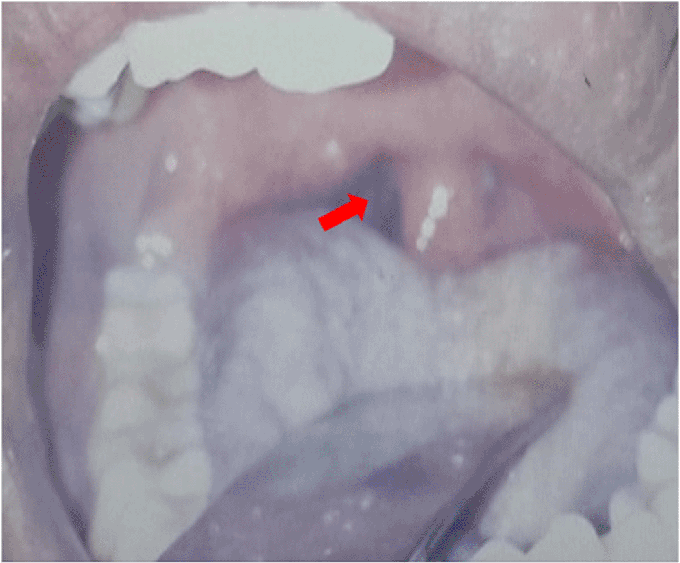
We performed an incision on the right and left peritonsillar using a scalpel number 11 in the most bomban area without topical anesthesia. It resulted in 6 ml of pus mixed with blood in the right peritonsillar and 13 ml of pus mixed with blood in the left. In addition, we carried out swab culture on the peritonsillar incision area. As a result, the isthmus faucium looked more spacious (Figure 3).

Chest X-ray suggested left paracardial infiltrates, and the patient was diagnosed with community-acquired pneumonia (CAP), which was then treated with levofloxacin injection at 500 mg every 24 hours. In addition, the patient was treated with 500 mg of paracetamol tablets every 8 hours and 200 mg of n-acetyl cysteine tablets every 8 hours related to chronic hepatitis B. Furthermore, we diagnosed him with bilateral peritonsillar abscess with CAP and chronic hepatitis B. The patient was also treated with 500 mg of metronidazole injection every 8 hours, 4 mg dexamethasone injection every 24 hours and gargle with 0.9% sodium chloride solution every 8 hours. We also gave the patient a soft diet because of the odynophagia.
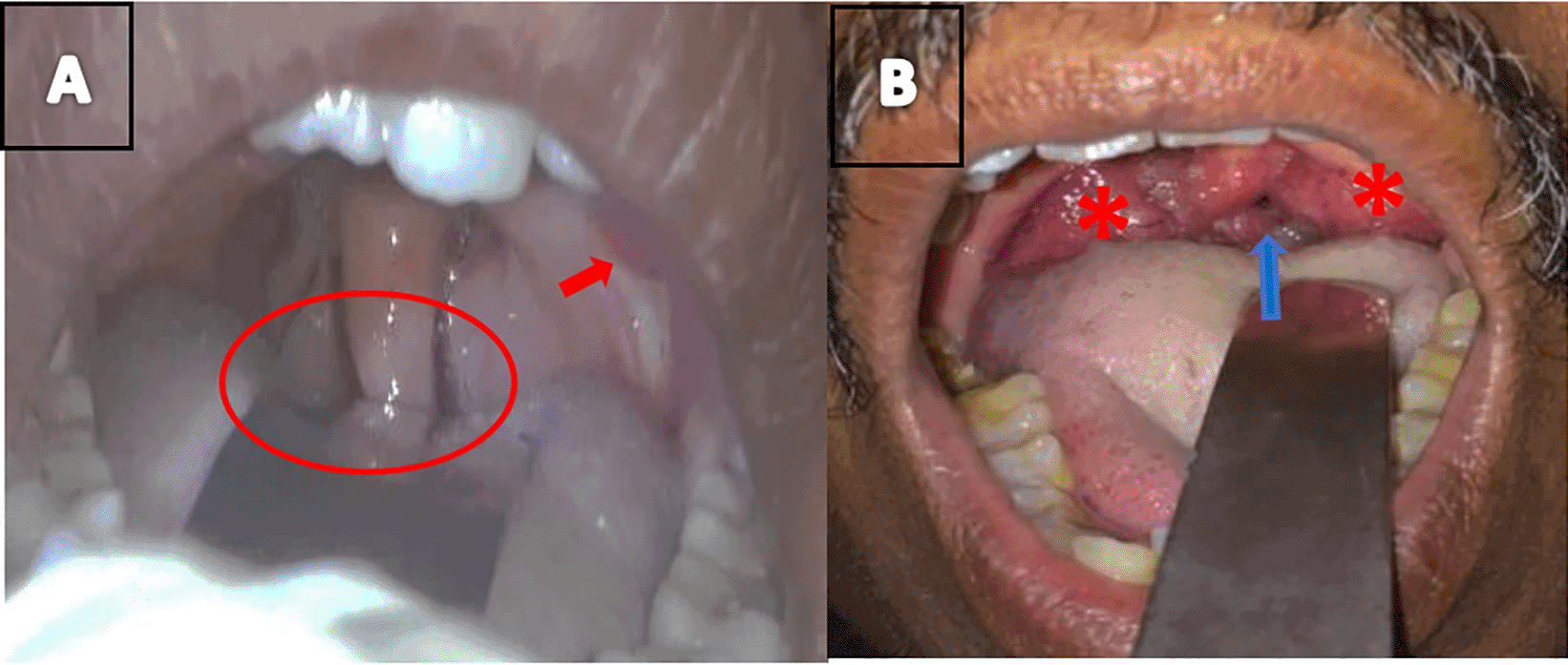
Evaluation on the first day showed reduced bombans, hyperemia, and edema in the right and left peritonsillar. We evaluated the incision area and obtained 0.2 ml of blood from the right peritonsillar and 0.5 ml of blood mixed with pus in the left peritonsillar (Figure 4A). On the second day of evaluation, the peritonsillar area returned to normal. There was a decrease in leukocytes on the third day, namely at 9.5 u/L and CRP 0.5 mg/L. We declared the patient cured and advised him to be outpatient based on the history, physical examination, and laboratory results. The patient was given medications in the form of 500 mg of levofloxacin antibiotics tablets every 24 hours, 500 mg of metronidazole tablets every 8 hours, 500 mg of anti-pain paracetamol tablets every 8 hours and 200 mg of n-acetyl cysteine tablets every 8 hours, and gargle with 0.9% sodium chloride every 8 hours until the eighth day after incision.

Patients came for a control checkup to the ENTHN Outpatient Unit of Dr. Soetomo General Academic Hospital on the eighth day after the incision. There were no complaints, the peritonsillar area was in good condition, and T3/T4 tonsils (Figure 4B). Swab culture results showed Streptococcus equi. We advised the patient to undergo a tonsillectomy, but he preferred first to cure his chronic hepatitis B and pneumonia.
Peritonsillar abscess is a disease often found in ENTHN services. In this case, we found it on a 20-year-old man. The main complaints included shortness of breath for two weeks, accompanied by complaints of swallowing pain, difficulty swallowing, and a feeling of a lump in the throat. There was a fever within two weeks and a cough for one month accompanied by thick phlegm. This disease is found at the age of 10–40 years, and the majority are diagnosed at the age of 22–28 years old with a male to female ratio of 2:1.2,4 Complaints with peritonsillar abscess include difficulty swallowing, fever, pain, and a lump in the throat.6,7 Complaint of bilateral peritonsillar abscess is longer than in unilateral peritonsillar abscess. Shortness of breath can be caused by narrowing the upper airway due to bilateral peritonsillar abscesses.5
The patient has a history of recurrent sore throat with a frequency of two to three times a year. The patient came in an acute state characterized by complaints of a sore throat for two weeks. A 2017 Brazilian study reported acute tonsillitis was the most common predisposing factor for peritonsillar abscess.3 Peritonsillar abscess can evolve from acute tonsillitis to peritonsillitis, then progress to peritonsillar cellulitis, and finally peritonsillar abscess or a consequence of Weber’s gland (salivary gland) abscess located at the supratonsil space.3–5
The patient has a history of frequent drinking and smoking. The reactive results on HBsAg examination and the CAP indicate the possibility of being immunocompromised, which makes it easier for peritonsillar abscesses to occur. Smoking and drinking alcohol are predisposing factors for peritonsillar abscess due to the adverse effects of smoking on the normal flora in the oropharynx.8 Immunocompromised conditions such as diabetes mellitus and HIV carry a more atypical and challenging risk of head and neck infections.3,9
Clinical examination revealed muffled voice, odynophagia, difficulty swallowing, trismus, and fever. T4-T4-sized tonsils, palate, and right and left peritonsillar areas to appear turgid and hyperemic. The uvula is in the middle, pressed by the bomban peritonsillar and the edema on the right and left tonsil area. The faucium isthmus becomes narrow, with about 30% remaining. Unilateral or bilateral peritonsillar abscess signs include muffled voice, odynophagia, difficulty swallowing, otalgia, trismus, hypersalivation, and fever. Unilateral peritonsillar abscess shows the asymmetric peritonsillar area, bulging, and uvula deviation, whereas there are no classic signs in bilateral cases.1–3,10 A midline uvula with bilateral mole bomban palate is a characteristic feature of a bilateral peritonsillar abscess. Trismus due to disruption of the lateral pterygoid muscles can result from either peritonsillar or parapharyngeal abscess.10
The location of the peritonsillar abscess, in this case, was at the superior right and left peritonsillar spaces. The most common location of peritonsillar abscess was in the superior peritonsillar space (61%), in the middle (33%), and below the peritonsillar space (6%). This unique abscess location is due to the failure of suppurative inflammatory drainage by blockade of crypts on acute tonsillitis, which causes infection to the peritonsillar space.1
Right and left peritonsillar abscesses, in this case, occurred at different levels. The left peritonsillar abscess is more mature, whereas the right peritonsillar is a transition from infiltrates to abscesses, seen in the right-side discharge that is less than the left. Research in Greece in 2011 reported that tonsillitis is an infection of the tonsils that can cause complications in the form of bilateral peritonsillar abscesses with different levels of development on each side.2 Bilateral peritonsillar abscess cases can occur when tonsillitis and abscess develop bilaterally.1–4 There was still be a discussion on whether peritonsillar abscess caused by a complication of acute tonsillitis or the blockage of the common duct of Weber’s gland. It has been postulated that the two bacteria infect the tonsillar mucosa (including the crypt mucosa) and spread to the peritonsillar space via the salivary duct system, where an abscess is formed if the immune system does not overcome the bacteria.11
Blood laboratory examination showed an increase in leukocytes of 34.020 u/L, 83.8% of neutrophils, and a CRP of 86.54 mg/L, indicating that bacteria caused the infection. Aerobic and anaerobic organisms mainly caused peritonsillar abscesses. Increased leukocytes and CRP are found in unilateral peritonsillar abscesses and even higher in bilateral peritonsillar abscesses.3,7
CT scan showed hypodense lesions (26–29 HU) with clear boundaries, irregular edges at the right and left peritonsillar, and showed rim enhancement on contrast. This outcome indicates the presence of fluid in the form of pus in the peritonsillar. Narrowing of the lumen in the oropharynx at the level of 2–3 cervical vertebrae with a diameter of ±0.1 cm gives a clinical picture of upper airway obstruction, a complication of bilateral peritonsillar abscesses.5 A CT scan determined the density of the mass and the remaining isthmus, and the abscess spread to the surrounding area. Head and neck CT scans also can detect deep neck abscesses that are not clinically visible.2,3,10,12 Upper airway obstruction is a possible diagnosis due to inspiratory stridor and the peritonsillar area being bombans on both sides, pushing the tonsils medially and leaving a small gap between them. A contrast-enhanced CT scan is preferred when the symptoms and signs of a peritonsillar abscess are atypical.5
Swab culture results showed Streptococcus equi. The majority of organisms in peritonsillar abscess are aerobic and anaerobic organisms. Aerobic organisms that often coexist are Streptococcus pyogenes, Streptococcus milleri, Haemophilus influenza, and Streptococcus viridans. Meanwhile, anaerobic organisms that often coexist are Fusobacterium sp. and Bacteroides sp.2,3,6,10 We can’t find the anaerobic organism might because of the antibiotic given before the swab.
We performed peritonsillar punctures at the most bomban point to make the diagnosis. We suggest a drainage aspiration or incision if pus occurs at the puncture.13 Immediate tonsillectomy is a definitive procedure when the incision and drainage of the abscess cannot properly evacuate the pus. However, it might cause an increased risk of postoperative bleeding. We recommend interval tonsillectomy 4–6 weeks after the incision and drainage of the peritonsillar abscess, indicated in patients who experience recurrent tonsillitis snoring leading to obstructive sleep apneu in the same year after peritonsillar abscess.2,3
The diagnosis of bilateral peritonsillar abscess is challenging because we can’t find the typical examination findings of unilateral peritonsillar abscess. In this case, the diagnosis was established with a contrast-enhanced CT scan, revealing bilateral peritonsillar abscess with airway obstruction. Bilateral peritonsillar bulge may masquerade other diseases such as acute tonsillitis, infectious mononucleosis, lymphoma, and retropharyngeal abscess. When in doubt, the clinician must perform an imaging examination as a guide. Contrast-enhanced CT scan can reveal the presence of an abscess and also its extension. The most emergent and important step of treatment is ensuring the safety of the airway. The patient had to undergo incision and drainage due to the airway obstruction caused by bilateral peritonsillar abscess.
We found CAP at this patient. At first, we thought that the pneumonia is caused by the bilateral peritonsillar abscess (aspiration pneumonia) then we found that is what was caused by CAP. Aspiration pneumonia is one of the complications of peritonsillar abscesses that can also be life-threatening, but it didn’t found in this case.
We treated the patient having a peritonsillar abscess with 500 mg of levofloxacin injection every 24 hours and 500 mg of metronidazole injection every 8 hours as antibiotics. The intravenous route was chosen for the better effect on the patient. The treatment choice is due to Streptococcus sp., the organism that causes the most common peritonsillar abscesses. Penicillin is the first-line antibiotic for peritonsillar abscesses and is not different from other broad-spectrum antibiotics.2,4,10 Meanwhile, broad-spectrum quinolones are third-line therapy for peritonsillar abscess treatment and first-line therapy for CAP.14,15 Therefore, antibiotic therapy for this case is a combination of quinolone and metronidazole. Furthermore, the administration of metronidazole depends on the frequency of anaerobic organisms in the peritonsillar abscesses.
We report a bilateral peritonsillar abscess with a complication of upper airway obstruction. Bilateral peritonsillar abscesses are rare cases. Puncture in the peritonsillar area resulted in pus. We performed a CT Scan to determine the extent of the abscess to the surrounding area. Then, we carried out incision and drainage to treat the patient. Antibiotic therapy of levofloxacin, metronidazole, and dexamethasone injections, oral anti-pain paracetamol and N-acetyl-cysteine, and 0.9% sodium chloride gargle is given to the patient with bilateral peritonsillar abscess after incision and abscess drainage.
Written informed consent for publication of clinical details and clinical images was obtained from the patient.
All data underlying the results are available as part of the article and no additional source data are required.
Repository: CARE checklist for ‘Case report: Bilateral peritonsillar abscess with complications of upper airway obstruction’. https://doi.org/10.5281/zenodo.6519444.16
Data are available under the terms of the Creative Commons Attribution 4.0 International license (CC-BY 4.0).
| Views | Downloads | |
|---|---|---|
| F1000Research | - | - |
|
PubMed Central
Data from PMC are received and updated monthly.
|
- | - |
Is the background of the case’s history and progression described in sufficient detail?
Partly
Are enough details provided of any physical examination and diagnostic tests, treatment given and outcomes?
Yes
Is sufficient discussion included of the importance of the findings and their relevance to future understanding of disease processes, diagnosis or treatment?
Yes
Is the case presented with sufficient detail to be useful for other practitioners?
Partly
Competing Interests: No competing interests were disclosed.
Reviewer Expertise: Otorhinolaryngology, Head and neck surgery, Rhinology, Thyroid and parathyroid surgery, Ear surgery, Laryngology
Competing Interests: No competing interests were disclosed.
Reviewer Expertise: Otolaryngology, Bronchoesophagology, Endoscopy
Is the background of the case’s history and progression described in sufficient detail?
Partly
Are enough details provided of any physical examination and diagnostic tests, treatment given and outcomes?
Partly
Is sufficient discussion included of the importance of the findings and their relevance to future understanding of disease processes, diagnosis or treatment?
Partly
Is the case presented with sufficient detail to be useful for other practitioners?
Partly
Competing Interests: No competing interests were disclosed.
Reviewer Expertise: Otolaryngology, Bronchoesophagology, Endoscopy
Alongside their report, reviewers assign a status to the article:
| Invited Reviewers | ||
|---|---|---|
| 1 | 2 | |
|
Version 3 (revision) 14 Sep 23 |
||
|
Version 2 (revision) 28 Jul 22 |
read | read |
|
Version 1 19 May 22 |
read | |
Provide sufficient details of any financial or non-financial competing interests to enable users to assess whether your comments might lead a reasonable person to question your impartiality. Consider the following examples, but note that this is not an exhaustive list:
Sign up for content alerts and receive a weekly or monthly email with all newly published articles
Already registered? Sign in
The email address should be the one you originally registered with F1000.
You registered with F1000 via Google, so we cannot reset your password.
To sign in, please click here.
If you still need help with your Google account password, please click here.
You registered with F1000 via Facebook, so we cannot reset your password.
To sign in, please click here.
If you still need help with your Facebook account password, please click here.
If your email address is registered with us, we will email you instructions to reset your password.
If you think you should have received this email but it has not arrived, please check your spam filters and/or contact for further assistance.
Comments on this article Comments (0)