Keywords
Adrenal adenoma, Adrenal protocol, Atypical, Cushing’s syndrome, Imaging findings, Ultrasonography.
This article is included in the Global Public Health gateway.
Adrenal adenoma, Adrenal protocol, Atypical, Cushing’s syndrome, Imaging findings, Ultrasonography.
ACTH: Adrenocorticotropic hormone
APW: Absolute percentage washout
CECT: Contrast-enhanced computed tomography
CT: Computed tomography
DCT: Delayed contrast enhanced CT
DEXA: Dual energy X-ray absorptiometry
HU: Hounsfield Unit
MDCT: Multi-detector computed tomography
MRI: Magnetic resonance imaging
RDW: Red cell distribution width
RPW: Relative percentage washout
USG: Ultrasonography
Adrenal tumors are common in humans, occurring in 9% of autopsy series. The prevalence of adrenal adenoma is reported to be age-related; the frequency of undiagnosed adenoma is 0.14 percent in patients aged 20–29 years and 7% in those older than 70 years.1 Primary adrenal tumors can be hyper-functioning, producing excess hormones and resulting in clinical symptoms, or non-functioning which is more common. However, 10-15% of patients with adrenal adenoma might exhibit clinical features of Cushing's syndrome.2 In the evaluation of adrenal tumors, abdominal ultrasound has a reported sensitivity of 96% for tumors smaller than 2 cm and 100% for tumors larger than 2 cm. Multi-detector computed tomography (MDCT) is the single most useful modality for identification and characterization. Magnetic resonance imaging (MRI) and functional imaging modalities such as nuclear scintigraphy are useful for a thorough evaluation when in doubt.3 Computed tomography (CT) imaging can detect adrenal masses with diameters greater than 5 mm. Adrenal adenomas with typical imaging characteristics account for a vast majority of these benign adrenal masses. However, distinguishing between atypical adrenal adenomas and adrenal malignant lesions is critical, because a small percentage of these are potentially malignant lesions.4 Furthermore, because imaging cannot reliably distinguish between functioning and non-functioning adenomas, the biochemical profile must be used. To identify abnormal adrenal function in an adenoma, NP-59 scintigraphy can supplement biochemical and radiological imaging results.3 This case report highlights atypical findings in a functioning adrenal adenoma as well as possible differential diagnoses that could lead to a misdiagnosis.
We report a 16-year-old South Asian female who presented at the out-patient service of the Endocrinology department of Bir Hospital, Kathmandu with a chief complaint of increased weight for two years, primary amenorrhea and lethargy. The patient had not sought any prior medical attention or undergone any intervention in the past. She did not have any history of similar illness or genetic conditions in her family. Vitals taken showed blood pressure to be 180/130 mm Hg, pulse rate of 78 beats/minute, respiratory rate of 18 breaths/minute and temperature to be 98°F (36.7°C).
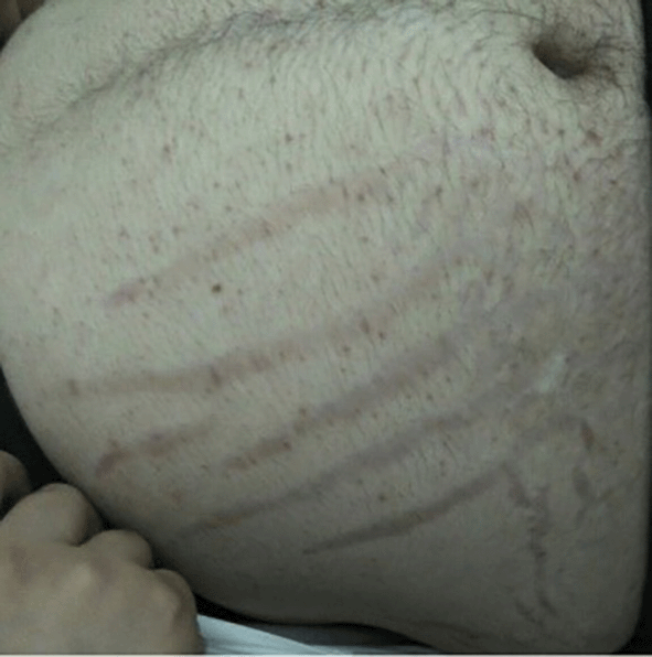
A thorough physical examination was performed: the patient had a moon-shaped face with facial hair, supra-clavicular hump, deepening of voice and purple abdominal striae were noted (Figure 1). However, axillary and pubic hair was present and breast development was normal (Tanner stage 5). A suspicion of Cushing's syndrome was made and further investigations were sent (Table 1). In addition, a bone densitometry scan was performed by dual energy X-ray absorptiometry (DEXA) showing a Z score of -2.8 in the spine along with a Z score of -0.8 and -1.5 in the right and left femur neck, respectively.
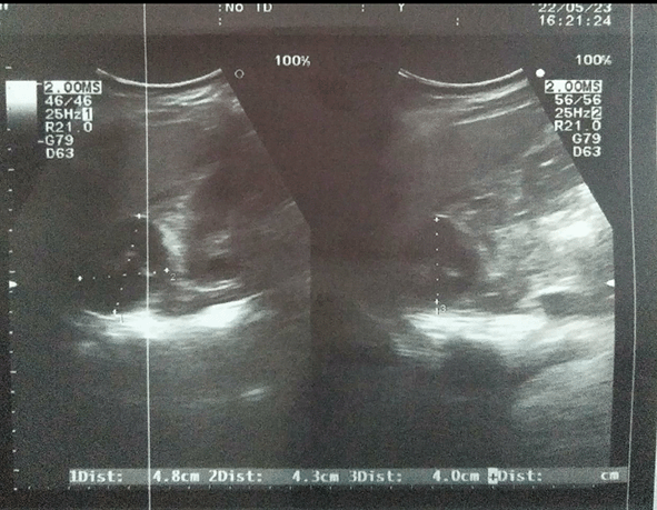
Ultrasonography (USG): Well-defined hypoechoic lesion measuring 48×43×40 mm in the right suprarenal region. Tiny hyperechoic focus (likely calcification) was noted within the lesion. On color Doppler, the lesion did not show blood flow (Figure 2).
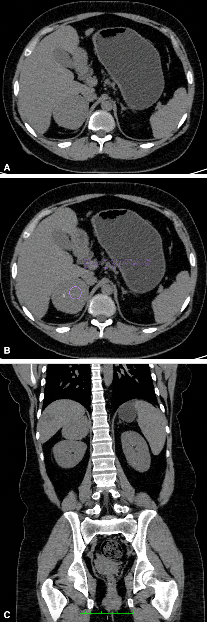
Contrast-enhanced computed tomography
(CECT) (adrenal protocol) findings: Well-defined soft tissue density lesion measuring 56×49×45 mm was noted in the right suprarenal gland region, without separate visualization of the right adrenal gland. The lesion showed tiny calcified foci in its posterior aspect. There was no evidence of fat-attenuating foci within the lesion. In non-contrast images, the lesion showed (Hounsfield unit (HU)+41) (Figure 3). On post-contrast images taken 60 seconds after contrast injection, the lesion showed significant homogenous enhancement with (HU+109). On delayed images taken 15 minutes after contrast injection, the lesion showed (HU+56) (Figure 4). The absolute percentage washout of 77.9% (which is >60%) and relative percentage washout of 48.6% (which is >40%) was noted, suggesting lipid-poor adrenal adenoma.

The patient was counselled about the need for surgery but was non-compliant. She was managed with anti-hypertensive and antidiabetic medications. Her blood pressure and blood glucose has been well under control. She visited the hospital for a follow up one month later in which she had her blood pressure and blood glucose under control. She was again advised of the need for surgery during follow up which is planned for a later date when the patient is compliant.
Adrenal tumors that are incidentally found on imaging are known as adrenal incidentalomas, which comprises benign adrenal masses to metastatic tumors. They are found during imaging of areas other than adrenal gland. The prevalence of adrenal incidentalomas is between 0.35% to 1.9% on CT scan. Among them, around 54 percent are found to be adrenal adenomas.5 However, our case was imaged for the proven diagnosis of Cushing's syndrome. In the case of Cushing's syndrome in adults, only 20% of the cases are due to adrenal causes; however, in the first decade of life adrenal causes predominate majority of cases. Among the cases of adrenocorticotropic hormone (ACTH)-independent Cushing's syndrome, it was found that 95% of cases were due to adrenal adenomas or carcinomas, among which 65% consisted of hyper-functioning adrenal adenoma, similar to our case, while primary pigmented nodular adrenal dysplasia and ACTH-independent macronodular hyperplasia accounts for the rest of the cases.6 On imaging, adrenal adenomas are usually well-defined ovoid to round nodules measuring 1-5 cm, with homogenous or slight heterogenous attenuation. Imaging cannot differentiate between functional and non-functional adenomas: this needs adrenal venous sampling.7
An ultrasonography imaging was performed, which found a homogenous and hypoechoic lesion, consistent with the ultrasound imaging criteria for adrenal adenoma. However, in our case we also noted a tiny hyperechoic focus, which was likely a calcification within the mass. The size of the mass on USG was 4.8 cm and comparable to the range of 1.0-6.3 cm seen in an ultrasound study by Fan et al. in China.8
Since MDCT is the imaging of choice in adrenal adenoma, an MDCT was performed and showed well-defined soft tissue density; density evaluation is highly sensitive and specific, as 70% of the cases consist of high lipid density in which <10HU is considered 71% sensitive and 98% specific. However, our case showed a HU of +41 and an adrenal CT washout was performed, showing an absolute percentage washout (APW) and relative percentage washout (RPW) of >60% and >40% respectively, highly suggestive of adenoma.9 Moreover, it was also seen that RPW was more accurate than APW and a 15-minute delayed contrast enhanced CT (DCT) was more accurate than a 10-minute DCT for the diagnosis of adrenal adenoma. Compared to other modalities, washout CT provides the highest accuracy for characterization of adenoma.1 In our case, we calculated RPW and a 15-minute DCT practicing evidence-based medicine at our center. Contrary to the typical findings of adrenal adenoma, this case featured atypical findings which might hinder the accurate identification of the case. Adrenal adenomas with calcifications have been reported in about 14% of cases. This necessitates its differentiation from various other etiologies like adrenocortical carcinomas, myelolipomas and metastases. It was found that calcified adrenal adenomas represent 15% of calcified adrenal masses second only to adrenal cyst.4
In our patient, bone densitometry scan showed a lowered Z score. The skeletal system's structural and functional impairment is a significant cause of morbidity and disability in Cushing’s syndrome patients, particularly given the high prevalence of vertebral fractures. Glucocorticoids reduce bone collagenous matrix synthesis while increasing its degradation. The reduction in osteoblast number and function appears to be central to the bone loss caused by glucocorticoid excess. Glucocorticoid-induced osteoporosis is reversible, but the recovery of bone loss is slow and takes about ten years. Fractures are common in patients with Cushing’s syndrome who have severe osteoporosis.10
Adrenal adenomas can present with features of hormonal excess (functioning adenoma). Diagnosis of adrenal adenoma needs a multidisciplinary approach of clinical, biochemical and imaging studies. Cushing's syndrome is one of the presenting clinical feature in functioning adrenal adenoma. Adrenal adenoma presents a diagnostic puzzle when presented with atypical imaging findings in CT. It is essential to have the knowledge of atypical findings and follow an adrenal protocol when dealing with adrenal adenomas to prevent a misdiagnosis.
I reside in a rural area of Nepal due to which I did not seek medical attention for my problems which seemed very minor to me, after visiting the hospital I came to realize that I had a bigger problem than I thought. I am happy that the doctors were able to come to a diagnosis and provided me medications. I will visit the hospital from now onwards without delay if I have any symptoms and will opt for surgery.
| Views | Downloads | |
|---|---|---|
| F1000Research | - | - |
|
PubMed Central
Data from PMC are received and updated monthly.
|
- | - |
Provide sufficient details of any financial or non-financial competing interests to enable users to assess whether your comments might lead a reasonable person to question your impartiality. Consider the following examples, but note that this is not an exhaustive list:
Sign up for content alerts and receive a weekly or monthly email with all newly published articles
Already registered? Sign in
The email address should be the one you originally registered with F1000.
You registered with F1000 via Google, so we cannot reset your password.
To sign in, please click here.
If you still need help with your Google account password, please click here.
You registered with F1000 via Facebook, so we cannot reset your password.
To sign in, please click here.
If you still need help with your Facebook account password, please click here.
If your email address is registered with us, we will email you instructions to reset your password.
If you think you should have received this email but it has not arrived, please check your spam filters and/or contact for further assistance.
Comments on this article Comments (0)