Keywords
acute inflammatory swelling; cellulitis; deep tissue injury; implant superstructures
acute inflammatory swelling; cellulitis; deep tissue injury; implant superstructures
During the prosthetic phase of dental implant treatment, patients may present with severe pain and acute swelling several days after definitive prosthesis delivery or reconnection of a loose healing abutment (HA) or prosthesis. Empirically, most patients recover within several weeks of medical care and follow-up. Unfortunately, the condition may deteriorate, in the form of cellulitis, in some cases, requiring referral to oral and maxillofacial surgery (OMS) specialists.
When patients report to the clinic or emergency room (ER) with these complaints, especially of severe pain and swelling around the implant, it is important to determine whether a bacterial infection has caused the signs and symptoms. However, empirically, the patients’ history, physical examination, and laboratory data may not help in conclusively distinguishing between infection and inflammation.1,2 Owing to the similarity in the clinical features of acute inflammatory swelling and cellulitis caused by infections, clinicians often overtreat such patients, and patients may suspect the previous prosthetic procedure to be the cause of the infection. Furthermore, uncertainty about why the (re) connection of the implant superstructures could cause acute symptoms creates a dilemma for clinicians. Exploring the cause and progress of the pathology is the first step to prevent and to resolve it.
In medicine, a pressure ulcer is the most well-known injury caused by mechanical pressure on the body.3,4 Deep tissue injury (DTI) has been proposed as a subtype of pressure ulcers by public health specialists.3–9 DTI develops when pressure and shear force caused by external mechanical load induce ischemia and tissue deformation in the subcutaneous tissues of the load-bearing area. Acute inflammatory edema develops and spreads to adjacent tissues such as connective tissue and muscle, following a bottom-up pathway, which means that the injury propagates from the inside toward the surface. The injury is initiated at the interface between bone and soft tissue.3,6 Once DTI is initiated, acute edema develops almost immediately and progresses over time as a result of the ischemia-reperfusion injury and obstruction of lymphatic vessels.
Based on the premise that acute swelling may result from pressure and external force applied on the peri-implant gingiva while connecting the implant superstructure, the concept of DTI could explain the pathogenesis underlying the clinical presentation observed in some patients. Furthermore, this could also explain why some patients show signs of infection. The progressive edema and impaired lymphatic system in the peri-implant tissue10,11 increase the risk of infection,12,13 and the transmucosal region of the implant can provide a pathway for microbial invasion.14–17 Therefore, the present case series study aims to propose a possible mechanism of how implant superstructure connections can cause acute swelling and pain, based on the concept of DTI.
Ten cases in nine patients, who exhibited acute swelling or suspected infection that developed within a month of placing a definitive prosthesis or reconnecting loosened or exfoliated HAs or prosthesis, were reviewed. The patients were treated at the prosthodontic department of the Seoul National University Bundang Hospital (Seongnam, South Korea) between March 2013 and March 2021. The following data were retrieved from the patients’ medical records: sex, age, medical history, reasons for superstructure connection, physical examination, laboratory examination result (if laboratory examinations were performed), treatment, and recovery time. In addition, the clinical findings, patient factors, and duration of the signs and symptoms were analyzed based on the conceptual framework and physiological process of DTI. The institutional review board at the Seoul National University Bundang Hospital approved this study (IRB No. B-2104-681-101), and the study protocol conforms to the Declaration of Helsinki. The institutional review board waived the need for informed consent due to the retrospective nature of the research.
Patient data are summarized in Table 1.18 There were seven women and two men, and their median age was 66 years (55–84 years). Cases 4-1 and 4-2 (Table 1) were of the same patient. Six patients underwent pathological blood investigations.
| Case 1 | Case 2 | Case 3 | Case 4-1 | Case 4-2 | Case 5 | Case 6 | Case 7 | Case 8 | Case 9 | |
|---|---|---|---|---|---|---|---|---|---|---|
| Age-ranges/Sex | 80s/F | 60s/F | 60s/M | 60s/F | 80s/F | 60s/F | 80s/F | 50s/M | 60s/M | |
| Underlying diseases | Osteopenia | Diabetes Hypertension | Diabetes Hypertension | Hypothyroidism Renal cell carcinoma | None | None | Diabetes Hyperlipidemia | Diabetes Hyperthyroidism | Diabetes | |
| Reasons for connecting superstructures1 | #24i-26i Fixed dental prosthesis delivery | #36i HA reconnection due to loosening | #46i crown with abutment exfoliation 10 days earlier due to abutment screw fracture | Replacement with wider HA | HA reconnection due to loosening | #37i Crown delivery | #35i-36i splinted crown delivery | #16i crown with abutment exfoliation 7 days earlier due to abutment screw fracture | #46i crown with abutment exfoliation 14 days earlier due to abutment hex fracture: HA connection instead | #36i Crown delivery |
| Onset of symptoms according to patient statements | On the day of the connection | On the day of the connection | On the day of the connection | On the day of the connection | Unknown | On the day of the connection | On the day of the connection | On the day of the connection | On the day of the connection | |
| Time interval between connection day and emergency visit | 6 days | 5 days | Within 24 h | 2 days | 3 days | 21 days | 2 days | 2 days | Within 24 h | Within 24 h |
| Route of admission | Unscheduled visit | Unscheduled visit | ER | Unscheduled visit | Unscheduled visit | Unscheduled visit | Unscheduled visit | Unscheduled visit | ER | ER |
| Vital signs2 | N/A | N/A | SBP: 156 DBP: 107 RR: 15 BT: 36.5 | N/A | N/A | N/A | N/A | N/A | SBP: 133 DBP: 83 PR: 90 BT: 37.2 | SBP: 124 DBP: 83 PR: 75 BT: 36.0 |
| Emergency visit laboratory test results3 | N/A | HbA1c: 7.2 | HbA1c: 6.0 CRP: 4.58 WBC: 8.90 | BUN: 29 Uric acid: 9.4 WBC: 7.79 | Glucose: 128 BUN: 35 Uric acid: 11.6 WBC: 8.55 | N/A | N/A | HbA1c: 7.2 Glucose: 168 CRP: 8.57 WBC: 15.86 | Glucose: 124 CRP: 9.61 WBC: 11.74 | Glucose: 179 CRP <0.40 WBC: 10.83 |
| Mental state | Alert | Alert | Alert | Alert | Alert | Alert | Alert | Alert | Alert | |
| Signs and symptoms | Pain; Gingival swelling around the implant | Pain; Gingival swelling around the implant; Left cheek and submandibular swelling; Warmth; Trismus; Dysphagia | Pain; Gingival swelling around the implant; Right cheek and submandibular swelling; Warmth; Dysphagia | Pain; Tenderness; Gingival swelling around the implant; Both submental and submandibular swelling on the affected side; Trismus; Dysphagia | Pain; Gingival swelling around the implant; Left cheek and submandibular swelling; Pus from gingival sulcus; Dysphagia; Trismus | Pain; Gingival swelling around the implant | Pain; Gingival swelling around the implant; Right cheek swelling; Pus from gingival sulcus; Warmth; Dysphagia | Pain; Tenderness; Gingival swelling around the implant; Right cheek swelling; Pus from gingival sulcus; Warmth; Chills | Pain; Tenderness; Gingival swelling around the implant; Left cheek swelling | |
| Treatment | CHX irrigation, F/U | Peri-implant curettage, F/U | Pain control, Prosthesis disconnection, I&D, F/U | CHX irrigation, F/U | Prostheses disconnection, I&D, F/U | Prosthesis disconnection, CHX irrigation, F/U | Prosthesis disconnection, CHX irrigation, F/U | Pain control, F/U | Pain control, Prosthesis disconnection, CHX irrigation, F/U | |
| Antibiotic treatment | N/A | + (Oral) | + (Oral) | + (IM, Oral) | + (IM, Oral) | N/A | + (Oral) | + (IM, Oral) | + (IM, Oral) | |
| Duration | 3 weeks | 10 days | 2 weeks | 3 weeks | 3 weeks | 2 weeks | 2 weeks | 2 weeks | 2 weeks | |
| Other tests | N/A | N/A | N/A | Titanium allergy test: Negative | N/A | N/A | N/A | N/A | N/A | |
| Implant system4 (Connection type) | Superline (Internal) | Luna (Internal) | Superline (Internal) | CMI (Internal) | CMI (Internal) | Osstem TS III (Internal) | Osseotite (External) | Superline (Internal) | Luna (Internal) | |
2 Normal ranges of vital signs: BP, 90/60 mmHg-120/80 mmHg; RR, 12-18 per minute; PR, 60-100 beats per minute; BT, 36.5°C–37.3°C.
Most patients stated that the swelling developed within a day of superstructure connection placement, except for one patient who could not recall the time of the onset of symptoms (case 5 in Table 1). Five patients underwent superstructure reconnection during the visit preceding symptoms due to loosening, and the other five underwent definitive prostheses deliveries.
Most patients showed increasing swelling and pain within a certain time, regardless of whether the connected superstructures were retrieved and exchanged with narrower ones. Except for two cases with only localized gingival swelling around the implants, antibiotics were prescribed for all patients based on a cellulitis-like appearance. In three cases (5 [Figure 1a], 7, and 8; Table 1), pus discharge from the peri-implant sulcus was observed. None of the patients developed fever. The symptoms subsided within three weeks in all patients, and there was no relapse. None of the patients showed any significant radiographic change in the alveolar bone after symptom onset. Representative radiographs are presented in Figure 2.
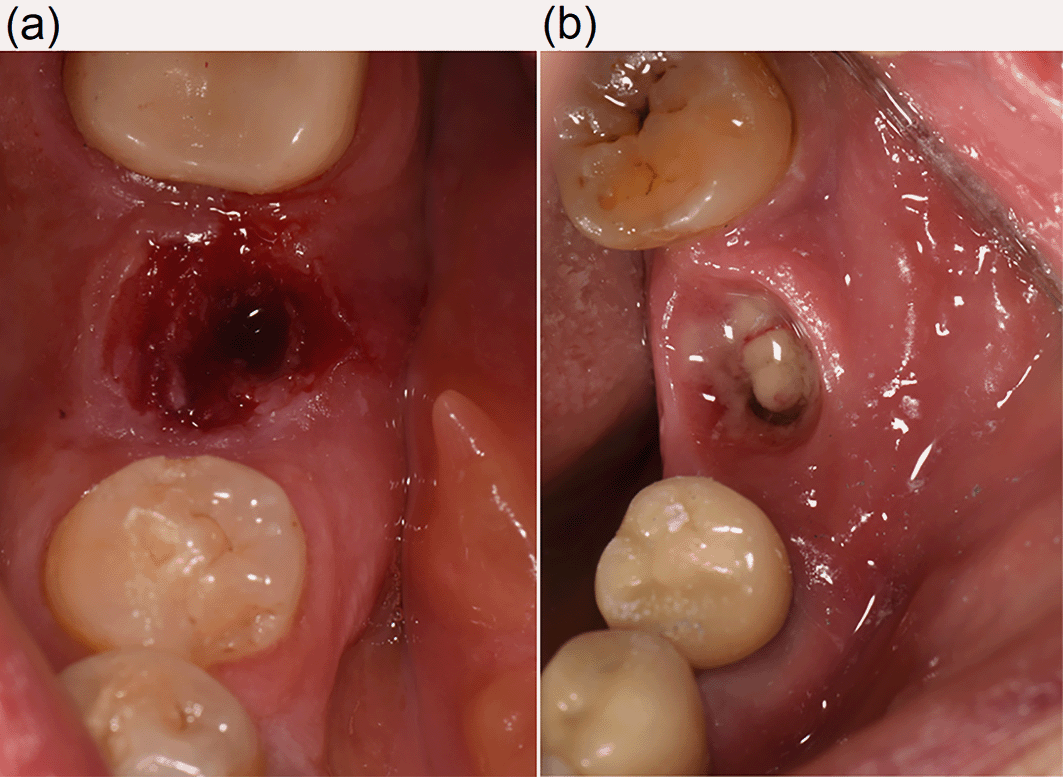
(a) Pus discharge from the peri-implant gingival sulcus (case 5). (b) Swollen, uneven, and bleeding peri-implant gingiva (case 3).
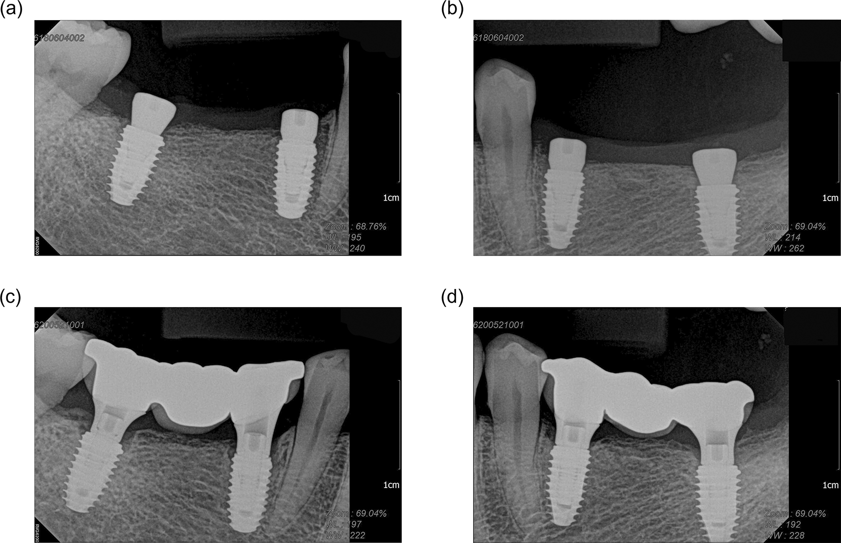
No significant changes in terms of the crestal height was seen in all presented cases.
In terms of the patients’ medical history, five of the nine patients were undergoing treatment for diabetes. Moderate to high glycated hemoglobin (HbA1c) and glucose levels were observed in six patients. An abnormal range of uric acid and blood urea nitrogen levels was observed in the patient presenting with two episodes of acute swelling (cases 4-1 and 4-2). This patient was undergoing treatment for renal cell carcinoma and hypothyroidism.
Elevated white blood cell (WBC) levels were observed in three patients. Two of these patients showed high C-reactive protein (CRP) levels, while in the other patient, the CRP level was low.
A woman in her 60s, undergoing treatment for diabetes and hypertension, underwent implant placement in the mandibular left first molar region. After the healing period, a fixture-level impression was made for crown fabrication. Ten days after the impression was recorded, the HA loosened and exfoliated. Therefore, a staff member reconnected the loosened HA.
Five days after the reconnection appointment, the patient revisited the clinic complaining of pain, difficulty in swallowing, and swelling in the gingiva around the implant and the left cheek. The patient reported that the swelling had developed several hours after the reconnection of the HA. Physical examination showed the following: tenderness and edema in the peri-implant gingiva; dysphagia; redness, tenderness, and warmth involving the left cheek and left submandibular region; and mouth opening limitation with a soft end feel. The patient was apyrexial, and there was no pus discharge in relation to the implant.
After the HA was disconnected, chlorhexidine irrigation was performed at the implant site. The HA was then replaced with one that was narrower in size. The patient was transferred to the OMS department to confirm a diagnosis of cellulitis. Peri-implant curettage was performed, and an antibiotic (Mesexin: methylol cephalexin lysinate, Hanlim Pharm. Co., Seoul, South Korea) and a non-steroidal anti-inflammatory drug (NSAID; Ketoprofen) were prescribed. Fourteen days after the loosened HA was reconnected, the symptoms and clinical signs had completely subsided. Six days after the onset of symptoms, an HbA1c level of 7.2% was measured during a routine diabetes check-up at the endocrine internal medicine department.
A man in his 60s, undergoing treatment for diabetes and hypertension, visited the clinic because an implant crown, placed on the implant in the right mandibular first molar region two years previously, had fallen out 10 days earlier. Intraoral assessment revealed that the abutment screw was fractured, so the screw remnant was removed from the fixture. After disinfecting the exfoliated abutment and crown, the prosthesis was reconnected with a new abutment screw.
At 6:48 am the following day, the patient was transferred by ambulance to the Seoul National University Bundang Hospital ER with severe pain around the implant and adjacent teeth. On arrival, the numerical rating scale score for self-reported pain intensity was 8–9. Vital signs were recorded as systolic blood pressure, 156 mmHg; diastolic blood pressure, 107 mmHg; respiratory rate, 15/min; and body temperature, 36.5°C. Laboratory tests showed HbA1c level, 6.0%; CRP level, 4.58 mg/dL; and WBC count, 8.90 × 10³/μL. In the ER, the patient’s dental history was established, pain control was initiated, and the patient was transferred to the prosthodontics department.
During the intraoral evaluation performed on the same morning, gingival swelling and redness were observed around the implant. While disconnecting the crown from the implant, sulcular bleeding and uneven and swollen gingiva were observed (Figure 1b). Chlorohexidine irrigation and minocycline ointment were applied in the sulcus, and an HA was connected to the implant. The patient was dismissed after scheduling a follow-up appointment.
Three days later, the patient was transferred to the OMS department due to severe pain, dysphagia, and swelling and tenderness on the right cheek and neck. The patient did not present with fever or chills. Incision and drainage were performed, and an antibiotic (Amoclan duo: amoxicillin and clavulanate) and NSAID (Aclofen: Aceclofenac) were prescribed. Most of the symptoms and signs subsided within 15 days of onset.
DTI, a subclass of pressure ulcers, is often unfamiliar to dentists. The pressure-induced injury initiated at the bone and soft tissue interface resolves rapidly after relieving the mechanical load.6,9 Blanching of the peri-implant gingiva is commonly observed during connection of implant superstructures. Sometimes, patients also report intense pain. The blanching and pain resolve within several minutes in most cases. Physiologically, gingival blanching is an induced ischemic condition of the subcutaneous tissue caused by the pressure applied during the connection. The associated pain indicates forcibly overstretched peri-implant tissues by the connected components. Therefore, it is reasonable to attribute the acute pain and swelling to the pressure and force applied to the peri-implant soft tissue during the connection process. Nevertheless, the mechanism explaining the acute inflammatory response and infection following connection/reconnection of a superstructure to an implant remains unclear.
From our review of the experience of patients in this study and previous studies on DTI, we concluded that the development of symptoms after connection or reconnection of implant superstructures follows the physiological process of DTI. There are several reasons for considering DTI as the underlying pathogenesis of acute swelling or suspected cellulitis developing after the connection of implant superstructures.
First, the act of connecting a superstructure through the transmucosal region of an implant meets the requirements for the development of pressure ulcers, which are external mechanical load, pressure and shear, friction, and tissue and patient tolerance.3,4 Figure 3 shows the events occurring during the abutment connection to the implant. The friction, pressure, and combined force cause tissue deformation and ischemia in the surrounding gingiva of the implant. As a result, strain develops within the gingival connective tissue. The resultant shear force can unleash bone and connective tissue interface injury.
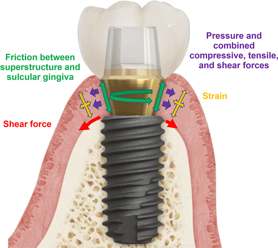
The induced strain in the connective tissues and shear forces initiate the injury at the bone-connective tissue interface. The arrows represent the direction and distribution of forces. The forces are represented by arrows of the corresponding color.
Second, the onset and progress of symptoms and signs follow a time frame similar to that in DTI. Most patients stated that the pain and swelling developed on the day of superstructure connection. In cases 3, 8, and 9, the patients visited the ER within 24 h from the previous visit for superstructure connection. In the physiological process of DTI, acute inflammatory edema commences within two to four hours after external mechanical loading,6,7,9 and the injury progresses rapidly and can cause severe tissue damage.5,8 According to previous studies on DTI progression, acute inflammatory edema grows and intensifies by the fifth day after onset, and recovery occurs after approximately 14 days.9 As described in Table 1, most patients presented within six days of the superstructure connection, and resolution of symptoms occurred in two to three weeks.
Finally, the clinical features resemble an inflammatory response more closely than signs of infection. Rapid swelling was the major confounding feature that hindered differentiation between inflammation and infection. The most challenging aspect is determining whether the inflammation was the result of infection. Therefore, antibiotic therapy was administered empirically in most patients in this study. As summarized in Table 1, the symptoms, signs, laboratory data, and time frame of the presentation were not necessarily consistent with the general progression of odontogenic infections in the head and neck region.1 Above all, it was difficult to identify the infection source other than the implant superstructure connection performed several days previously. It can be postulated that a loose superstructure favors microbial invasion through the transmucosal region. However, this does not apply to patients who underwent insertion of a definitive prosthesis for the first time without previous superstructure loosening.
Laboratory data were retrieved to explore the cause of the events. Though both WBC count and CRP level are inflammatory markers and indicators of infection, clinical correlation is important to ensure an accurate diagnosis. The laboratory data in cases 3, 7, 8, and 9 showed elevated WBC count or CRP level (Table 1). In case 3, CRP level was elevated, WBC count was normal, and the respiratory rate and body temperature were within the normal range, despite slight hypertension. In cases 7 and 8, both elevated WBC count and CRP level were increased. Case 9 showed elevated WBC count and normal CRP level. Most patients did not exhibit fever/chills or toxic appearance, which are the clinical signs of cellulitis in the head and neck regions.1,2 A prospective study on facial cellulitis reported that 91% of the patients presented with chills or rigors before or at admission.2 Only one patient (case 8) described chills, although the patient’s body temperature was only slightly elevated within the normal range. Mild tachycardia and elevated WBC count and CRP level were also observed.
Even if the symptoms and signs are caused by infection, it is assumed that infection represents a secondary development resulting from the progressive swelling and impaired lymphatic drainage.13 The lymphatic drainage in the peri-implant tissue10,11 can be obstructed or occluded primarily by pressure from the superstructure connection and the rapidly growing swollen area. Obstruction of lymphatic drainage increases the risk of infection.13 Moreover, the transmucosal region of the implant can be an infection route for microorganisms.15,16
The rare instances of development of this acute inflammatory condition during routine implant prosthetic treatments seem to be related to the patients’ conditions. Presence of underlying diseases may increase the susceptibility to inflammation and infection.3,4 Diabetes mellitus is a disease that is well known to predispose patients to various infections.12 In the present study, five patients were being treated for diabetes, and laboratory tests revealed moderate to high levels of HbA1c or glucose. Furthermore, the patient who experienced acute swelling twice (cases 4-1 and 4-2) had impaired renal function and hypothyroidism. These underlying conditions may have facilitated the development of acute swelling due to pressure-induced injury.19
Another contributing factor that could be the triggering of the local defense mechanism in the peri-implant tissue by neurogenic regulation leading to an inflammatory response. The distribution of substance P-containing sensory nerve terminal and neurokinin-1 receptors in the peri-implant epithelium has been reported.17 The pain impulse derived from the implant superstructure connection and the secondary pain caused by the growing DTI is afferently transferred via the substance P-neurokinin-1 receptor pathway. Additionally, substance P itself is known to be involved in antimicrobial activity.14 As a result, the afferent signal via the substance P-neurokinin-1 receptor pathway efferently stimulates neutrophil infiltration, immune regulation, vasodilation, and plasma leakage of endothelial cells.16
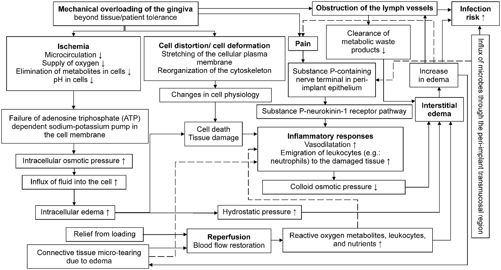
Based on the analysis and previous studies on DTI, we suppose that the most likely pathogenic mechanism of acute inflammatory swelling or suspected cellulitis after connection of implant superstructures is the physiological process of DTI and peri-implant defense mechanisms. Figure 4 presents a schematic diagram of the possible pathogenesis of an acute inflammatory swelling caused by mechanical overloading of the gingiva beyond tissue/patient tolerance based on the DTI conceptual framework.3–9 The diagram also describes the relationship between lymphatic obstruction and infection risk,10,11,13 which includes characteristic permeable biological sealing of the peri-implant tissue14–17 and infection.2–4,12,19 In summary, the external mechanical loading exerted by implant superstructure connections can activate the DTI chain reaction as follows: 1) cell deformation and ischemia in the peri-implant gingiva; 2) induced strain on the gingiva and shear force to initiate intra-/inter-cellular edema and inflammatory responses from the interface between the peri-implant connective tissue and bone; and 3) extensive swelling resulting from the ischemia-reperfusion injury and lymphatic obstruction in the peri-implant tissue. The neurogenic defense mechanisms in the peri-implant tissue can also increase the inflammatory responses. This evolving process continues until leukocytosis, particularly neutrophilic leukocytosis, decreases. Additionally, in medically compromised patients, this condition can increase the risk of infection resulting from the combination of lymphatic obstruction and the inherent permeability of the barrier system of the transmucosal region of the implant.
From our review of the severity of symptoms, we found that those patients who underwent reconnection of the exfoliated HAs or prosthesis tended to exhibit more severe symptoms than those receiving superstructures for the first time. The relatively stronger pressure applied to the constricted gingiva, which results from the absence of a superstructure, may cause a more intense inflammatory response during super-structure reconnection because the extent of the injury is known to be proportional to the load applied.9 Bacterial infection may be initiated through the transmucosal region of the implant.
In such cases, clinicians must first reassure the patient by explaining the possible causes of the symptoms and signs depending on the timeframe of the likely pathogenesis. A careful review of the patient’s medical history may aid diagnosis. Then, the superstructure should be disconnected and replaced with a narrower-sized HA to prevent sustained loading and relieve obstruction of the lymphatic drainage system. If the patient presents with acute swelling, fever, chills, tachycardia, elevated respiratory rate, and a toxic appearance, the patient should be immediately transferred to an OMS specialist. In less severe cases, prescription of anti-inflammatory agents and follow-up may be adequate.
The limitations of this study include the small number of cases, limited availability of blood test results, and lack of bacterial cultures to confirm cases of infection. Further in vivo studies are needed to corroborate the suggested pathogenesis. Structured prospective studies are also needed to trace the duration of symptoms, changes in symptoms and signs, and laboratory test results.
Acute swelling with or without infection may develop due to acute inflammatory responses triggered by connecting or reconnecting implant superstructures. Clinicians need to be aware of the causes and physiological process of pressure-induced injury. Based on the pathogenesis of the injury, clinicians should evaluate the patient carefully and provide adequate treatment or refer the patient for specialist care.
Figshare: Underlying data for ‘Deep tissue injury as possible pathogenesis of acute inflammatory swelling or cellulitis after connecting implant super-structures: a case series study’, https://doi.org/10.6084/m9.figshare.20133998.v1.18
Additional data not provided in Figshare are not publicly available due to the privacy of the patients but are available from the corresponding author on reasonable request (Yang-jin Yi, [email protected]).
Figshare: STROBE checklist for ‘Deep tissue injury as possible pathogenesis of acute inflammatory swelling or cellulitis after connecting implant super-structures: a case series study’, https://doi.org/10.6084/m9.figshare.20134202.v1.20
Data are available under the terms of the Creative Commons Attribution 4.0 International license (CC-BY 4.0)
| Views | Downloads | |
|---|---|---|
| F1000Research | - | - |
|
PubMed Central
Data from PMC are received and updated monthly.
|
- | - |
Provide sufficient details of any financial or non-financial competing interests to enable users to assess whether your comments might lead a reasonable person to question your impartiality. Consider the following examples, but note that this is not an exhaustive list:
Sign up for content alerts and receive a weekly or monthly email with all newly published articles
Already registered? Sign in
The email address should be the one you originally registered with F1000.
You registered with F1000 via Google, so we cannot reset your password.
To sign in, please click here.
If you still need help with your Google account password, please click here.
You registered with F1000 via Facebook, so we cannot reset your password.
To sign in, please click here.
If you still need help with your Facebook account password, please click here.
If your email address is registered with us, we will email you instructions to reset your password.
If you think you should have received this email but it has not arrived, please check your spam filters and/or contact for further assistance.
Comments on this article Comments (0)