Keywords
Wolf’s isotopic response, herpes zoster, post herpetic isotopic response, Ruocco’s immunocompromised district, comedone
This article is included in the Datta Meghe Institute of Higher Education and Research collection.
Wolf’s isotopic response, herpes zoster, post herpetic isotopic response, Ruocco’s immunocompromised district, comedone
The term Wolf’s isotopic response is described as the occurrence of a new skin disorder at the exact site of another, unrelated and healed skin disease. It is usually noticed after herpes zoster infection caused by varicella-zoster virus; hence, it is also known as post-herpetic isotopic response.1 This is a rare and unique phenomenon as much still needs to be understood about the compromise of regional immunocompetence post herpes infected dermatomes making them susceptible to future dermatoses. Herpes zoster is a condition that causes painful, grouped vesicular eruptions, which are unilateral and only affect a dermatome innervated by a single sensory ganglion. This is caused by reactivation of varicella-zoster virus present in dormant form within the sensory ganglia.2 The most widely accepted hypothesis at play behind Wolf’s isotopic response is that of neuro-immune destabilization.3 Langerhans’ cells, a key mediator in neuro-immune balance are decreased in post-herpetic lesions. Sensory nerve fibers not only conduct sensorial stimuli but also modulate the dermal immune response by secreting neuromodulators such as substance P, vasoactive intestinal peptide and calcitonin gene-related peptide which interact with membrane receptors of immune cells.3 Thus, post herpetic viral damage to these sensory nerve fibers alters the neuro-immune homeostasis making the site involved susceptible to other dermatoses.
A 36-year-old male presented to the Dermatology Outpatient Department of the Datta Meghe Institute of Higher Education and Research affiliated tertiary care teaching hospital at Sawangi, Wardha, Maharashtra on 28th March 2023 with complaints of multiple pigmented papular eruptions extending from the midline of the chest to the right upper limb and right side of his back, for the last three months [Figures 1(A) and (B)]. The lesions were not associated with any burning, pain or itching sensation. He had no history of significant medical or surgical comorbidity. Past medical history revealed that he had an episode of herpes zoster involving the same region four months before for which he was treated with Valacyclovir 1 g three times a day for five days along with topical calamine lotion.
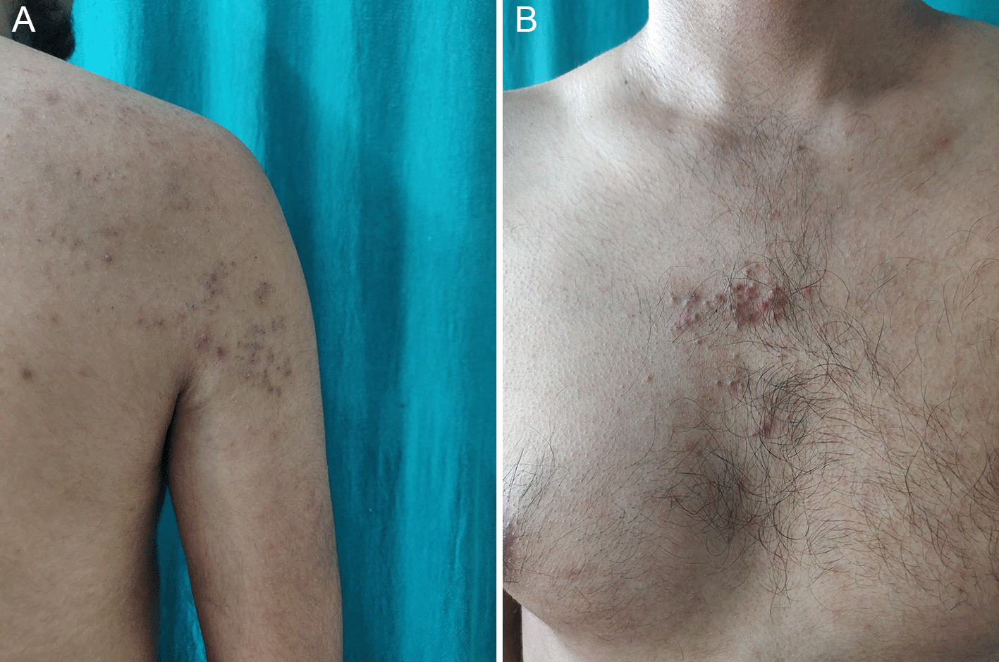
(A) Multiple, grouped comedones on the right side of back. (B) Multiple, grouped comedones on the midline of chest.
On cutaneous examination, multiple, grouped open and closed comedones were noted on the midline of the chest, and the right side of the upper chest and back. The lesions were distributed in accordance with the thoracic T2 dermatome. Dermoscopic examination revealed multiple, round, brown-colored follicular keratinous plugs with peri-lesional erythema (Figure 2). A skin biopsy was performed, and histopathological analysis showed a large dilated follicular canal containing orthokeratotic stratum corneum consistent with closed comedone formation (Figure 3). Based on these findings, the patient was diagnosed with post-herpetic zosteriform comedones. The patient was prescribed oral isotretinoin 20 mg, topical tretinoin 0.05% cream with a moisturizer and asked to follow up regularly. The lesions gradually healed over a five-month period [Figure 4(A) and (B)].
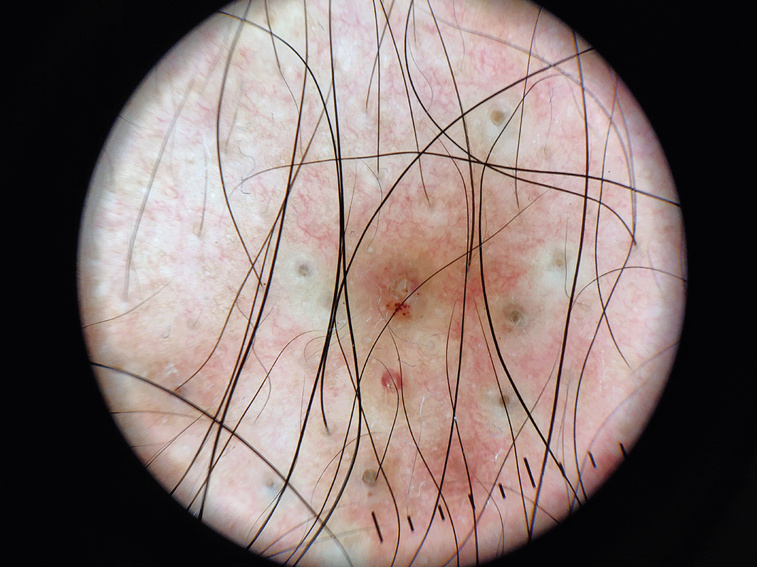
Polarized dermoscopic image showing multiple, round brown colored follicular keratinous plugs with peri-lesional erythema.
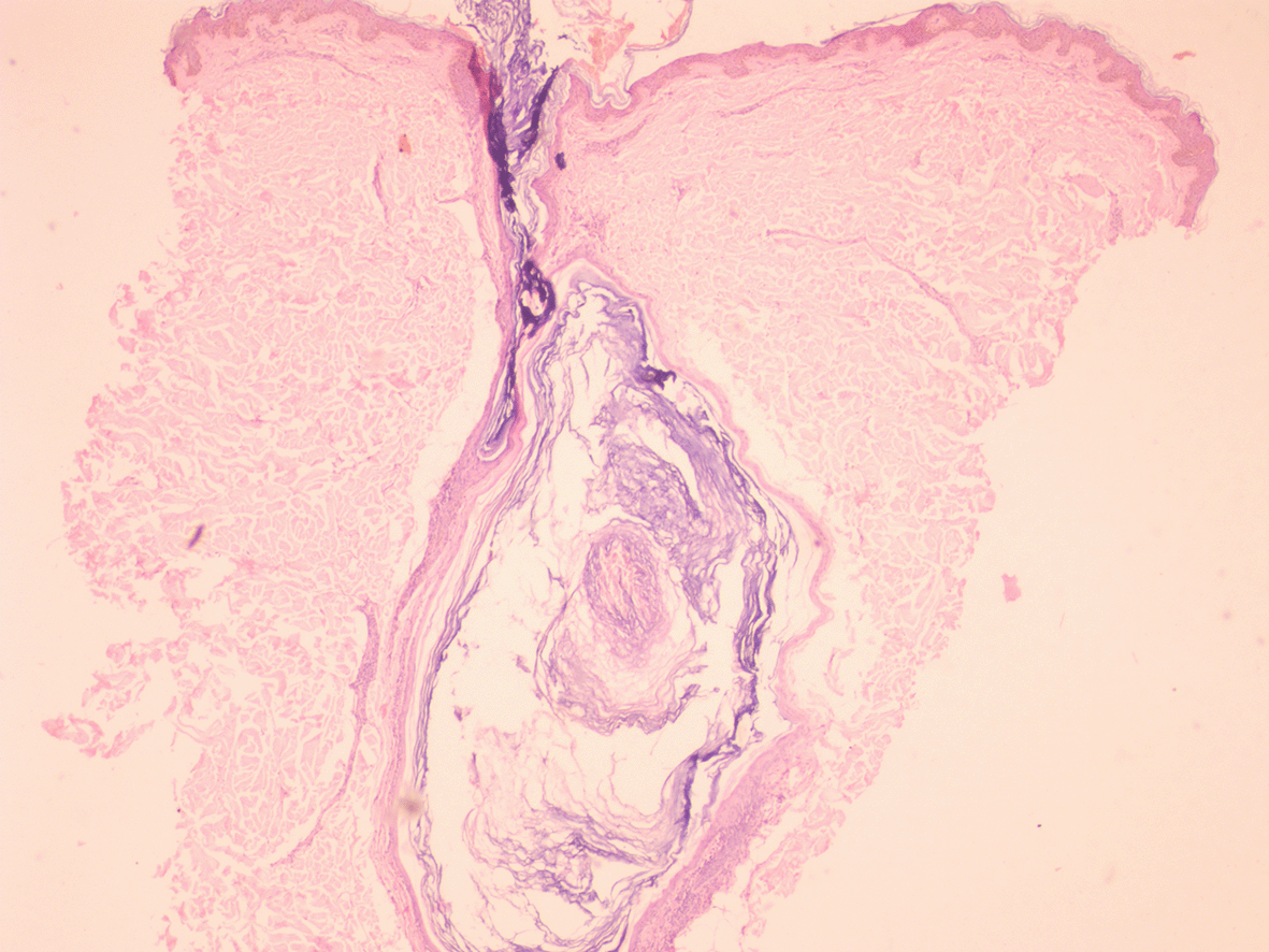
(H&E, ×40)
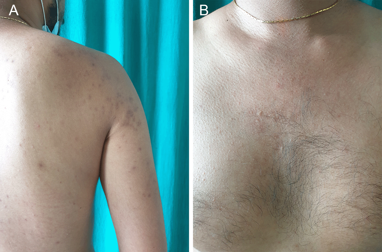
Gradual resolution of lesions over five months. (A) Right side of back. (B) Midline of chest.
Post-herpetic isotopic response was first described in 1955 by a British neurologist, Wyburn Mason, who reported twenty-six patients having developed skin lesions at the locations of previously treated herpes simplex or herpes zoster infections.2 The majority of these secondary lesions were breast, squamous or basal cell carcinomas. Wolf et al. in 1985, described the first non-cancerous skin disorder as tinea corporis occurring at the site of previously healed herpes zoster infection in two patients and subsequently devised the term “isotopic response” in 1995 from the previously known “isoloci response”.2,4 At present, the most widely accepted term Wolf’s post-herpetic isotopic response is used when describing this phenomenon since herpes infection was reported to be the primary disease in the majority of the cases studied.3 The time elapsed between the primary and secondary diseases can be anywhere between a few days to several years.5 This phenomenon also falls under the umbrella term immunocompromised districts described by Ruocco et al. as a skin area more vulnerable than other sites as a result of either acquired (chronic lymphatic stasis, herpetic infections, UV radiations, burns, trauma, tattooing, intradermal vaccinations etc.) or genetic (primary lymphedema and skin mosaicism) etiology resulting in regional immune dysregulation. Failure in function of blood or lymphatic circulation, cytokines, immune-competent cells, neuropeptides or peripheral nerve fibers compromises the local immune homeostasis making the area susceptible to other infections, tumors or disorders of keratinization. Therefore, immunocompromised districts can develop even in an immunologically stable individual.6
There are multiple theories proposed behind the etiology of the Wolf’s post-herpetic isotopic response including viral, immunological, neural and vascular hypotheses.4 An interplay between these different factors leads to neuro-immune destabilization caused by viral damage to the sensory nerve fibers causing release of various neuropeptides and immune modulators that alter the local immune cells (macrophages, lymphocytes and Langerhans’ cells) making the involved site more susceptible to subsequent skin diseases.2,3 Substance P, a neuropeptide released from the damaged nerve endings post herpes infection can stimulate lipogenesis of the sebaceous glands causing increased activity of Propionibacterium acnes leading to the formation of comedones.7
In our case, timely medical treatment along with regular follow up and histopathological analysis enabled us to manage the disease effectively by preventing its further progression. Since this was a single case study, similar case series involving multiple patients will be required for further investigation to improve our understanding of this phenomenon. The primary takeaway lesson from our case is as follows: Since Wolf’s isotopic response is a rare clinical phenomenon, physicians need to be highly observant and take a comprehensive clinical history especially when encountering any dermatoses in a dermatomal distribution pattern in order to make the diagnosis and initiate treatment. Patients need to closely monitor the affected site on a regular basis to assess the development of any new lesions. Patients should be followed up regularly for effective and timely medical management.
| Views | Downloads | |
|---|---|---|
| F1000Research | - | - |
|
PubMed Central
Data from PMC are received and updated monthly.
|
- | - |
Provide sufficient details of any financial or non-financial competing interests to enable users to assess whether your comments might lead a reasonable person to question your impartiality. Consider the following examples, but note that this is not an exhaustive list:
Sign up for content alerts and receive a weekly or monthly email with all newly published articles
Already registered? Sign in
The email address should be the one you originally registered with F1000.
You registered with F1000 via Google, so we cannot reset your password.
To sign in, please click here.
If you still need help with your Google account password, please click here.
You registered with F1000 via Facebook, so we cannot reset your password.
To sign in, please click here.
If you still need help with your Facebook account password, please click here.
If your email address is registered with us, we will email you instructions to reset your password.
If you think you should have received this email but it has not arrived, please check your spam filters and/or contact for further assistance.
Comments on this article Comments (0)