Keywords
Giant cell arteritis, systemic vasculitis, autoimmune disorder, pyrexia of unknown origin, stroke
This article is included in the Datta Meghe Institute of Higher Education and Research collection.
Giant cell arteritis, systemic vasculitis, autoimmune disorder, pyrexia of unknown origin, stroke
Giant cell arteritis (GCA) is a systemic vasculitis that primarily affects large and medium-sized arteries, including the aorta. It can lead to serious complications such as aortic aneurysm and dissection. The presentation of GCA includes constitutional symptoms such as fatigue, malaise and fever, as well as headache, scalp tenderness and jaw claudication (pain with chewing). Vision changes, such as transient or permanent visual loss, double vision or diplopia can also occur due to involvement of the ophthalmic artery. Other symptoms may include weight loss, joint pain and proximal muscle weakness. In this case report, we discuss a 70-year-old female patient with a 2-month history of intermittent fever associated with temporal headache, which was relieved on taking over the counter medications. She was investigated thoroughly, but due to a lack of any significant findings, a positron emission tomography CT (PET-CT) scan was done to rule out any malignancy which guided us to the path of our diagnosis of giant cell arteritis.
The diagnostic criteria of GCA are based on a combination of clinical features, laboratory findings and histopathological findings. Table 1 shows the commonly used diagnostic criteria of GCA.
The presence of three or more of these criteria has a high specificity and sensitivity for the diagnosis of GCA. Clinical judgement is necessary when making the diagnosis. Additionally, the absence of these criteria does not rule out the possibility of GCA and further testing may be required. Early diagnosis and prompt treatment with high-dose steroids can prevent serious complications, such as vision loss, associated with this condition.
Diagnosis of giant cell arteritis typically involves a combination of clinical evaluation, blood tests, imaging studies and biopsy of affected tissue. A biopsy of the temporal artery is the gold standard for diagnosis, as it can show characteristic inflammatory changes in the arterial wall. Blood tests may also reveal elevated levels of inflammatory markers such as erythrocyte sedimentation rate (ESR) and C-reactive protein (CRP). A definitive diagnosis of GCA is made through biopsy of the affected artery, typically the temporal artery.1
Treatment for giant cell arteritis involves the use of corticosteroids, such as prednisone, to reduce inflammation and prevent further damage to affected arteries. The dosage and duration of treatment may vary depending on the severity of the disease and the individual patient’s response. In some cases, immunosuppressive drugs may also be used to help control the autoimmune response.
Pyrexia of unknown origin (PUO) is a condition characterized by persistent fever of at least 38.3°C for more than three weeks, with no apparent cause despite extensive diagnostic workup. The etiology of PUO is varied, with infectious diseases accounting for approximately one-third of cases, followed by inflammatory and malignant conditions.2 PUO can be challenging to diagnose and manage, as it requires a systematic approach in ruling out potential causes. The diagnosis involves a thorough medical history, physical examinationand laboratory tests, including blood cultures, serological testing, imaging studies and biopsy, if necessary. Despite its rarity, PUO poses a significant diagnostic challenge. Its early recognition and prompt intervention can prevent significant morbidity and mortality.2
A 70-year-old female patient, a teacher by profession and currently retired, presented to the outpatient department with chief complaints of fever on and off for two months along with headache, loss of appetite and weight loss. Patient was apparently alright two months ago when she started experiencing fever which was insidious in onset, high grade intermittent, associated with chills and rigour. Fever was relieved on taking anti-pyretics. Patient complained of headache which was localised to the temporal region, squeezing type, not associated with photophobia, phonophobia, nausea or vomiting. There were no aggravating factors for headache and it was relived on taking over the counter medications. Patient also complained of generalised weakness and loss of appetite for the last 2 months. Patient gives history of weight loss of around 12 kilograms which accounts to significant weight loss.
Patient was diagnosed with hypothyroidism 10 years ago, taking Tabthyroxine 100mcg once daily. Patient also had a history of tubercular lymphadenitis 50 years ago and had taken the full course of anti-tubercular therapy, however no documents were available. Apart from these, the patient had no other complaints. Patient had no history of any constitutional symptoms prior to two months ago, no history skin rash, oral ulcers, joint pain or swelling.
On examination patient was vitally stable, there were no mucocutaneous lesions, and all peripheral pulses were well felt. General examination also did not reveal any abnormalities.
Patient was investigated on the line of pyrexia of unknown origin as she was symptomatic for more than two months. All routine investigations, as mentioned in Table 2, were normal, fever profile was normal. ESR and CRP were on the higher side. Autoimmune profile including anti-nuclear antibodies, anti-dsDNA antibodies, anti-Smith antibodies, anti-Ro and anti-La were all negative. Chest X-ray and chest CT also did not reveal any abnormalities. Bone marrow examination was also normal. Table 1 shows various diagnostic modalities used to screen for PUO.
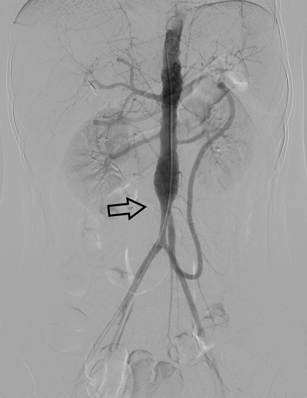
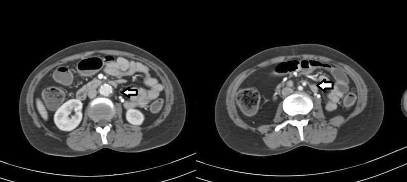
In view of no significant findings in examination and investigations, 3 days after admission to the hospital, it was decided to do a positron emission tomography CT (PET-CT) scan to rule out any malignancy. Due to the hospital not having a FDG-PET scan available, the patient was referred to another hospital for this procedure and then returned to our hospital with the results of the PET-CT. The results showed a small uptake in aorta and a differential of giant cell arteritis was considered. A CT aortogram and contrast enhanced CT abdomen were done, which revealed circumferential thickening of distal descending part of aorta as shown in Figures 1 and 2 respectively. Temporal artery biopsy was done, and the biopsy report showed giant cell arteritis. Patient was started on high dose steroids (oral prednisone at 1mg/kg/day) and was started on supportive management. Serial monitoring of ESR and CRP showed the values to be decreased 3-4 days after the initiation of steroids and the patient improved drastically. Patient was eventually discharged with follow up every two months.
Giant cell arteritis is a systemic inflammatory disorder that predominantly affects medium to large-sized arteries, commonly the temporal arteries. GCA usually presents with headache, scalp tenderness, and jaw claudication, but it can also present as pyrexia of unknown origin in some cases.
When GCA presents as PUO, the patient has a persistent fever of at least 38.3°C for more than three weeks, with no apparent cause despite extensive diagnostic workup. In such cases, the patient may have constitutional symptoms such as fatigue, malaise, and weight loss. The diagnosis of GCA should be considered in patients over the age of 50 with PUO, especially in the presence of other symptoms of GCA such as headache, scalp tenderness, and jaw claudication.
Laboratory tests may reveal elevated inflammatory markers such as erythrocyte sedimentation rate and C-reactive protein. Biopsy of the temporal artery can confirm the diagnosis of GCA, showing characteristic findings such as granulomatous inflammation and giant cells.3
PET imaging with 2-fluoro-2-deoxy-D-glucose (FDG) allows for non-invasive visualization and quantification of metabolic activity in tissues, which can be used to differentiate between normal and inflamed or malignant tissues. In GCA, FDG-PET imaging can detect increased metabolic activity in affected arteries, including the aorta, and can provide valuable information about the extent and severity of disease involvement.4
Diagnosis of GCA can be challenging, as the disease can present with nonspecific symptoms and physical examination findings, and laboratory tests may be inconclusive. PET imaging with FDG has emerged as a valuable tool in the diagnosis and management of GCA. Several studies have demonstrated the utility of FDG-PET imaging in the diagnosis and management of GCA. In a study by Blockmans et al.,5 FDG-PET imaging had a sensitivity of 87% and a specificity of 100% in the diagnosis of GCA, and was able to detect early disease involvement in patients with negative temporal artery biopsy results. In another study by Nuenninghoff et al., FDG-PET imaging had a sensitivity of 78% and a specificity of 91% in the diagnosis of GCA, and was able to detect aortic involvement in patients with clinically suspected but biopsy-negative disease.
In our case, PET scan came out to be the major modality which helped us in investigating a diagnosis of GCA which was later confirmed by biopsy and CT aortogram.
Aortic involvement in GCA is an important predictor of disease severity and risk of complications, including aortic aneurysm, dissection, and rupture. However, diagnosis of aortic involvement can be challenging, as the aorta is a deep-seated structure that is not easily accessible to physical examination or biopsy.
FDG-PET imaging can be a valuable tool in the diagnosis and management of aortic involvement in GCA. In a study by Meller et al.,6 FDG-PET imaging had a sensitivity of 83% and a specificity of 90% in the detection of aortic involvement in patients with GCA, and was able to differentiate between active and inactive disease in the aorta. In another study by Salvarani et al., FDG-PET imaging was able to detect aortic involvement in 67% of patients with clinically suspected GCA and negative temporal artery biopsy results.
A high index of suspicion for GCA should be maintained in patients presenting with PUO, especially in those over the age of 50 with additional symptoms of GCA. Prompt diagnosis and treatment with high-dose steroids are crucial to prevent irreversible complications associated with GCA, such as vision loss.
FDG-PET imaging is emerging as a valuable tool in the diagnosis and management of GCA, particularly in patients with suspected aortic involvement. FDG-PET imaging can detect early disease involvement, differentiate between active and inactive disease, and provide valuable information about disease extent and severity. However, FDG-PET imaging should be used in conjunction with other diagnostic modalities, such as clinical evaluation, laboratory testing, and imaging studies, to confirm the diagnosis of GCA and guide appropriate treatment.
| Views | Downloads | |
|---|---|---|
| F1000Research | - | - |
|
PubMed Central
Data from PMC are received and updated monthly.
|
- | - |
Provide sufficient details of any financial or non-financial competing interests to enable users to assess whether your comments might lead a reasonable person to question your impartiality. Consider the following examples, but note that this is not an exhaustive list:
Sign up for content alerts and receive a weekly or monthly email with all newly published articles
Already registered? Sign in
The email address should be the one you originally registered with F1000.
You registered with F1000 via Google, so we cannot reset your password.
To sign in, please click here.
If you still need help with your Google account password, please click here.
You registered with F1000 via Facebook, so we cannot reset your password.
To sign in, please click here.
If you still need help with your Facebook account password, please click here.
If your email address is registered with us, we will email you instructions to reset your password.
If you think you should have received this email but it has not arrived, please check your spam filters and/or contact for further assistance.
Comments on this article Comments (0)