Keywords
Schatzker fracture, tibia, fibula, range of motion, therapeutic rehabilitation, balance exercises, proprioceptive training, muscle energy technique, case report.
This article is included in the Datta Meghe Institute of Higher Education and Research collection.
Schatzker fracture, tibia, fibula, range of motion, therapeutic rehabilitation, balance exercises, proprioceptive training, muscle energy technique, case report.
The primary long bone in the lower leg is termed the tibia. It is referred to as the “shin bone” and can easily be felt sliding down the anterior (front) of the shin between the knee and the ankle. Located laterally to the tibia, the fibula is a shorter long bone that aids in ankle rotation and stability. The tibia is the longest bone that is most likely to fracture out of all the bones in the lower extremity.1 There are many different morphological presentations of tibial plateau fractures, and several categorization schemes have been suggested to reflect this variance.2 Fractures of the tibia that occur close to the articular surface are referred to as tibial plateau fractures. The prevalence of these fractures comprises around 1.2 percent of all fractures and is often higher among young individuals who have had high-energy trauma.3 Most older people who have had low-energy trauma are also affected by them.3 According to the Schatzker classification, there are six different types of tibial plateau fractures: lateral plateau fractures without depression (type I), lateral plateau fractures with depression (type II), compression fractures of the lateral or central plateaus (type IIIA or IIIB), medial plateau fractures (type IV), bicondylar plateau fractures (type V), and plateau fractures with diaphyseal discontinuity (type VI).4 The fibula is one of the most often fractured long bones, mostly as a result of its anatomical location and function. Fibular fractures frequently occur with tibial fractures as a result of a high-energy pattern of damage.5 Post-operative physiotherapy rehabilitation shows beneficial improvement in pain score, mobility activity, range of motion, increased muscle strength, gait pattern, and activities of daily living, etc.6 This case report describes a complex Schatzker type 6 proximal tibial fracture with the head of a fibula fracture right side following a road traffic accident who underwent surgical intervention and subsequently underwent physiotherapy rehabilitation. This article is reported in line with CARE guidelines.10
Here we are reporting a case of a 41-year Indian male who is desktop worker and was apparently alright four months back. On the same day of the accident, he was brought to hospital casualty by relatives with an alleged history of a road traffic collision near Wardha sustaining an injury to his right knee and leading to inward rotation of the leg. On admission, the patient was conscious and complaining of knee pain, swelling, and inability to get up and bear weight on his right leg. The pain was sudden in onset and severe in intensity. Pain gets aggravated with movement and gets relieved with rest. Diffused swelling was present over the right knee and ankle. There was no history of head trauma and ear, nose, or throat (ENT) bleeding. After radiological investigation, it revealed that proximal tibial fracture with a fibular head fracture on right side. The patient underwent surgical intervention for proximal tibial fracture right side. The patient visited physiotherapy outpatient department on 11 April 2023. At that time the patient’s chief complaints were pain in the right leg, unable to bend the knee completely, and difficulty in walking. To the present day, physiotherapy is continued. Patient had no relevant past, medical, psycho-social and family history.
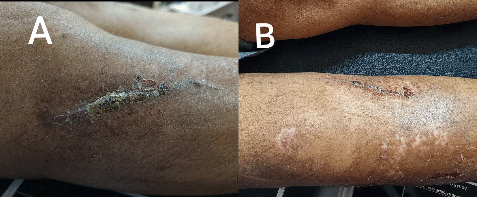
Proper informed consent was taken from the patient prior. Physical examination was done in the supine position. On inspection and palpation, scar marks (Figure 1A and 1B) were present on the anteromedial and anterolateral aspects of the proximal tibial and fibular regions. The scar was hyperpigmented, vascularity was intact. Shiny skin was present along the course of the tibial shin. An 8 cm long scar was present on the anterior-medial aspect of and a 6 cm long scar mark on the anterolateral aspect of the proximal leg. There was obliteration of the hollowing which was present at the medial and lateral edge of the patella. Diffused swelling was present in the knee joint, which was extending beyond the limit of the joint cavity. Wasting and loss of the bulk of quadriceps muscle as compared to non-affected limb which was confirmed on girth measurement. Grade 2 tenderness was present: over the inferior pole of the patella, tibial tuberosity, along the tibial shin proximal to distal. All sensation and reflexes were intact. There was no limb length discrepancy. Manual muscle testing is shown in Figure 2.

(A): Scar mark on the anterolateral aspect of proximal leg. (B): Scar mark on the posterolateral aspect of proximal leg.
The timeline of the event is mentioned in Figure 3 which shows the sequence of events from when the patient was admitted to the hospital till the start of the rehabilitation.
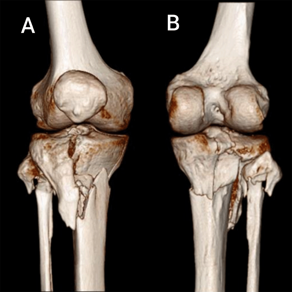
The radiological evaluation indicates a pre-operative computed tomography scan, as illustrated in Figure 4A and 4B, which revealed a right-sided Schatzker type 6 proximal tibial fracture with fibular head fracture. The patient underwent surgical intervention open reduction and internal fixation with plate osteosynthesis for proximal tibial fracture right side mentioned in Figure 5A and 5B.

(A): Shows tibial plateau and fibular fracture in anterior aspect of knee joint. (B): Shows tibial plateau and fibular fracture in posterior aspect of knee joint.
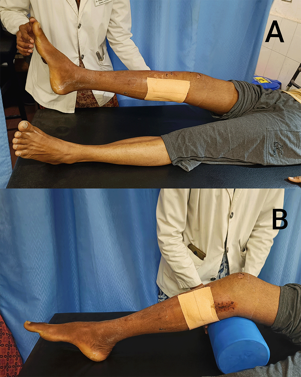
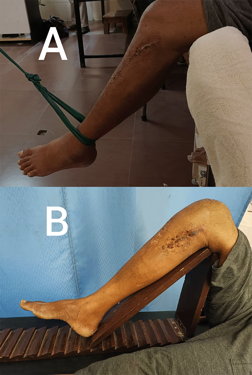
Physical therapy intervention is essential in the restoration and functional activities of patients with Schatzker type 6 fractures, which usually requires a multidisciplinary approach. Tables 1 and 2 show some broad ideas and elements of rehabilitation for Schatzker type six with fibular head fracture. Smarts objectives include patient and his family education: Inform the patient about their illness, teach them how to take care of themselves, and provide them tips on how to avoid getting injured or relapsing in the future. Teach the patient about ergonomics, correct body mechanics, and ways to prevent the worsening of their existing ailment. Pain management: Lower pain levels using a variety of methods, including physical treatment, therapeutic exercises, and techniques like heat or cold therapy. Joint flexibility and range of motion (Figure 6A): increase the range of affected joints and mobility by using an angle frame (Figure 7B) to stretch specific structures. Strengthening of lower limb muscles with a bolster (Figure 6B), theraband (Figure 7A), and weight cuff (Figure 8A): substantially improves muscular strength and structural stability of joints. Enhance overall posture and balance to decrease the risk of falls and perform better in daily activities. Gait training (Figure 8B): Focus on correcting locomotor inefficiencies, improving steps, and encouraging balance, and load redistribution in order to return to a normal walking pattern. Help the patient recover independence by doing activities of daily living (ADLs) including dressing, using the bathroom, and taking care of themselves. Functional independence: Allow the patient to restore the greatest amount of functional independence and carry out their prior level of activities, including jobs, hobbies, and leisure activities. Enhance the patient’s entire quality of life by enhancing physical function, minimizing restrictions, and fostering psychological wellness. Long-term self-management: Provide the patient with the information and abilities to autonomously manage their disease, including self-care, exercise, and continuous maintenance techniques. No adverse effect to the intervention was noted.

(A): Shows active assisted straight leg raise. (B): Vastus medialis strengthening with bolster.

(A): Strengthening exercises with theraband. (B): Knee flexion using angle frame.
The outcome measures were assessed on the first day of physiotherapy rehabilitation and follow-up was taken on the last day of the physiotherapy intervention. Results are shown in Table 3.
The rehabilitation strategy for bi-condylar tibial plateau fractures is extremely challenging because they frequently occur from high energy trauma and are complicated by soft tissue injuries.3 Tibial plateau fractures are a significant source of morbidity. In situations of high velocity injuries, treatment should be dependent on the fracture pattern, soft tissue condition, and overall health of the patient.7 In order to restore full knee flexion, manage swelling, ensure appropriate gait training, and improve the strength of quadriceps muscle, muscle strength should be maintained throughout the healing process using isometric, isotonic, and isokinetic muscle contraction.6 The muscle energy technique is a the type of manual therapy that increases range of motion and strength of the muscles. Physiotherapy can assist in preserving and enhancing mobility and strength in stressful post-operative situations.8 The aim of the treatment sessions was to preserve muscular integrity while enhancing the lower extremity activities, non-weight-bearing walking with a walker and little help for everyday activities. To start and improve the knee range of motion, electrotherapy modalities including continuous passive motion were applied.9 A structural rehabilitation programme is always advised for the post-operative patient’s recovery based on their physical condition and functional requirements to provide favourable prognostic results. The patient in our case was given written protocol, urged to schedule follow-up appointments, and instructed to complete all exercises as part of home workouts. The objective of this case study was to highlight the need of quick surgical intervention and crucial physical therapy rehabilitation to achieve the functional goals regarding the patient and its prognosis.
In this study, rehabilitation of a patient with a Schatzker type VI tibial plateau fracture were emphasized. The patient had open reduction and internal fixation (ORIF), which was followed by a thorough rehabilitation regimen. Several phases made up the rehabilitation programmed, including initial immobilization, pain management, range-of-motion exercises, muscle strengthening, balancing training, and functional status. The patient progressed through these stages based on their level of comfort, functional capacity, and the results of the radiographs. The patient demonstrated remarkable improvements in pain levels, range of motion, muscular strength, and functional skills throughout the rehabilitation procedure. They were fully capable of performing daily tasks on their own including activities related to the lower limb. The positive results obtained in this instance serve as a guideline for future clinical work and additional study in the area of musculoskeletal rehabilitation.
Written informed consent for the publication of their clinical details and clinical images was obtained from the patient.
All data underlying the results are available as part of the article and no additional source data are required.
Zenodo: Standard rehabilitation protocol for complex tibial plateau fracture associated with fibular head fracture managed with plate osteosynthesis: a case report, https://doi.org/10.5281/zenodo.8186763. 10
Data are available under the terms of the Creative Commons Attribution 4.0 International license (CC-BY 4.0).
We would like to thank the patient and their relatives for providing us with detailed information about the condition and supporting us during the study.
| Views | Downloads | |
|---|---|---|
| F1000Research | - | - |
|
PubMed Central
Data from PMC are received and updated monthly.
|
- | - |
Provide sufficient details of any financial or non-financial competing interests to enable users to assess whether your comments might lead a reasonable person to question your impartiality. Consider the following examples, but note that this is not an exhaustive list:
Sign up for content alerts and receive a weekly or monthly email with all newly published articles
Already registered? Sign in
The email address should be the one you originally registered with F1000.
You registered with F1000 via Google, so we cannot reset your password.
To sign in, please click here.
If you still need help with your Google account password, please click here.
You registered with F1000 via Facebook, so we cannot reset your password.
To sign in, please click here.
If you still need help with your Facebook account password, please click here.
If your email address is registered with us, we will email you instructions to reset your password.
If you think you should have received this email but it has not arrived, please check your spam filters and/or contact for further assistance.
Comments on this article Comments (0)