Keywords
Periodontitis, Infrabony defects, Bioresorbable Collagen Membrane, Xenograft, Advanced platelet rich fibrin, periodontal regeneration, CBCT, scaling and root planning.
This article is included in the Datta Meghe Institute of Higher Education and Research collection.
Periodontitis, Infrabony defects, Bioresorbable Collagen Membrane, Xenograft, Advanced platelet rich fibrin, periodontal regeneration, CBCT, scaling and root planning.
Periodontitis is a chronic multifactorial inflammatory disease characterised by a dysbiotic dental biofilm that leads to gradual loss of periodontal attachment and bone which ends with a variety of negative consequences on speech, aesthetics, masticatory function, psychological well-being, as well as quality of life.1
Regeneration of periodontium is defined as “the reproduction or reconstitution of a lost or injured part to restore the architecture and function of the periodontium. Regenerative periodontal therapies aim with the repairment of the periodontal structure and function.”2,3 With loss of attachment apparatus due to periodontal diseases, the main goal is to regenerate the attachment of periodontium to involve both development of newer cementum on the root of the tooth and newer periodontal attachment between recently formed bone and cementum.
Even though autologous bone grafts are still the preferred method for bone augmentation surgeries, heterologous bone substitutes are successfully used to address the limited supply of autologous tissue, reducing morbidity of donor site, potential resorption, mismatch of size, insufficient graft material, operating time, and costs.4,5 Yet, to prevent site of a second surgical harvest and medical issues such as ankylosis and root resorption, people resort more to alternate choices for periodontal treatment.
Xenografts are bone grafts that are transplanted in humans from different species (usually porcine and bovine). Anorganic bovine bone graft (ABM) is deproteinized bovine bone mineral that is naturally generated. Hard tissue repairs intrabony defects in humans and clinical attachment level have been improved using anorganic bovine bone. The most prevalent source of xenografts in dentistry is deproteinized bovine bone and is utilised extensively for regeneration of alveolar bone and has a high rate of success, particularly in intraoral treatments.6
An open flap debridement alone is less effective than guided tissue regeneration (GTR). Based on biological basic principles, the membrane is positioned to prevent epithelial cells of gingiva from moving apically. Deproteinized bovine bone matrix xenograft (DBBM), a deproteinized form of bovine bone mineral, is routinely added to GTR.7,8 An X-ray beam with a conical form utilised in cone-beam computed tomography (CBCT) makes a 2-dimensional array of image detectors more sensitive. Studies that evaluated infrabony abnormalities using CBCT indicated greater precision and accuracy.9 Because CBCT readings are more accurate, it is possible to detect anomalies in buccal and lingual regions of the teeth.
To the best of our knowledge, this is the first clinical trial for infrabony defects that will compare collagen membrane combined with deproteinized bovine bone matrix and A-PRF combined with deproteinized bovine bone matrix.
This protocol is reported in line with the Standard Protocol Items: Recommendations for Interventional Trials (SPIRIT) checklist.16
To evaluate the effectiveness of biodegradable collagen membrane in combination with xenograft in the treatment of human infrabony defect.
The objectives of this study are:
1. To evaluate the effectiveness of Collagen Membrane combined with Deproteinized Bovine Bone Matrix regarding increase in clinical attachment level, probing pocket depth reduction and radiographic bone fill in human infrabony defects at 6 months after surgery.
2. To evaluate the effectiveness of A-PRF combined with Deproteinized Bovine Bone Matrix regarding increase in clinical attachment level, decrease in probing pocket depth and radiographic bone filled, 6 months after surgery.
3. To compare the effectiveness of biodegradable Collagen membrane and A-PRF membrane in combination with Deproteinized Bovine Bone Matrix bone graft at 6 months after surgery.
Calculation of sample size was done with the use of previous study data by Gorkhali et al. in 2020.7 The result of the calculation is 12 in each group, thus around 24 samples will be undertaken for the entire study.
Ratio of sample size (Group 1/Group 2) = 1:1
Total sample size 24 with each 12 per group.
The outpatient department of periodontics, Sharad Pawar dental college, Sawangi, Wardha will choose 24 systemically healthy individuals with mild to advanced chronic periodontitis based on the following inclusion criteria:
Inclusion criteria
1) Presence of at least 1 or 2 radiographically detectable interproximal infrabony osseous defect with probing pocket depth ≥5 mm and clinical attachment loss ≥5 mm following initial therapy (assessed radiographically by CBCT).
2) Depth of intraosseous component of the defect ≥3 mm by clinical and radiographic means, which will be confirmed on intra-surgical measurement (assessed using UNC 15 probe).
3) A radiographic base of the defect at least 3 mm coronal to the apex of the tooth (assessed radiographically by CBCT).
4) Presence of at least 3 mm width of keratinized gingiva around test teeth to allow complete soft tissue coverage of the defect (assessed using UNC 15 probe).
Exclusion criteria
1) Evidence of localized aggressive periodontitis.
2) Patients with unacceptable oral hygiene (Plaque Index >1).10
3) Smokers (recent history of smoking more than 10 cigarettes/day) or who used any of tobacco products.
4) Tooth with inadequate endodontic/restorative treatments.
5) Tooth with mobility exceeding grade II and exhibiting a class III or class IV furcation defect.
6) History of periodontal surgical therapy of the selected quadrant.
7) Women who are pregnant or lactating.
In a specially created chart, information about food status, oral hygiene practises, systemic history, gingival and also periodontal state, in addition to standard clinical information, will be documented.
Supragingival and subgingival scaling and root planning will be done as a part of the initial therapy. If required, a coronoplasty will be carried out. Patient will receive repeated plaque control instructions until they have a plaque score of less than 1. Following the conclusion of the initial therapy, a re-evaluation will be conducted to do assessment of the patient towards therapy. If during re-evaluation test after 6 weeks, the periodontal pocket depth is more than 3 mm, plaque score is less than 1 then only we will take the patient for surgery.
Each patient will be given an explanation of the study’s goal and design prior to getting started, and their informed consent will be obtained.
The probe angulations and position will be standardised using a specially designed occlusal acrylic stent. A cast model made from an alginate impression will be used to create an occlusal stent. The occlusal stent will be stretched to encompass coronal one-third of the teeth. A reference point or slot will be created on the deepest site of affected tooth. Linear, fixed reference point will be the apical border.
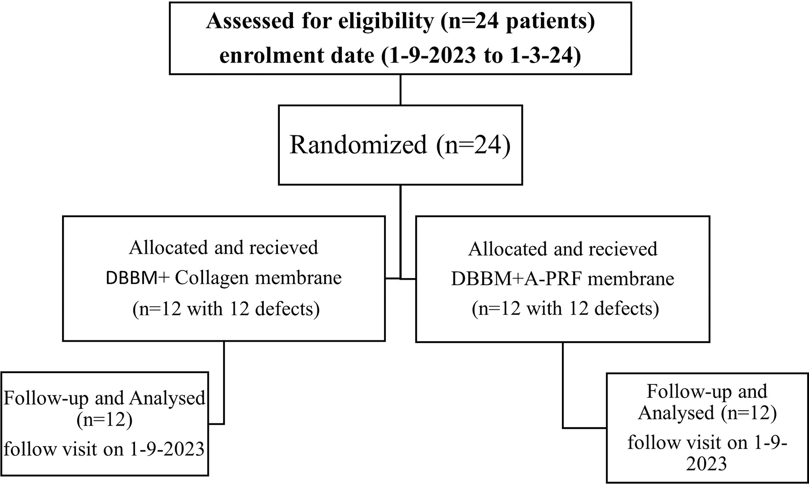
After receiving initial therapy, a total of 24 systemically healthy individuals will be chosen for the study. A coin flip will be used to randomly distribute the participants, in keeping with a randomised parallel design, each of which will have 12 defects, prior to surgery (Figure 1).
The clinical parameters that will be documented like Plaque index, papillary bleeding index, probing pocket depth, relative clinical attachment level, and relative gingival marginal level on the day of surgery as baseline and then after six months. Moreover, additional data will be collected including intraoral periapical radiographs, intraoral clinical pictures, and periodontal charting on a specially prepared form.
Indices
Plaque index, a measurement of the buildup of full mouth supragingival plaque, will be used in order to assess the patient’s oral hygiene health. Papillary bleeding index will be used to evaluate inflammation of gingiva. These are:
The best score will be less than 1. Worst score will be more than 1.
Probing measurements
A UNC-15 probe will be inserted into the slot of acrylic stent at the gingival margin level, with the tip positioned at that level. The measurement will be taken up to the bottom edge of the stent, will be recorded as the relative gingival margin level. A relative attachment level will then be determined. The depth of the probing pocket will be measured (PPD).
With a Periodontal Probe calibrated to UNC-15, width of keratinized gingiva will be determined. At baseline, three months, all probing measurements will be recorded.
Radiographic analysis
CBCT
Using CBCT, osseous defect sites will be examined at baseline and six months post-surgery. The lowest discontinuous point of the periodontal ligament will serve as landmark for the location of the base of defect. Root surface will be intersected by a line that runs perpendicular to the alveolar crest (AC). AC is the point of intersection along the root surface.
Rinsing the patients mouth using 0.2% chlorhexidine gluconate solution at the start of the surgical procedure is required for the patients. The condition of asepsis will be preserved during the entire process. The area undergoing for surgery will be anaesthetized using a local anaesthetic solution (2% w/v Lignocaine adrenaline solution (1:100,00) will be used. Maximum of 15 ml I.M. dosage will be given).
Flap design (incisions)
Using BP blades number 12 or 15, intracrevicular (sulcular) incisions will be made to include periodontal access flap on the buccal and lingual sides. To retain the complete interdental papillae and accomplish primary wound closure, the incisions will be carried. The flap will include a tooth mesially and distally to the tooth and is connected with the defect. If additional access is needed, divergent vertical relief incisions will be made one tooth away from the problem.
Flap reflection
A periosteal elevator will be used for reflecting a full thickness mucoperiosteal flap to expose alveolar bone inside the osseous defect. By removing granulomatous tissue, extreme care will be taken to prevent perforation of the reflected periodontal flap.
Debridement and root surface management
Hand tools (Hu-friedy Gracey curettes) and ultrasonic tools (EMS, piezo 150) will be used to remove granulation tissue from the osseous defect. It will be taken care of to ensure that the flap is not extremely thin. Plaque and calculus on the root surfaces will be removed. The root surfaces will be planed until they have smooth and firm surface.
At this point, UNC15 probe will directly assess the vertical bone defects and quantity of the bony walls present. If the vertical depth of the bone defect is less than 3 mm, the subject’s eligibility will be determined in the end. If the base of the defect doesn’t bleed enough, a half-round bur will be used to perform intra-marrow penetration. At this time, deproteinized bovine bone matrix graft will be used in combination with porcine collagen membrane to correct the test site defects.
Preparation of APRF
Venous blood (10 ml) will be drawn under aseptic conditions from antecubital fossa (10 ml venous blood will be withdrawn in 18 gauge needle) and moved to sterile test tubes before being centrifuged (REMI, (R-8C)) at 1500 rpm for the next 14 minutes to prepare A-PRF, according to Ghanaati et al.12 In the test sites, the infrabony defect will first get a Porcine Collagen Membrane with DBBM.
Procedure for test group
It will be possible to completely isolate and hemostasize the defect. To enable quick flap closure after the implantation of the graft material, the flap will be presutured without making a knot. Deproteinized Bovine Bone Matrix bone graft will be inserted inside the osseous defect in the test site until maximal coronal level is reached of the osseous wall by raising the flap, on which Collagen membrane will be placed over the defect. Membrane will be applied so that it extends 3 mm or more. The flap will then be coronally repositioned and then sutured accordingly.
Now the reflected flap will be held in place combining vertical mattress sutures and interproximal sutures. After two minutes, saline-soaked gauze will be used to close any gaps that could allow a clot to develop and prevent reattachment. Periodontal dressing will be applied.
Procedure for control group
The surgical process on selected control site and the test site will be the same, with the exception that the osseous deficiencies at control sites will be filled with Deproteinized Bovine Bone Matrix graft and then APRF membrane will be applied on the top of the filled bone graft.
Antibiotics including Amoxicillin 500 mg thrice a day and analgesics with the combination of Ibuprofen 325 mg and Paracetamol 400 mg thrice a day will be prescribed post-surgical for 5 days. Patients will be instructed to rinse daily with 0.2% chlorhexidine gluconate twice for 6 weeks. Periodontal dressing and sutures will be removed after 8-10 days post-surgery. Patients will be instructed to clean the treated site with cotton pellet saturated with 0.12% chlorhexidine gluconate for additional 2-3 weeks in an apico-coronal direction and later using a soft toothbrush. After this period, patients will be again instructed to resume mechanical oral hygiene measures, use of interdental cleaning aids such as interdental brush and to discontinue chlorhexidine.
Monitoring
The study will be monitored till completion by data monitoring committee (DMC) which includes PG Guide, head of the department, research convener and chief advisor of university research cell.
The principal investigator (PI) will have access to these interim results and make the final decision to terminate the trial.
Data will be collected, assessed, and spontaneously reported during adverse events and other unintended effects of trial interventions or trial conduct.
The project management group meet will review the trial conducted every month. The trial steering group and the independent data monitoring and ethics committee meet will review and conduct the trial period till the trial is complete.
Patients will be recalled back at the first, third, and sixth months after surgery. All patients will be given oral hygiene instructions together with oral prophylaxis (supragingival scaling). In the first six months following surgery, neither probing nor subgingival instrumentation will be done.
At six months, during follow-up visit a thorough post-operative evaluation will be carried out. Plaque index, gingival index, probing pocket depth, relative clinical attachment level, and relative gingival marginal level are the main clinical parameters that will be recorded. Radiographs and CBCT will also be required.
After 6 months, we are expecting increase in clinical attachment level and decrease in periodontal pocket depth with radiographic bone fill in the test group.
Data analysis
The data collected from patients will be entered on Microsoft excel by PI ensuring the quality checks. Software for statistical analysis will be SPSS version 15.0, SPSS, Chicago, USA). Student’s unpaired t-test will be done.
We expect both surgeries to lead to significant reductions in probing pocket depth and clinical attachment gains, and treatment with open flap debridement with bovine-derived xenografts and collagen membrane will lead to significantly higher probing pocket depth reduction and clinical attachment gain than treatment with open flap debridement alone.
The reconstruction of periodontal osseous defects is a continuous challenge with periodontal therapy. The periodontal wound healing after periodontal therapy occurs by periodontal repair. In recent years, efforts have been concentrated towards periodontal regenerative procedures aimed at restoring the vanished periodontal support. The present investigation aims to evaluate the effectiveness of bioresorbable collagen membrane together with bovine xenograft for periodontal regeneration in human infrabony defects. The current discussion is based on effectiveness of combination of xenograft with bioresorbable collagen membrane in comparison to combination of xenograft with A-PRF for periodontal regeneration in human infrabony defects (Periodontal defects).
Xenograft has been shown to exhibit good biocompatibility and osteoconductivity in both animal and human study. In addition it has been found that resorption rate of xenograft corresponds to the formation of newer bone.7 According to the findings of the study by Gorkhali et al.,7 patients with chronic periodontitis who underwent surgical therapy with open flap debridement alone or open flap debridement (OFD) with bovine-derived xenograft and collagen membrane experienced a clinically significant PPD reduction and CAL gain, but only in the case of CAL at six months across both groups. Various investigations have been done for the evaluation of bone grafts and collagen membrane in adjunct to surgical therapy for regenerative procedure.
Sculean et al.13 had a similar study where they compared the treatment of deep intrabony defects with bovine-derived xenograft and a bioresorbable collagen membrane to open flap debridement. Bio-oss showed better regenerative properties in infrabony defects than open flap debridement.14,16 According to Luepke et al.,15 resorbable barrier membrane combined with DFDBA produced superior results than resorbable barrier membrane used alone.
Procedures will be performed in keeping with the ethical standards of the institutional. This protocol was permitted by the Institutional Ethics Committee of Datta Meghe Institute of Higher education and Research, Sawangi, Meghe, Wardha (approval number DMIHER (DU)/IEC/2023/576) on 6th February 2023. This trial has been registered at the Clinical trial registry of India (CTRI) (REF/2023/05/066732; registration pending).
Case histories will be taken for each patient. A unique I’d number will be provided to each case histories which will be stored in password protected folder in desktop and while sharing it will be ensured that identifying information such as age, address, IPD number and OPD number will be deleted. PI and institutional authorities will be accessible to the final trial dataset.
| Views | Downloads | |
|---|---|---|
| F1000Research | - | - |
|
PubMed Central
Data from PMC are received and updated monthly.
|
- | - |
Provide sufficient details of any financial or non-financial competing interests to enable users to assess whether your comments might lead a reasonable person to question your impartiality. Consider the following examples, but note that this is not an exhaustive list:
Sign up for content alerts and receive a weekly or monthly email with all newly published articles
Already registered? Sign in
The email address should be the one you originally registered with F1000.
You registered with F1000 via Google, so we cannot reset your password.
To sign in, please click here.
If you still need help with your Google account password, please click here.
You registered with F1000 via Facebook, so we cannot reset your password.
To sign in, please click here.
If you still need help with your Facebook account password, please click here.
If your email address is registered with us, we will email you instructions to reset your password.
If you think you should have received this email but it has not arrived, please check your spam filters and/or contact for further assistance.
Comments on this article Comments (0)