Keywords
Pleural effusion, lung boost device, Pulmonary function test, chest expansion, conventional chest physiotherapy.
This article is included in the Datta Meghe Institute of Higher Education and Research collection.
Pleural effusion, lung boost device, Pulmonary function test, chest expansion, conventional chest physiotherapy.
The pleura is the tissues that name as thin layer that covers single lung and turn up backwards to create the border of the cavity of chest. Pleural effusion is a medical term for an collection of fluid more than normal in the spaces of pleura. It may be difficult for the treating physician to diagnose because it is related to systemic disorders, pleural diseases, and lung diseases. first step in examine the patients with a effusion of pleura is to decide whether it’s an exudates or a transudate.1
The causes are highly varied, differ from effusion which are benign related with pleuritis caused due to viral infection that are critically decisive in terms of prognosis because of congestive heart failure or malignancy. One-year death rates for people with non-malignant pleural effusions range from 25% to 57%. The average rate of the hydrostatic and oncotic pressure differential between the systemic and pulmonary circulation and the pleural space, as well as their generation and absorption. This equilibrium has been disturbed in pleural effusion, likely as a result both have production which is excessive and resorption which is decreased. Factors that are pathological and effective to contribute to significance that is clinically correlated and characteristics that distinguish effusion of transudate of pleura vs. exudates-include pressure that is low oncotically, elevated pressure of pulmonary capillary, elevated permeability, obstruction of lymphatic system, and diminished negative pressure between the pleura.2
Pleural effusion, a frequent symptom of the critically ill, is frequently thought by clinicians to be intrinsically lung-restrictive, worsening overall respiratory system complaisance as it stiffens wall of the chest and compresses the lung. Studies accomplished on individuals who were breathing on their show that intrapleural liquid may reduce lung capacity by only a small portion of the total liquid volume when effusions separate the quantity of the lung and chest wall.3 In contrast, there is no usefulness in using B-mode ultrasound as an imaging modality to determine whether a PE is malignant. Computed tomography (CT) is a common method of overview imaging for this use.4 On the other hand, determining the malignancy of a PE using B-mode ultrasonography as an imaging tool is useless. For this function, computed tomography (CT) is a popular overview imaging technique.5
Shivering, increase in heart rate, dyspnoea, pain in chest, retained sputum, auscultatory alterations, and changes in radiological findings are all symptoms of the postoperative chest. According to recent research, the illness is caused by muscle spasms, abdominal pain, and other reactions that limit the movement of the diaphragm wall of the chest. In patients with postoperative discomfort, the continuous constriction they experience keeps the lungs' size from expanding during normal tidal breathing, which lowers FRC. Studies have shown that airways, particularly dependent on regions of lung, may close for part or the entirety of the respiratory cycle. As a possible adaptive reaction to the worsening pressure-volume relationships brought on by atelectasis, tidal volume falls and respiratory rate rises.6
At the ground level of lungs, the breath sounds are heard unilaterally or bilaterally that are reduced or non-existent, and the percussion is indistinct at the base. If the effusion is significant, tachypnoea might be present. When a par pneumonic effusion is first begins, a pleural rub may be audible. Large quantity of fluid in pleural effusions inspire space in the chest that pulmonary parenchyma typically fills, resulting in a reduced in total lung capacities.2 Pleural effusion may cause in the decrease in expansion of chest and it leads to atelectasis of the lung, because capacity of the lung in the thorax is limited and extra fluid leads to collapse to the lungs. Hypoventilation does occur in certain areas in the lungs because of the pain and muscle guarding after surgery, atelectasis and pleural effusion.7
The space of pleura is often a space which is that is present in middle of the mesothelium of the parietal and visceral pleura, with the measurement of 10 to 20 millimetres wide. It may hold a little amount of lubricating fluid (0.3 mL/kg body mass) with a minor amount of protein (around 1 g/dL).8 The inward recoiling of the lung and the outward recoiling of the chest wall are typically balanced at the FRC, and there is a contemporaneous negative pleural pressure.9
According to some series, lung cancer accounts for roughly half of all occurrences of benevolent pleural effusion. In all Up to 15% of lung cancer patients may have an MPE when they first arrive, and up to 50% may develop one throughout the course of the illness.10 MPE can be develop directly from invasion at tumour or spread that is heterogenous to the pleura that is parietally located, but the most common causes are direct or hematogenous dissemination of benevolent cells to the visceral pleura with secondary seeding to the parietal pleura.11 A pleural effusion can also be brought on by malignancy without any involvement of the pleura. This disorder, known as a Para malignant effusion, can result from a number of different processes, including superior vena cava syndrome, obstructive pneumopathy, pulmonary embolism, lymphatic mediastinal blockage, and pulmonary embolism.12 Muscle guarding at the ICD site brought on by this pain results in hypoventilation. Therefore, it's crucial to emphasise the reduction of discomfort and expansion. Total number of 266,000 Medicare managed care patients were evaluated in 2010 and 2012 for this large cohort study, and discovered that pain with dyspnoea frequently co-occurred, developed, and resolved simultaneously.13 An important method for enhancing lung function is respiratory physiotherapy. Dyspnoea, localised pain at ICD, improper breathing sign, poor briefing, decreased lung expansion, activity intolerance, and anxiety are some of the most prevalent impairments associated with pleural effusion.14 Chest physiotherapy mainly reduced the duration of hospital stay and improved recovery are the effects of combining respiratory physiotherapy, which includes breathing exercises, posture corrections exercises and mobilisations, sputum clearance exercises, and patient education, with medical treatment and drainage for effusion of pleura. This combination therapy causes intrathoracic pressure changes that improve drainage and hence improve expansion.15
This study's objective is to assess and evaluate the effectiveness of lung boost device in patients who underwent post icd removal of pleural effusion in a two-arm parallel superiority/equivalence based randomized control trial (RCT). The end point results will be compared on a marginal basis to determine effectiveness.
➢ To assess and evaluate the subjects underwent pleural effusion for the change in pulmonary function parameters (FEV1, FVC AND FEV1/FVC ratio) treated with lung boost device and with conservative physiotherapy management, if it can improve pulmonary functioning in the entire populace.
➢ To assess and evaluate the subjects underwent pleural effusion for the change in chest expansion treated with lung boost device and with conservative physiotherapy management, if it can improve pulmonary functioning in the entire populace.
➢ To analyse the efficacy over the treatment of lung boost device along with conventional physiotherapy management and along with conventional physiotherapy management for bringing on change in pulmonary function and chest expansion for the population underwent post icd pleural effusion.
Trail design – single centric, two arm parallel randomized controlled trial.
Methodology: This study will be conducted with written informed consent from all participants. Participants will be chosen from inpatient and outpatients from respiratory medicine department at AVBRH Sawangi Meghe, Wardha, Maharashtra, following acceptance from the institutional ethics committee of DMIHER. Participants in the study will be split into two groups. Group-A (conservative physiotherapy management) & Group-B (lung boost devices and conservative physiotherapy management) allotted randomly with 1:1 allocation with intent to treat purpose. All The participants whoever participating will be screened for the study as per inclusion and exclusion criteria, and then followed by randomization by using a computer-generated list. Allocation will be done by sequentially numbered opaque sealed envelopes Allocation and participant enrolment will be done by primary investigator. Selection for the will be based on the cut-off values at baseline parameters when engaging participants through inclusion and exclusion criteria. Throughout the six-month recruitment phase, a second source of recruitment will be used if more study participants are needed. We will ensure that the patients adhere well to the recommended treatment through regular treatment sessions. If needed, patients will be counselled or contacted telephonically for a reminder about the therapy sessions. To compare improvements in pulmonary function capacity and chest expansion at the end point, the interventional group will receive lung boost device and conservative physiotherapy management (Table 1), and the control group will receive conservative physiotherapy management (Table 1). The total number of participants will be enrolled and evaluated with the several intervals, including their first visit and second visit for subject enrolment and the screening will be done on baseline and two weeks respectively, after treatment the follow-up are taken, where the primary and secondary parameters will be assessed. The study design is depicted in Figure 1.
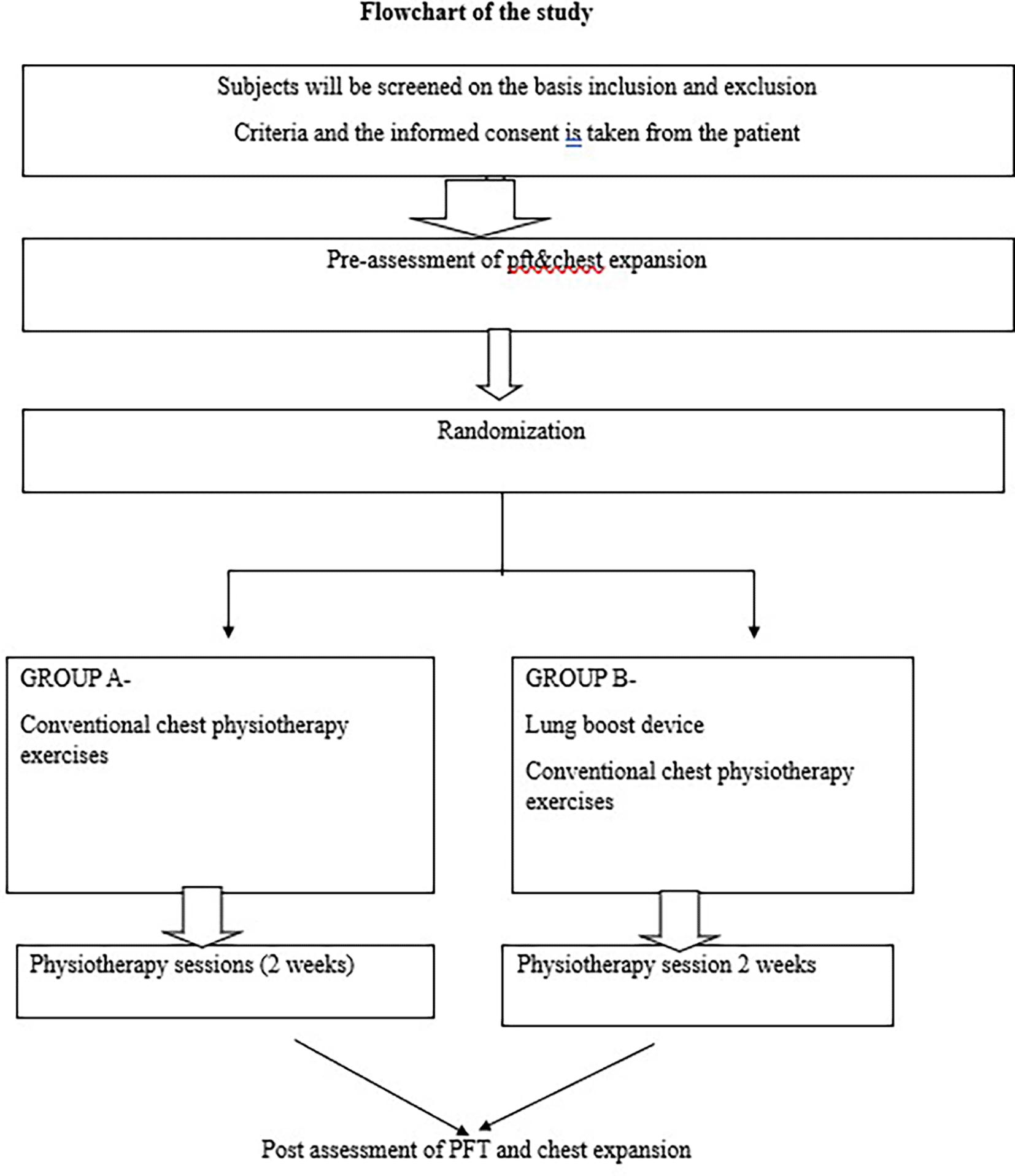
All the patients enrolled into the study are required to complete the whole two weeks of rehabilitation protocol after enrolment in the study. The assessment will be performed at initial and at the end of the session. Follow up will be taken after one week.
Group A- Group A will receive conservative physiotherapy management which will start from POD-3. At the beginning of the training, the patient will give with thoracic expansion, deep breathing exercise, incentive spirometry, postural correction. The session of treatment will be given for two sets of 15 repetitions for six days/week daily for two weeks. Table 1 explains the conventional treatment that will be administered to the patients.
Group B- Group B will receive lung boost device along with conservative physiotherapy management which will start from POD-3. At the beginning of the postoperative training, the patient has started with the stage one settings with the patient and the difficulty level will be increasing till five stages after that a small valve is applied that will apply resistance which will lead to start again from level 1. Participants will be instructed to take breathe deeply and forcefully for 2-3 seconds, slight pause, then exhale forcefully for 2-3 seconds for 15 repetition two sets. The treatment will be taken for 6 days in a week for two weeks twice a day. The time period for each session will be approx. about 15-20 minutes in which the rest period is also includes. Table 2 explains the treatment protocol of interventional group that will be administered to the patients.
Safety outcomes: Adverse events are reported at each time.
Primary outcome measures: -
1. Pulmonary function test.
This is the specific measurement was computed by an instrument called Spirometry (RMS HELIOS401). Following are the parameters:
Forced Vital Capacity (FVC) - Forced Vital Capacity (FVC) it is the maximum volume of gas that can be expired when the patient exhales as forcefully and rapidly as possible after a maximal inspiration. This procedure often referred to as the FVC maneuver.
Normal value- FVC and VC (vital capacity) should be within 200 ml of each other.
Forced Expiratory Volume in One Second (FEV1)-FEV 1 measures the volume expired over the first second of an FVC maneuver.
Normal values: Forced expiratory volume in persons with normal respiratory functions is as follows:
FEV 1 = 75%-85% of total vital capacity.
FEV 1/FVC Ratio - The ratios may be derived by dividing predicted FEV1 by predicted FVC.
Peak expiratory flow rate (PEFR) - PEFR is the maximum flow attained during an FVC maneuver. It is expressed in liter per second.
Normal range for adults- 100-850 L/min
2. Chest expansion.
The changes between Maximum inspiration and maximum expiration is known to be chest expansion. Chest expansion will be measured with the help of tape method at different stages of the chest which measures symmetry and extent of expansion. It will perform at three levels, for three different lobes of the lungs from top to bottom. This method is active for measuring chest expansion in unilateral lung disease. The levels of measurements are at sternal notch and at xiphiod process.
As because of pain over the suture site the patient was not able to maintain the proper posture. So, after the treatment the patient was analyse with the good and bad posture and had given the exercises for the good posture.
Sample size calculation resulted at 5% level of significance considering both the sides at 5% error probability with total 10 % for Z(1-α) value =1.64 & (1- β) at power of 80 % = 0.84 measuring the mean difference (effect size) of = 0.34 & standard deviation ( = 0.102
Primary Variable (Forced vital capacity)
Mean ± SD. (Pre) result on forced vital capacity for conventional chest therapy (Control group) = 1.25 ± 0.28.
Mean ± SD. (Post) result on forced vital capacity for conventional chest therapy (Control group) = 1.59 ± 0.38.
Difference. = 0.34 ± 0.16. (As per ref.article)
Clinically relevant superiority = 30 % = (0.34*30)/100 = 0.102.
Total samples required = 31 per Group.
Considering 10% drop out = 4
Total sample size required = 35 per group
Ref Article: - Gunjan SB, Shinde NK, Kazi AH et. al. Effectiveness of deep breathing versus segmental breathing exercises on chest expansion in pleural effusion. Int J Health Sci Res. 2015; 5(7):234-240
All the results will be calculated using R software version 4.3. Descriptive statistics over the categorial variable will be tabulated & presented with frequency & percentage. For the quantitative assessment over the continuous variable will be expressed & presented with Mean±SD, maximum and minimum. For inferential statistics. The outcome assessment variable over continuous scale (quantitative) will be tested following normal distribution using Shapiro- Wilk test. If data comes under non normal distribution will be transformed into normal distribution using mathematical algorithms. For finding significance over mean parametric test will be used as paired t-test for intra assessment & independent t-test will be used for inter-group assessment. Alternative Wilcoxon Test as a non-parametric test will be used if the data persist with non- normal distribution. Similarly, for unpaired-t test Mann Whitney test will be using as an alternative test. Association analysis for finding significance of cofounding parameters will be evaluated by using Chi-squared test or Fisher’s exact test or by using multi-variant analysis. Sensitivity and specificity of the device will be tested over primary outcome (MIP). AUC (area under curve) will be calculated based on observational values for finding accuracy of the device.
Dissemination- Planning for presenting my study in conference preceding.
Study status- The study is yet to be started.
The study is going to be done in comparison with the pulmonary functions and the chest expansion in the control as well as in the experimental group. Mainly we are doing the study to find out the effective result in both the group. Another study which is done as taking the experimental group in abdominal surgery has got the significant change in pulmonary functions and in the chest expansion. As we are doing by taking the experimental group just for evaluating in pleural effusion and finding the results from it. The experimental group is expected to show more effective results and as the device is useful to treat the patient in the short duration of time period as compared to control group. Tiwari et al studied about Physical Therapy Rehabilitation for a Chronic Alcoholic Patient with Loculated Pleural Effusion. In this study the author has studied about the effects of pursed lip breathing along with the thoracic expansion exercise in patient with pleural effusion. The study concluded that there is a significant improvement in chest expansion in pleural effusion patient.1 Amisha. A. Alande et al studied the effect of lung boost device on inspiratory muscle strength in abdominal surgery patients. 30 subjects with abdominal surgeries were enrolled in the study and were divided into two groups. the study concluded that g lung boost device showed significant improvement in inspiratory muscle strength and in 6 minute walk distance.16
| Views | Downloads | |
|---|---|---|
| F1000Research | - | - |
|
PubMed Central
Data from PMC are received and updated monthly.
|
- | - |
Provide sufficient details of any financial or non-financial competing interests to enable users to assess whether your comments might lead a reasonable person to question your impartiality. Consider the following examples, but note that this is not an exhaustive list:
Sign up for content alerts and receive a weekly or monthly email with all newly published articles
Already registered? Sign in
The email address should be the one you originally registered with F1000.
You registered with F1000 via Google, so we cannot reset your password.
To sign in, please click here.
If you still need help with your Google account password, please click here.
You registered with F1000 via Facebook, so we cannot reset your password.
To sign in, please click here.
If you still need help with your Facebook account password, please click here.
If your email address is registered with us, we will email you instructions to reset your password.
If you think you should have received this email but it has not arrived, please check your spam filters and/or contact for further assistance.
Comments on this article Comments (0)