Keywords
Wandering spleen, Hepatomegaly,VSD, Ovarian cyst.
This article is included in the Datta Meghe Institute of Higher Education and Research collection.
Wandering spleen, Hepatomegaly,VSD, Ovarian cyst.
Wandering spleen with torsion is a very uncommon condition, with a reported incidence rate of less than 0.2%, and accounting for only 0.002% of splenectomies.1 There can be associated hepatic complications. Liver dysfunction is one of the major non-cardiac complications associated with congenital heart disease (CHD) and has been reported in 6% of deceased patients with CHD.2 The development of hepatic complications may result from persistent chronic passive venous congestion or reduced cardiac output, either due to the underlying cardiac disease or as a consequence of palliative cardiac surgery.2
With respect to the recent data of epidemiology of atrial septal defects (ASDs), there is a prevalence of 1.6 per 1000 cases.3 Among these ASDs, the third most common type is ostium primum. This occurs when the septum primum fails to fuse with the endocardial cushions, leading to atrio-ventricular complications.4 In most of the cases of ASDs, lengths smaller than 5 mm undergo spontaneous closure in their first year of birth. However, defects larger than 1 cm require medical or surgical procedures to help in the closure of the defect.5
This case report describes a rare presentation of a female patient having wandering spleen with ostium primum type of atrial defect along with an enlarged liver. The patient was undiagnosed till she was of this age before she visited the Cardiology department of the Shalinitai Meghe Superspeciality Centre, Wardha, India. This is a rare presentation involving several clinical conditions and organ systems which makes the treatment more challenging.
A female patient in her early 20s, who is a resident of Wardha and is a student, came to the outpatient department, in a conscious state with chief complaint of decreased appetite for one month. This was associated with weight loss of 5kg within two months. There is an associated history of hematemesis, syncopal attacks lasting for a few seconds and continuous palpitations and irregular menstrual cycles with passage of clots and hot flashes. Patient is a known case of ventricular septal defect (diagnosed at the age of 16 years). In a routine health checkup, the physician heard an ejection systolic murmur due to septal defects. The clinician referred the patient to the Cardiology department of Acharya Vinoba Bhave Rural Hospital which comes under Shalinitai Meghe Super Specialty Center, where the patient was immediately advised to undergo routine investigations, chest x-ray, electrocardiography (ECG), ultrasonography (USG), computed tomography (CT) of the abdomen and pelvis, 2-dimensional echocardiography and CT abdominal angiogram. There was no significant family history.
On general examination, the patient was conscious and well oriented with time, place and person. The patient seemed afebrile, pulse rate was around 76/min, respiratory rate was recorded at 18/min, and blood pressure was measured and was found to be lower than normal (100/70 mm of Hg). Estimated weight was 40kg, height was 138 cm and body mass index (BMI) was 21. The patient’s oxygen saturation was 79% (SpO2) on admission. On further examination, pallor and cyanosis were seen. There were no other significant findings in the general examination.
A clinical examination revealed normal findings of the central nervous system (as per Glasgow coma score and reflex examinations), respiratory system (air entry bilaterally equal) and renal system. Cardiovascular system and gastrointestinal system showed abnormalities; the examination is detailed below.
On cardiovascular assessment, apex impulse was visible, whereas no precordial bulge, dilated veins, scars, and sinuses were visible. On palpation, the presence of a thrill was heard on the apex beat over the 5th intercostal space and anterior axillary line. On auscultation, S1 and S2 were heard best at the mitral area along with the ejection systolic murmur {grade 4 (loud and associated with a palpable thrill)} radiating to the anterior axillary line. Loud P2 was heard with a wide variable split. Abdominal assessment showed a soft abdomen. There was mild tenderness over the epigastric region without any distension. Liver was palpable and the spleen was not palpable.
Lab findings showed normal sodium levels (134 mEq/dl), normal potassium (4.5 mEq/dl) and low serum creatinine (0.6 mg/dl). Hematological findings showed normal hemoglobin levels (13.4g%) and normocytic normochromic red blood cells with few microcytes. However, the platelet count was reduced on smear with giant platelets. The absolute platelet count is 40000 cells per cubic millimeter. No haemoparasites were seen.
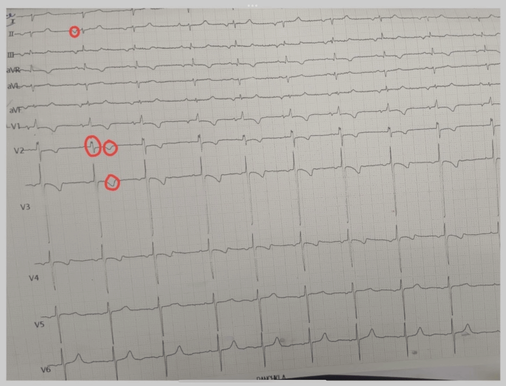
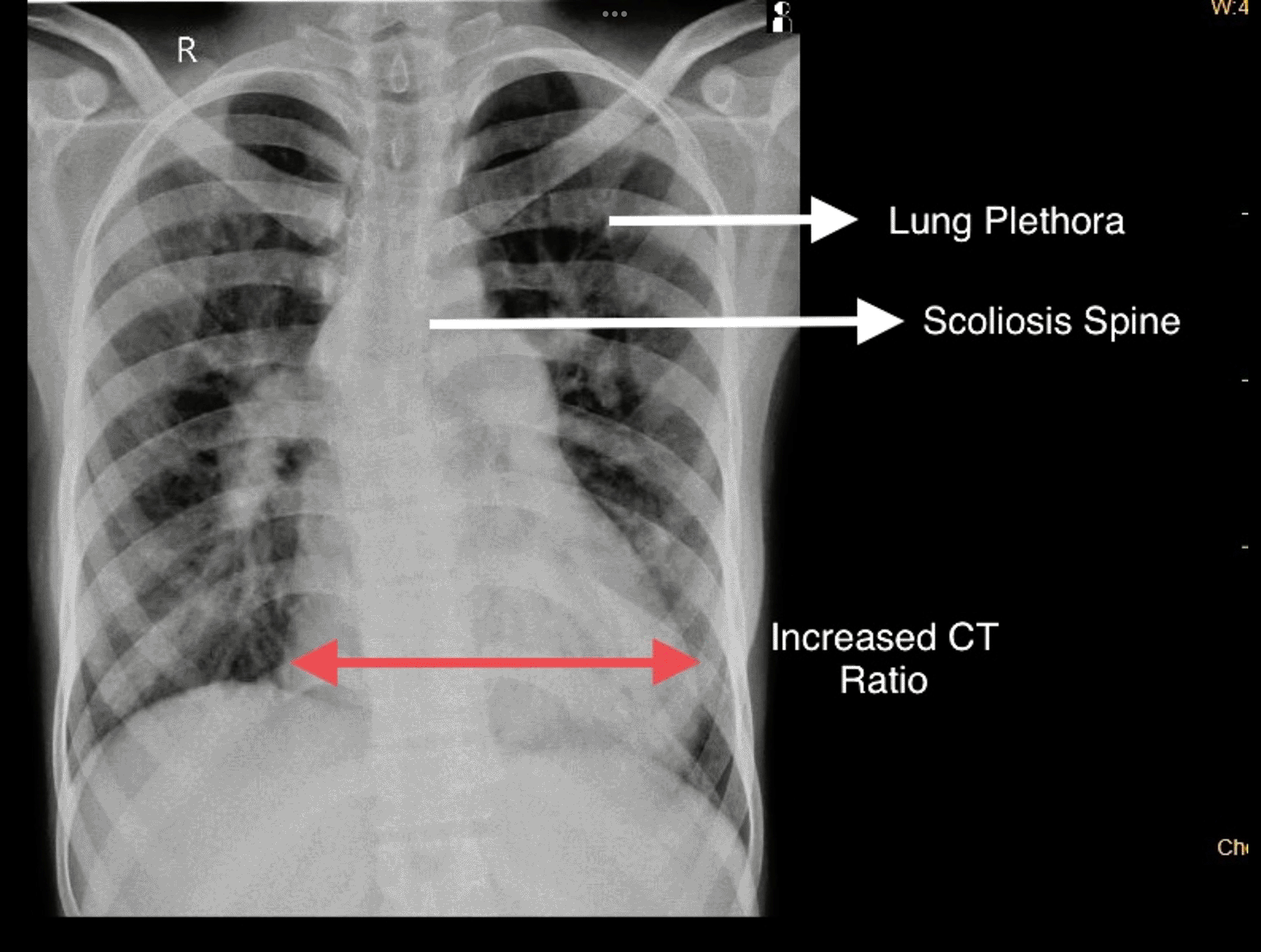
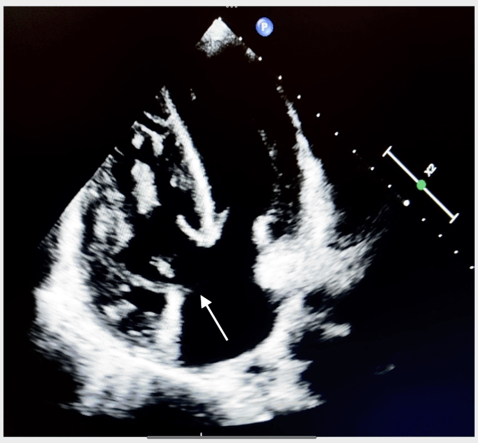
On recording ECG, there were biphasic waves seen in V2 V3, right axis deviation (right ventricular hypertrophy) along with the presence of inverted P wave in lead 2, 3, as shown in Figure 1. On chest x-ray, plethora of lungs, cardiomegaly (increased CT ratio), and scoliosis of the spine were found, as shown in Figure 2. A 2D echo was performed, which showed ostium primum type of ASD. The observations are shown in Figure 3.
CT: cardiothoracic.
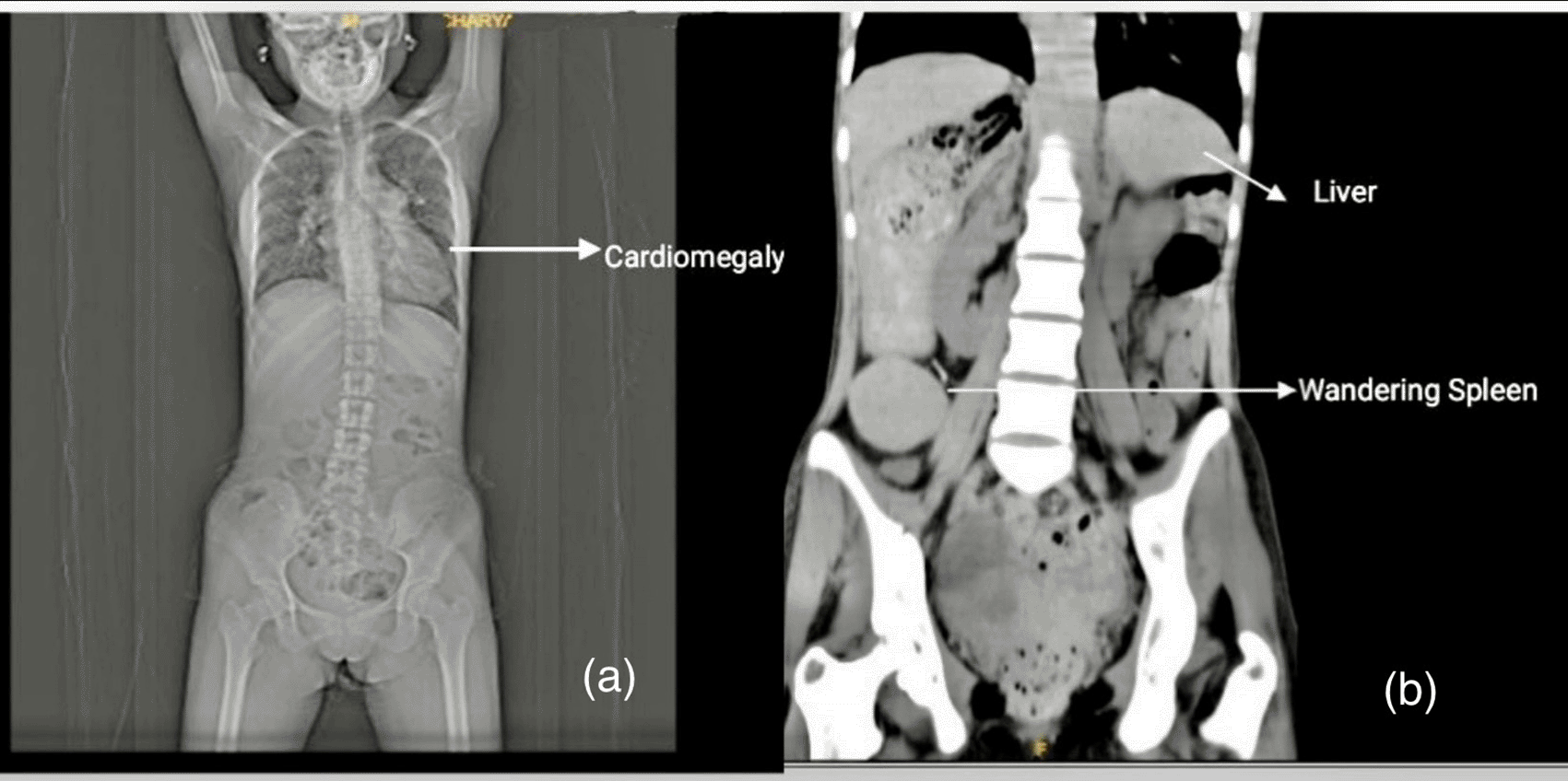
Upon CT abdomen and pelvis plane, it was observed that there was a large central liver, simple right ovarian cyst, mild bulky pancreas and the spleen could not be visualized at its normal position. A wandering spleen was seen. There was a cystic mass of 5.1 by 4.1 centimeters in the right ovary. Left ovaries appeared normal. Cardiomegaly was also noted from the CT. Renal system was normal. Observations are shown in Figure 4a and 4b.
Abdomen angiography was also done which suggested the presence of approximately 46 by 5 mm well defined homogeneously enhancing splenunculi -like structure noted in the right lumbar region with splenic vessels which directed us to the possibility of wandering spleen. The accessory splenic artery directly arises from abdominal aorta at T 11 level and measures 1.8 mm at origin. Early opacification of inferior vena cava and hepatic vein noted in arterial phase.
Surgery was not performed in this patient as the patient did not consent to the surgery. Deranged coagulation profile and low platelet count also contributed towards the fact that surgery could not be performed. Surgeries which this patient needs to undergo are for ostium primum, and the percutaneous closure of VSD. Patient was discharged with advice to continue drugs which were prescribed to her like sildenafil 25mg (thrice day dose), spironolactone 20mg (once daily), and Met XL 25mg (once daily). The medications were prescribed till the next follow up visit at 3 months.
Wandering spleen with torsion and splenic infarction causing acute abdomen is an extremely rare clinical entity. Patient was advised to attend regular follow-ups and the practitioner explained to her the potential for pregnancy related complications. CECT is the preferred diagnostic modality for the wandering spleen. Surgery was not performed in the patient as the patient did not consent to the surgery. The patient’s platelet count was much lower than normal levels which would have made surgery difficult. Surgery could be performed for ostium primum, and the percutaneous closure of VSD could also be done in the patient. The spleen did not cause any symptomatic difficulties, so it was not indicated for surgery. The cardiac anomalies along with hepatomegaly have been the cause of the severe growth abnormalities and clinical conditions like early satiety, abdominal fullness, and lethargy. The case becomes more important because if the patient had been diagnosed earlier, these clinical conditions could have been averted, and the patient could have had a better quality of life.
While there is no gender difference in cases under the age of ten, after the first decade, the prevalence of wandering spleen is higher in females than in males, with a ratio of 7:1.1 Wandering spleen can present in various ways, ranging from no symptoms to acute abdominal pain caused by torsion and infarction. In our patient, there was no complaint of abdominal pain. Wandering spleen is a condition where the spleen exhibits abnormal mobility and moves from its usual position in the left hypochondrium. This displacement is attributed to the absence of proper fixation and an elongated splenic pedicle. Normally, the spleen is held in position by gastrosplenic and lienorenal ligaments. It is a congenital condition or an acquired one. Congenital cases are due to the failure of ligament development, resulting in long splenic mesentery. Some acquired cases like prune belly syndrome and hypermobile colon can be associated with the condition.6 Splenic torsion can be acute, mimicking other abdominal conditions like appendicitis, peritonitis, or bowel obstruction.7
Different imaging techniques such as ultrasonography, Doppler ultrasound, plain radiography, contrast enhanced computed tomography (CECT), magnetic resonance imaging (MRI), scintigraphy, and angiography are diagnostic imaging techniques commonly used to identify wandering or ectopic spleen. These imaging modalities, along with a thorough physical examination, play a crucial role in accurately diagnosing the condition.1 Additional imaging examinations such as abdominal ultrasonography, Doppler and CECT abdomen are particularly important for a comprehensive diagnosis.
With respect to treatment modalities, surgical intervention is mostly favored for wandering spleen. There are two surgical options present depending on the condition of the spleen: splenopexy and splenectomy.8 The preferred method among these interventions is decided by the specific properties and requirements with respect to cases of patients. Laparoscopic or open splenopexy methods can be used to treat patients having uncomplicated cases of wandering spleen (WS).8 However, in the case of our patient with a significantly low platelet count, alternative considerations may be necessary. This can lead to complications, such as splenic infarction, rupture, hemorrhage, abscess, splenomegaly with mass effect, or signs of hypersplenism, for which the preferred treatment option is a splenectomy. This procedure can be performed using either an open or laparoscopic approach, depending on the specific circumstances and requirements of the patient.8
During the fourth week of gestation,9 atrial septation begins in the fourth gestational week9 as the primary atrial septum (septum primum). It extends from the roof of the primitive atrium to the endocardial cushions. The embryonic endocardium (the mesenchymal cap) is the source from which the mesenchymal cells are derived which enclose the caudal end of the septum primum. The septum primum extends and connects to the atrioventricular endocardial cushions, eventually closing and eliminating the ostium primum, which is the space between the mesenchymal cap and the atrioventricular cushions. In our patient, there is an ostium primum resulting in the formation of a single atrium.
Liver congestion caused by heart failure is a common problem that can lead to cardiac fibrosis and cirrhosis. To understand how venous congestion causes disease, it’s important to know about the unique blood supply of the liver. The liver has two vessel systems: the hepatic artery and the portal vein. About 70% of the blood supply to the liver comes from the portal vein. From the portal triads, both the portal venous system and the hepatic artery enter the acinus of the liver. Blood flows from both systems through the liver’s capillary system and toward the central vein, where it drains into the inferior caval vein. Hepatocytes near the portal triad receive oxygen- and nutrient-rich blood, while those near the central vein receive blood with fewer nutrients and less oxygen. There are defined metabolic zones in the liver that correspond to these differences in nutrient and oxygen supply. On histopathological evaluation, there were signs of venous congestion, such as sinusoidal dilation, hemorrhagic necrosis, and fibrosis, which are predominantly in the pericentral zone III.10 A typical pattern of fibrosis is seen in liver congestion, bridging the central veins.
Liver dysfunction in CHD is not solely caused by venous congestion, as various factors related to CHD can also affect the oxygen supply to the liver. These factors may include low cardiac output, cyanosis, or impaired oxygenation due to lung problems associated with CHD. Liver diseases in CHD can occur for reasons beyond just hemodynamic alterations. This is partly due to the fact that survival rates for CHD patients have significantly improved in recent decades, resulting in a growing population of CHD patients over the age of 65.11
The beneficial and functional management of this condition should consist of a team of a cardiologist, cardiovascular surgeon, gastroenterologist, and interventional radiologist. The cardiovascular surgeons and interventional cardiologists could not perform the surgery as the patient did not consent for an open and percutaneous approach of intervention. Thus, the patient was discharged with medical advice.
| Views | Downloads | |
|---|---|---|
| F1000Research | - | - |
|
PubMed Central
Data from PMC are received and updated monthly.
|
- | - |
Provide sufficient details of any financial or non-financial competing interests to enable users to assess whether your comments might lead a reasonable person to question your impartiality. Consider the following examples, but note that this is not an exhaustive list:
Sign up for content alerts and receive a weekly or monthly email with all newly published articles
Already registered? Sign in
The email address should be the one you originally registered with F1000.
You registered with F1000 via Google, so we cannot reset your password.
To sign in, please click here.
If you still need help with your Google account password, please click here.
You registered with F1000 via Facebook, so we cannot reset your password.
To sign in, please click here.
If you still need help with your Facebook account password, please click here.
If your email address is registered with us, we will email you instructions to reset your password.
If you think you should have received this email but it has not arrived, please check your spam filters and/or contact for further assistance.
Comments on this article Comments (0)