Keywords
Localized Scleroderma, Linear Scleroderma en coup de sabre, Morphea, Neurologic involvement, Neurologic manifestations, prefrontal syndrome
Localized Scleroderma, Linear Scleroderma en coup de sabre, Morphea, Neurologic involvement, Neurologic manifestations, prefrontal syndrome
Scleroderma is an immune-mediated and chronic connective tissue disease typified by a pathogenic triad encompassing vasculopathy due to endothelial dysfunction, dysregulation of innate and adaptive immunity, and progressive tissue fibrosis affecting the skin and multiple internal organs.1 This entity has traditionally been classified into systemic and localised forms.2 Systemic scleroderma, or systemic sclerosis (SS), can manifest at any age, mainly in adults. Like most autoimmune disorders, prevalence is higher in females.3 The SS epidemiological behaviour is notably variable due to its rarity, the broad clinical symptoms spectrum, the changing diagnostic criteria, and evolving classifications. These features complicate a comparative analysis plausibility across studies and an accurate prevalence trends estimation.4 In this vein, a recent meta-analysis encompassing 82 studies5 revealed that 83.9% of the world's countries had not reported systemic scleroderma epidemiological data. This study projected a global incidence of 8.64 affected individuals per 100,000 persons annually (range: 1.78 to 23.57). The prevalence was calculated as 18.87 per 100,000 (range: 1.55–25.28), approximating 1.47 million (range: 0.12–1.97 million) affected individuals globally. Incidence and prevalence were higher in females, adults, and high-income countries.5
Localised scleroderma (LS), or morphea, is delineated by skin lesions and involvement of underlying tissues (e.g., fascia, subcutaneous cellular tissue). Based on the extent and depth of fibrotic changes, LS is classified into limited, generalised, deep, mixed, and linear subtypes.6 LS incidence ranges from 0.3 to 3 per 100,000 individuals/year, affecting children and adults equally.7 Although it is primarily seen as a skin-limited disease, certain subtypes exhibit extracutaneous manifestations, including musculoskeletal (myositis, fasciitis, arthritis), central nervous system (headache, migraine, seizures, epilepsy), and ocular (uveitis) involvements.8 LS encompasses forms leading to progressive facial hemiatrophy (Parry-Romberg Syndrome) and those affecting facial and cranial regions without facial hemiatrophy (linear scleroderma en coup de sabre, LSCS), the latter initially described by Addison in 1854.6 In this sense, band-like sclerotic lesions located on the face and usually extended to the head, skin atrophy groove formation, underlying tissue changes, localised alopecia, and occasional changes in skin pigmentation are the typical presentation of LSCS. Though rare, neurological involvement has been described as epileptic seizures, cephalalgia, focal neurological deficits, and neuropsychiatric symptoms.9 Given the rarity of LSCS and the uncommon coexistence of neurological manifestations beyond epileptic seizures, we detail a case of LSCS associated with clinical neurological features compatible with a medial prefrontal syndrome.
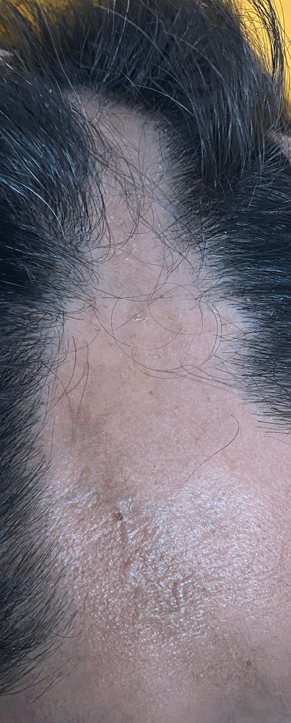
A 47-year-old professional truck driver, right-handed Hispanic male consulted our neurology department because of a 2-month history of behavioural changes. As reported by his spouse, these changes included apathy, anhedonia, abulia, decreased sociability, reduced spontaneous speech progressing towards mutism, irritability, and emotional lability. In addition, the patient refers to a right-pulsatile hemicranial headache with moderate intensity. There was no significant personal or familial past medical history. Initial physical examination revealed a sclerotic, depressed band with skin atrophy and alopecia extending from the forehead to the right parietal region. After inquiring with family members, this lesion progressed for over two years (Figure 1). Neurological examination demonstrated spatial disorientation, reduced spontaneous speech, decreased spontaneous motor activity, and reduced task-directed behaviours. Dysphoria was evident, but there were no speech or mnesic function disturbances. On the Montreal Cognitive Assessment (MoCA) scale, he scored 15/30, indicating impairments in executive function, cognitive flexibility, and mathematical reasoning. Cranial nerve examination, motor evaluation, sensory assessment, coordination, and gait were all within normal limits. The rest of the physical examination was unremarkable.
Laboratory tests showed elevated erythrocyte sedimentation rate (ESR): 24 mm/h (reference range: 0.0–15.0 mm/h), raised C-reactive protein (CRP): 32.50 mg/l (reference range: 0.00–10.00 mg/l), and decreased vitamin B12 levels: 140.8 pg/ml (reference range: 174.0–878.0 pg/ml). Blood analysis, kidney and liver function, ionogram, glucose levels, coagulation tests, veneral disease research laboratory (VDRL), ELISA HIV, tumor markers (prostate antigen, CA 19-9, alpha-fetoprotein, carcinoembryonic antigen), vitamin B1, folic acid, and autoimmune profile (anti-nuclear antibodies (ANA), anti-double stranded DNA antibodies (Anti-dsDNA), anti–Sjögren's-syndrome-related antigen A antibodies (AntiRo/SSA), anti–Sjögren's-syndrome-related antigen B antibodies (AntiLA/SSB), anti-Smith antibodies (Anti-SM), anti-ribonucleoprotein antibodies (Anti-RNP), antiphospholipid IgG and IgM, serum complement C3, C4, lupus russell viper anticoagulant venom test) were all within normal ranges.
Cerebrospinal fluid (CSF) macroscopic analysis showed a clear, transparent pH of 8 liquid. The cellular count was 0.003 cells/ul (leukocytes: 0–2 xc, erythrocytes: 0–3 xc), with 20% in polymorphonuclear cells, 80% in mononuclear cells and no bacteria observed at the microscope observation. CSF chemical analysis exhibited a glucose concentration of 74 mg/dl, 21.5 mg/dl proteins, and negative results for India ink and gram stains. Moreover, potassium hydroxide (KOH) test and culture stains showed no evidence of microorganisms.
Non-contrast and contrast-enhanced brain magnetic resonance imaging (MRI) revealed right frontal epicranial tissues and diploë thinning, a subcortical and deep white matter increased T2 signal, suggestive of vasogenic edema at the right cingulate gyrus, superior frontal gyrus, supramarginal gyrus and superior temporal gyrus. Underlying the high T2 area of vasogenic edema, there was a focal lesion which exhibited a low signal on the echo gradient sequence due to calcification at the right superior frontal gyrus with slight gadolinium lesional enhancement (Figure 2).

An eccentric hypointensity is evident, corresponding to the lesion area. (B) Vasogenic edema is observed with a central hypointense lesion adjacent to the temporal horn of the right lateral ventricle. (C) The lesion shows low signal in the Gradient Echo sequence and (D) homogeneous enhancement in the T1-weighted sequence following gadolinium administration.
The patient was hospitalised for further evaluation. General measures were implemented, including gastroprotection, thromboprophylaxis, and analgesia for the headache, and he was started on antiepileptic medication Valproic Acid 500 mg every 8 hours for seizure prevention and Sertraline 50 mg daily for mood management. Given the non-specific findings on MRI and the differential diagnosis considering a neoplastic lesion, it was decided to further the diagnostic workup with an electroencephalogram, spectroscopy analysis and scheduled for a stereotactic biopsy.
Lesions spectroscopy of the described revealed no elevated choline peaks and preserved n-acetyl-aspartate levels. On the eighth day of admission, the patient underwent a stereotactic biopsy. Post-procedure, the patient was transferred to an intermediate care unit for neurological monitoring and initiated on corticosteroid therapy with Dexamethasone 4 mg IV every 8 hours.
A follow-up non-contrast MRI of the brain, taken 24 hours post-surgery, showed minimal bleeding at the biopsy site and no evidence of vasogenic oedema progression compared to the initial neuroimaging assessment. Clinical examination remains unchanged from the evaluation at admission.
Lesional stereotactic biopsy showed normal-looking neurons, reactive gliosis areas, acute and chronic inflammatory infiltrates, and no evidence of microorganisms, granulomas, or malignancy at cerebral parenchyma. Immunohistochemical markers showed positivity for glial fibrillary protein (GFAP) and S-100 in the reactive astrocytes.
On the eleventh day post-admission, the patient experienced a sudden episode of retrosternal pain, dyspnea, desaturation, hypotension, and bradycardia, culminating in cardiac arrest. Despite immediately initiating basic and advanced cardiopulmonary resuscitation measures, the patient could not be resuscitated and was pronounced deceased.
LS, or morphea, is a rare immunoinflammatory disorder of the connective tissue affecting both adults and children,2 with a clear sex predominance in women with a 2–4:1 ratio.7 LS incidence ranges between 0.3 and 3 cases per 100,000 individuals annually,4 and although it occurs across all ethnicities, the White population exhibits the highest prevalence, followed by Latin American and other Hispanic-related groups.7
LSCS, also known as frontoparietal linear morphea or en coup de sabre scleroderma, is a rare variant of LS affecting the skin and bone in the frontoparietal region.10 LSCS is a very rare condition; most of the scientific literature indicates that it only accounts for 2–4% of linear scleroderma cases in adults, contrasting to the fourth-fold increase in LSCS prevalence in children with 3–17%.11 Based on an extensive and systematic literature review in Scopus, Web of Science, PubMed and Google Scholar, fewer than 100 case reports have been published on adult-onset LSCS,12–14 with only one case reported in Colombia.15 In Table 1 and Table 2, we present the case reports, cases series, and systematics reviews of patients with neurological manifestations identified during our literature search. Indeed, after this extensive search, we did not identify any studies specifically detailing the mediofrontal syndrome as a neuropsychiatric manifestation of neurological involvement in LSCS; thus, to our knowledge, this is the first report related to this association. In this regard, the accumulation of descriptive evidence from clinical case reports has enlightened previously considered an atypical manifestation, nervous system involvement in scleroderma is now increasingly recognised. Headache and seizures are the most frequently reported neurological manifestations in LSCS.16
| Study Type | Neurologic Manifestation | Number of Case Reports | Ref. |
|---|---|---|---|
| Case reports | Seizure | 9 | 17,18,19,20,21,22,23,17,24 |
| Focal Neurologic Deficit | 2 | 23,25 | |
| Cranial Nerve Involvement | 5 | 17,26,1,26,27 | |
| Headache | 4 | 20,22,28,29 |
| Study Type | Neurologic Manifestation | Frequency | Ref. |
|---|---|---|---|
| Case series | Headache | 8/26 | 30 |
| Seizure | 6/26 | ||
| Focal Neurologic Deficit | 2/26 | ||
| Cranial Nerve Involvement | 3/26 | ||
| Seizure | 4/9 | 31 | |
| Headache | 3/5 | 9 | |
| Seizure | 2/5 | ||
| Focal Neurologic Deficit | 3/5 | ||
| Headache | 5/12 | 32 | |
| Seizure | 3/12 | ||
| Focal Neurologic Deficit | 4/12 | ||
| Neuropsychiatric Symptons | 1/12 | ||
| Systematic review | Epilepsy | 140 | 33 |
| Headache | 19/92 | 16* | |
| Seizure | 42/92 | ||
| Focal Neurologic Deficit | 15/92 | ||
| Neuropsychiatric Symptons | 4/92 | ||
| Cranial Nerve Involvement | 8/92 |
The underlying pathophysiology of linear scleroderma remains only partially understood. It is believed to involve various factors, including genetics, environmental influences (such as trauma and infections), and disruptions in the immune system, primarily characterised by increased cytokine production. Based on current evidence, the pathophysiological process of morphea can be divided into three distinct phases: 1. early inflammatory, 2. sclerotic/fibrotic, and 3. late atrophic.34 In LSCS, both clinical and histopathological findings suggest a complex interaction between the vasculature and the immune system. Like SS, CD4 T lymphocytes appear to be associated with the fibrotic changes in these lesions. Initially, TH1 and TH17 inflammatory pathways predominate and later, fibrotic changes are believed to arise due to a shift in these inflammatory pathways, with a TH2 response dominance.35 Eventually, atrophy occurs, characterised by epidermal thickness, blood vessels, and inflammatory cell loss.34
LSCS typically presents as a solitary, linear, fibrous plaque that affects the skin and muscle, depressing the underlying bone and resembling the strike of a sabre – hence its name.10 Many authors consider extracutaneous manifestations of localised scleroderma extremely rare, with only approximately 20% of patients developing them. Among these manifestations, arthritis, uveitis, and epileptic seizures are predominantly observed, with neurological manifestations being the most frequent among them.22
Our patient exhibited atypical features and manifestations. One was a medial frontal syndrome as an initial clinical feature. According to the systematic review published by Amaral et al.,16 the most common neurological manifestations in linear scleroderma are epileptic seizures, affecting 41% of patients, followed by headaches in 18.8%. Only 1–3% exhibited cognitive decline and behavioural changes.
Additionally, another uncommon feature of our patient was his male gender. In the report by Taniguchi et al.,36 out of 16 cases of LSCS, only three were male, and none exhibited neurological manifestations. Among the 13 females, only five presented neurological manifestations, including headaches and abnormal findings in the electroencephalogram, akin to the observations reported by Amaral et al.16 None displayed behavioural or cognitive alterations.
Regarding complementary studies, abnormal neuroimaging findings exist in approximately 84% of patients. In a case report and literature review by Kister et al., only 11% of patients had normal brain MRIs. The remainder exhibited at least one T2 hyperintense lesion, primarily in the subcortical white matter and the corpus callosum, basal ganglia, and brainstem. These lesions were ipsilateral to the skin lesion in 88% of the patients. The predominance of these ipsilateral lesions to the skin manifestation suggests a potential pathogenic correlation between cerebral damage and the dermatological lesion, though the exact mechanism remains a topic of debate and investigation.22
On the other hand, it is relevant to consider that while a significant proportion of patients may exhibit abnormal brain MRI, not all of them experience overt clinical symptoms. This phenomenon underscores the need for regular neurological and radiological monitoring, allowing for early patient intervention in those who might develop complications. Some of these findings were identified during our patient evaluation, notably in subcortical and deep white matter hyperintensities ipsilateral to the skin lesion.
Regarding treatment, management guidelines recommend systemic therapy for localised scleroderma. Methotrexate, either as monotherapy or combined with corticosteroids, is suggested as a first-line treatment.37,38 Additionally, positive outcomes with mycophenolate and tocilizumab have been reported in other publications.39 Our patient received corticosteroid treatment consistent with descriptions in other case reports and as recommended in clinical practice guidelines. However, we could not monitor the treatment response and clinical progress due to the patient's demise from a non-neurological complication. Nevertheless, the follow-up imaging did not reveal the progression of the lesions noted in subsequent MRI scans.
| Views | Downloads | |
|---|---|---|
| F1000Research | - | - |
|
PubMed Central
Data from PMC are received and updated monthly.
|
- | - |
Provide sufficient details of any financial or non-financial competing interests to enable users to assess whether your comments might lead a reasonable person to question your impartiality. Consider the following examples, but note that this is not an exhaustive list:
Sign up for content alerts and receive a weekly or monthly email with all newly published articles
Already registered? Sign in
The email address should be the one you originally registered with F1000.
You registered with F1000 via Google, so we cannot reset your password.
To sign in, please click here.
If you still need help with your Google account password, please click here.
You registered with F1000 via Facebook, so we cannot reset your password.
To sign in, please click here.
If you still need help with your Facebook account password, please click here.
If your email address is registered with us, we will email you instructions to reset your password.
If you think you should have received this email but it has not arrived, please check your spam filters and/or contact for further assistance.
Comments on this article Comments (0)