Keywords
Rabbit models AVF, Intimal Hyperplasia, Umbilical Cord Mesenchymal Stem Cells, HIF-1α, eNOS, and MMP-2
Rabbit models AVF, Intimal Hyperplasia, Umbilical Cord Mesenchymal Stem Cells, HIF-1α, eNOS, and MMP-2
An arteriovenous fistula is a medical condition in which an abnormal connection exists between an artery and a vein. This connection causes blood to bypass the capillaries, increasing blood flow and blood pressure in the veins. Intimal hyperplasia is a common complication of AVF, characterized by the thickening of the inner layer of the blood vessel wall.1 Intimal hyperplasia is a major cause of arteriovenous fistula failure, contributing to the constriction of the AVF lumen and impaired outflow tract function. Studies have found that AVF hyperplasia can be attributed to factors such as HIF-1 alpha, MMP-2, and eNOS. A research conducted by Kim and colleagues analyzed the association between AVF hyperplasia and these factors.2
Hypoxia-inducible factor-1 alpha (HIF-1α) is a transcription factor that plays a crucial role in cellular responses to low oxygen levels. During hypoxic conditions, HIF-1α stabilizes and binds to other cofactors, activating the hypoxia response element of certain gene promoters.3
Endothelial nitric oxidase synthase (eNOS) is also important in the regulation of vascular tone and blood flow through the production of nitric oxide. Nitric oxide acts as a vasodilator, relaxing the smooth muscles in blood vessel walls and promoting increased blood flow. However, in conditions such as arteriovenous fistula, there is a dysregulation of eNOS activity, leading to impaired nitric oxide production and endothelial dysfunction.4 Matrix metalloprotease-2 (MMP-2) is a zinc-dependent enzyme that plays a crucial role in various biological processes, such as tissue remodelling, wound healing, and immune response regulation. However, increased MMP-2 activity can contribute to the development of intimal hyperplasia, inflammation, and plaque rupture in the context of arteriovenous fistulas.5
Mesenchymal stem cell (MSC) therapy involves the use of mesenchymal stem cells to treat vascular injuries, specifically in the context of arteriovenous fistula, and reduce the incidence of intimal hyperplasia. These stem cells have the ability to differentiate into specialized cells and promote tissue regeneration, making them a potential therapeutic option for AVF.6–8 Previous preclinical studies have shown that the topical administration of human adipose-derived MSCs to the adventitial surface of the outflow vein can attenuate the formation of venous neointimal hyperplasia and improve AVF patency.7 While surgery is typically recommended for AVF treatment, MSC therapy offers a promising alternative. Recent research has shown that mesenchymal stem cells can play a beneficial role in reducing intimal hyperplasia and promoting AVF maturation.8
This study aims to evaluate the effect of UC-MSCs administration on HIF-1α, eNOS, and MMP-2 levels in rabbit models with AVF. Once fistulas are established, aggressive anti-inflammatory therapy is important, but surgery is typically necessary.
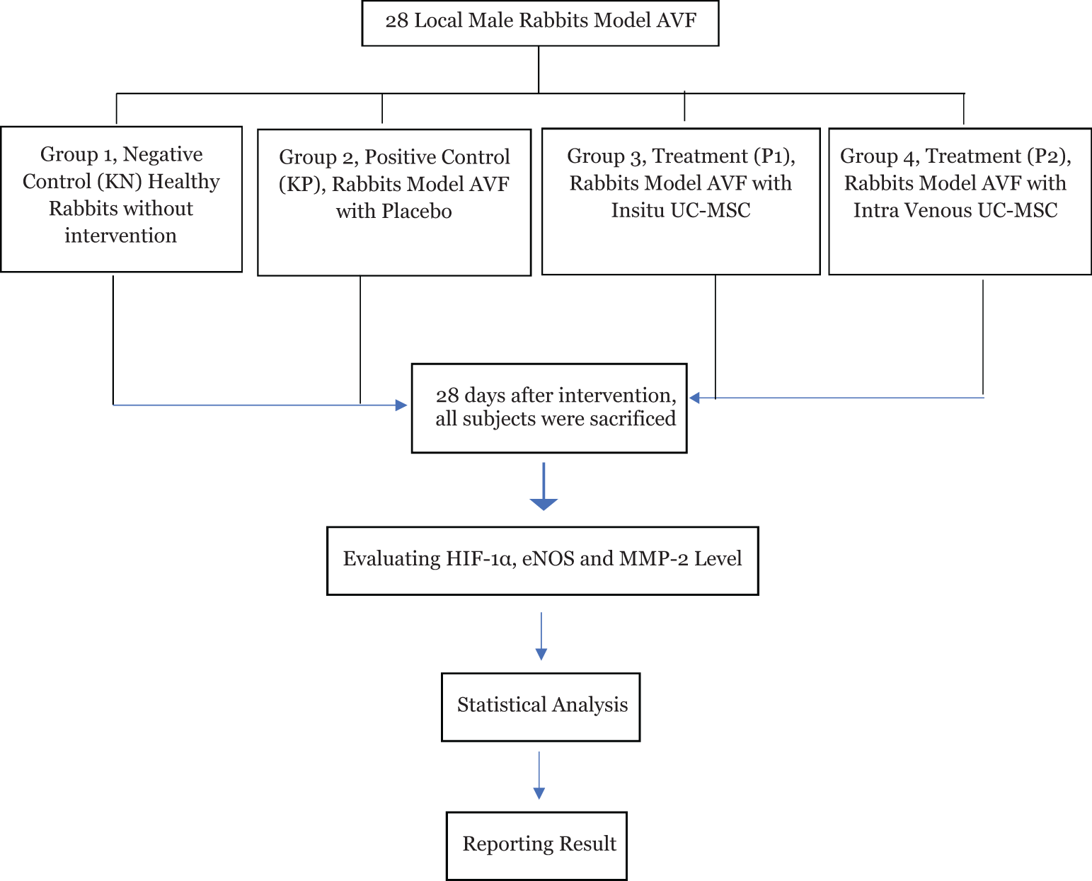
An experimental study using local male rabbits (Lepus Domestica) using a post-test design approach (randomized control trial post-test only design), conducted at Faculty of Veterinary Medicine experimental animal hospital Universitas Syiah Kuala, from January to March 2023. The research group was divided into 4, KN as the negative control group, KP as the positive control group of rabbit models AVF with placebo therapy, P1 as the treatment group of rabbit models AVF by giving in situ UC-MSCs, and P2 as the treatment group of rabbit models AVF by giving intra venous UC-MSCs.
The sample size was calculated using the sample formula from Federer. The minimum number of samples is rounded up to 6 subjects. The estimated number of subjects who dropped out was 10%, so the number of samples from the above formula was added by 10%; the minimum number of research subjects for each group is 7 samples. The total sample to be examined in this study was 28 subjects. The local male rabbits AVF were randomly divided into treatment or control groups. Ethical clearance approval was given by the Research Ethics Committee Faculty of Veterinary Medicine Universitas Syiah Kuala, date 5 December 2022, No.190/KEPH/XII/.
The research subjects were local male rabbit model AVF. The inclusion criteria were age 8-12 weeks, weight 2000-2500 grams, no visible anatomical abnormalities, no signs of previous infection, and no other diseases. Exclusion criteria were AVF model rabbits males note, drop out before 2 weeks post AVF period, sick or died since treatment and behavior changed during the study (limp and not agile).
The surgeon, who specialized in thoracic, cardiac, and vascular surgery, as well as a veterinarian, performed all of the anastomoses. This procedure was conducted at the experimental animal laboratory of the Faculty of Veterinary Medicine at Universitas Syiah Kuala. To ensure proper anesthesia, the animals were given 50 mg/kg of intramuscular ketamine and 5 mg/kg of intramuscular xylazine. The surgeon shaved the incision areas to improve visibility during surgery and disinfected them with povidone-iodine. To access the right carotid communis artery, a vertical incision was made in the neck. A dose of 100 IU/kg of intravenous heparinization was given before the right carotid communis artery was anastomosed end-to-side to the right internal jugular vein. The surgeon completed the anastomosis by suturing with a 7-0 polypropylene suture, one by one, and closed the tissues in an anatomical layer. The left internal jugular vein was left untouched.
The UC-MSCs were administered at a dosage of 1,000,000 cells per kilogram of body weight. The treatment group of rabbits with AVF received UC-MSCs in situ (P1) and intravenous (P2) injections. The UC-MSCs were obtained in Prodia Stemcell syringes, each containing 2,500,000 cells. The positive control group of rabbits with AVF were given a placebo solution (KP) consisting of 2.5 ml of normal saline solution. The negative control group consisted of healthy male rabbits without AVF procedure. After a period of 28 days, all groups of rabbit models with AVF were sacrificed. All groups of rabbits were evaluated for animal care and monitoring at the experimental animal laboratory, Faculty of Veterinary Medicine, Universitas Syiah Kuala, by a veterinarian.
Euthanasia is performed by slowly injecting an anesthetic dose of 150 mg/kgBW of ketamine and 15 mg/kg of xylazine through the intravenous route while monitoring the heart rate. The heart rate will gradually weaken until no heartbeat is audible, and the pupils will slowly dilate, and the eyes will lose their twinkle. Once this happens, the rabbit is considered dead.
This study assessed HIF-1α, eNOS and MMP-2 level after 28 days of intervention using ELISA sandwich methods.
Measurement of HIF-1α concentration in rabbits using a specific ELISA kit for rabbits, namely Rabbit Hypoxia Inducible Factor 1 Alpha (HIF-1α) (Cat. No. BZ-22179294-EB, Bioenzy). For eNOS concentration in rabbits using rabbit-specific ELISA kit Rabbit Endothelial Nitric Oxide (eNOS) (Cat. No. BZ-08172420-EB, Bioenzy), and MMP-2 in rabbits using the rabbit specific ELISA kit Rabbit Matrix Metalloproteinase 2 (Cat. No. BZ-22170591-EB, Bioenzy). The procedure for measuring HIF-1α, eNOS, MMP-2 levels was carried out following the procedure contained in the ELISA kit guide. The absorbance value and concentration of HIF-1α, eNOS, MMP-2 were measured using an ELISA reader at 450 nm wavelength (Biolegend, USA).
HIF-1α, eNOS, MMP-2 level data obtained were tested for normality using the Shapiro-Wilk test and obtained HIF-1α, eNOS, MMP-2 data were distributed normally (homogeneous) (P>0.05). To determine differences in HIF-1α, eNOS, MMP-2 concentrations between treatments, a test of variance (One Way ANOVA) was conducted and continued with Duncan’s post hoc test.
According to the study, the average level of HIF 1-α in healthy rabbits (KN) was 0.09±0.02 ng/mg. In AVF rabbits with placebo (KP), the level was 1.31±0.45 ng/mg, in AVF rabbits with in situ MSC (P1) it was 0.21±0.08 ng/mg, and in AVF rabbits with intravenous UC-MSC (P2) it was 0.33±0.12 ng/mg. The KN group had the lowest HIF 1-α level, followed by P1 and P2. KP had the highest level. P2 had a 15 times higher HIF 1-α level (1429%) than KN. KP and P1 had 3 times higher (286%) and 2 times higher (148%) levels than KN, respectively.
The statistical analysis showed a significant difference in the level of HIF 1-α between healthy rabbits and rabbits that received AVF treatment with placebo, in situ UC-MSC, and intravenous UC-MSC (Table 1). The P-value was <0.01.
| Intervention | Mean (ng/mg tissue) | Standart Deviation | P value |
|---|---|---|---|
| KN | 0.09 | 0.02 | 0.000** |
| KP | 1.31 | 0.45 | |
| P1 | 0.21 | 0.08 | |
| P2 | 0.33 | 0.12 |
The mean±standard deviation concentration/level of HIF 1-α in healthy rabbits, AVF, and AVF rabbits with UC-MSC addition (N=7) is presented in Table 1.
The study showed that rabbits with AVF who received UC-MSC treatment, either in situ or intravenously, had similar levels of HIF 1-α as healthy rabbits (KN) with no significant difference (P>0.05). However, rabbits with AVF who received a placebo had 15 times higher levels of HIF 1-α than healthy rabbits (KN) and the difference was statistically significant (P<0.05).
These findings indicate that both in situ and intravenous UC-MSC treatments can effectively decrease HIF 1-α levels to the same level as healthy rabbits. Moreover, in situ administration of UC-MSC was more effective at reducing HIF 1-α levels compared to intravenous administration. Refer to Figure 2 for a comparison of the mean (±standard deviation) HIF 1-α levels between treatments.
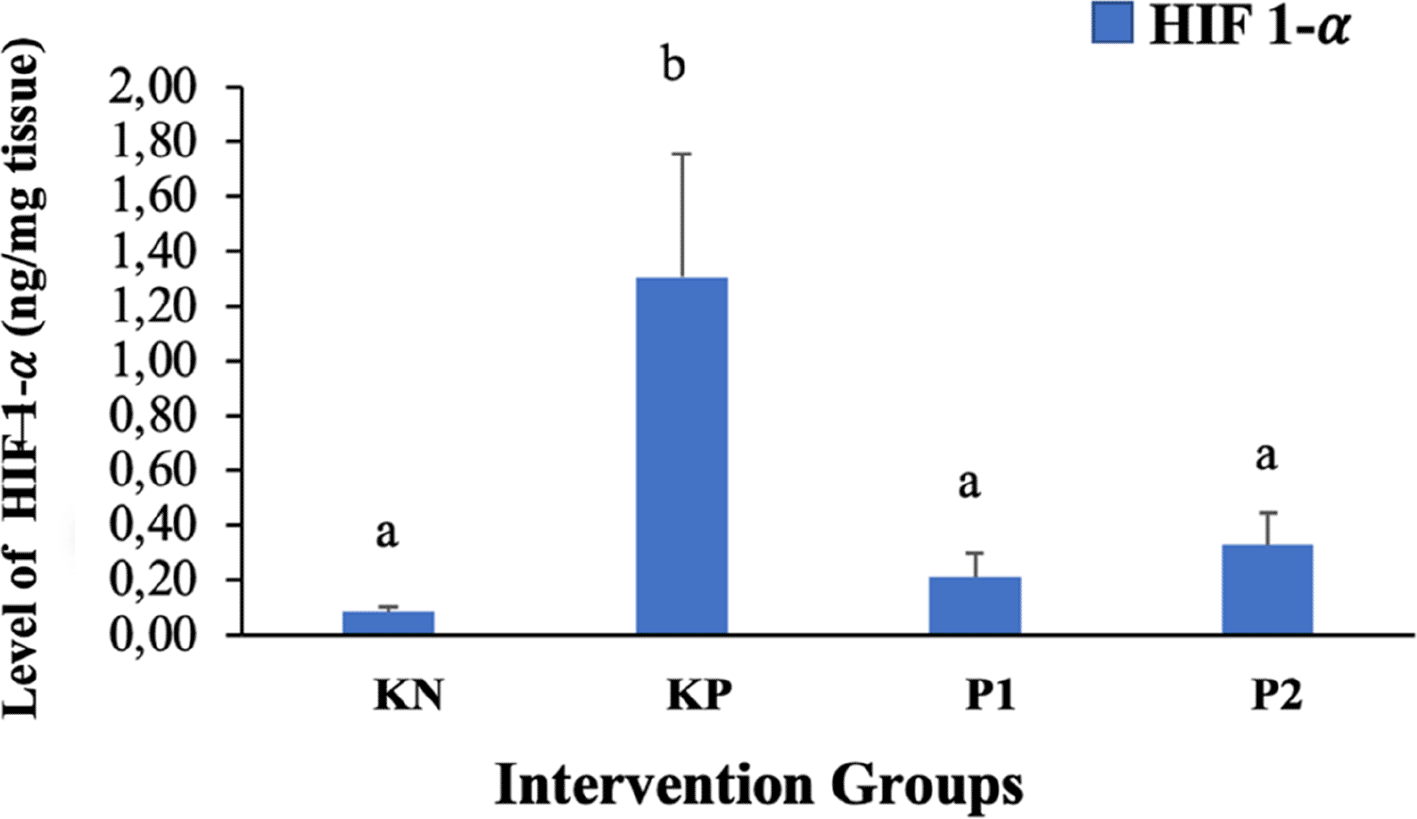
Different superscripts a,b above the histogram indicate significant differences (P<0.05).
The eNOS levels in healthy rabbits (KN), AVF rabbits with placebo (KP), AVF rabbits with UC-MSC in situ (P1), and AVF rabbits with intra-venous UC-MSC (P2) were recorded as 3.73±0.71 ng/mg, 24.77±4.93 ng/mg, 9.21±4.11 ng/mg and 13.79±3.36 ng/mg respectively. The lowest eNOS level was observed in the healthy rabbit group (KN), while the highest was seen in AVF rabbits with a placebo (KP). The eNOS levels in AVF rabbits with in situ UC-MSC (P1) and intra-venous UC-MSC (P2) were lower than AVF rabbits with placebo (KP), but higher than healthy rabbits (KN). The eNOS level in KP, P1, and P2 was higher by 554.07%, 146.91%, and 269.71%, respectively compared to healthy rabbits (KN).
Statistical analysis showed that there was a significant difference in eNOS levels between healthy rabbits, rabbits with AVF treatment followed by placebo, UC-MSC in situ, and UC-MSC intra venous (Table 5.10). The p-value was found to be less than 0.01.
The results of further tests (post hoc) showed that the concentration of eNOS in the KN group (healthy rabbits) was significantly lower (P<0.05) compared to other treatment groups (KP, P1, and P2). The highest eNOS concentration was obtained in treatment group 2 (KP) and was significantly higher than P1, P2, and KN (P<0.05). The eNOS concentration in treatment groups P1 and P2 decreased and was significantly lower than that in KP (P<0.05), while the eNOS concentration in P1 was significantly lower than that in P2 (P<0.05). These results show that stem cell treatment can reduce the concentration of eNOS in rabbits experiencing AVF. A comparison of the mean (±standard deviation) eNOS between treatments can be seen in Figure 3.
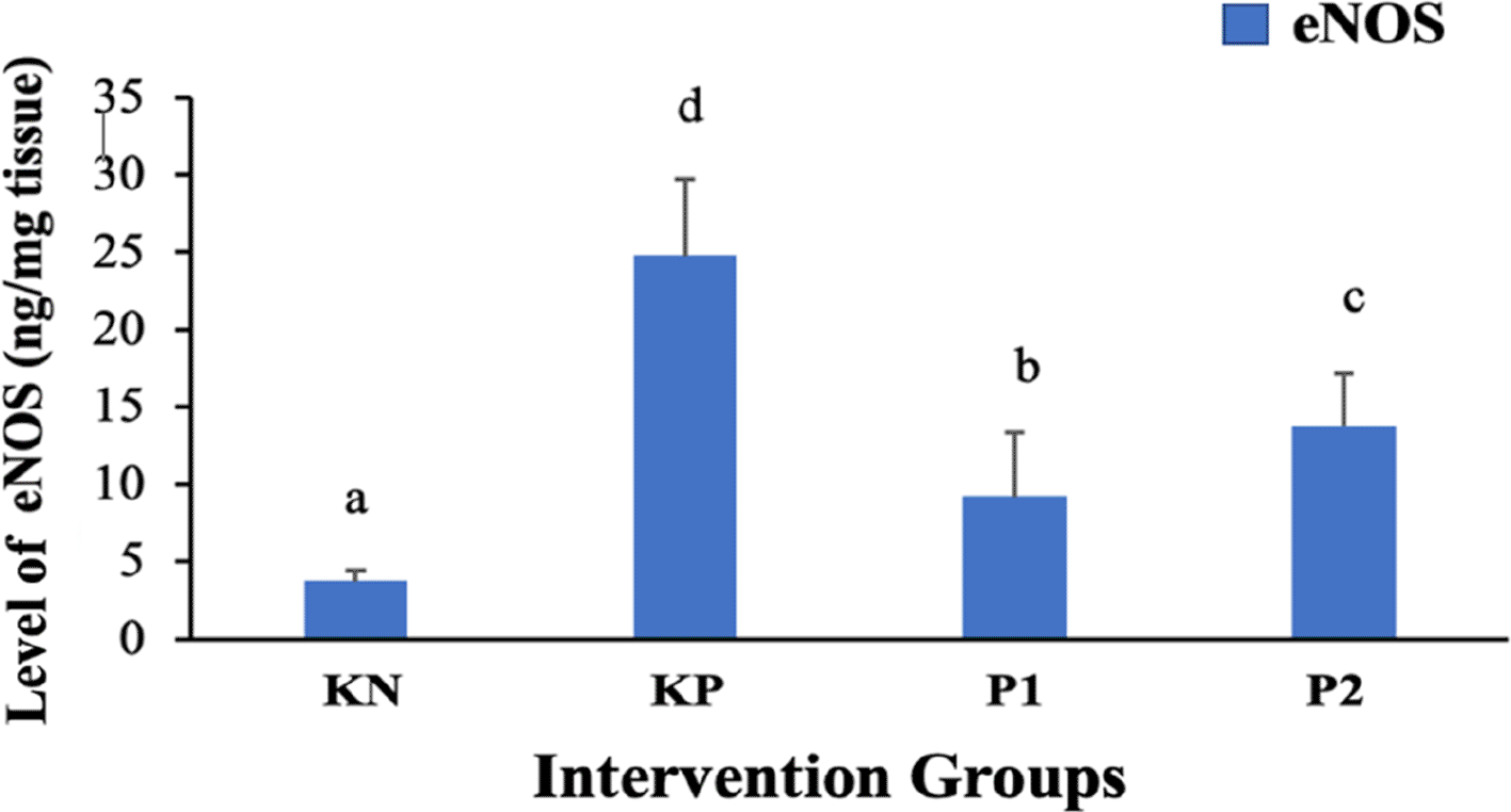
Different superscripts a,b,c d above the histogram indicate significant differences (P<0.05).
We collected data on the levels of MMP-2 in healthy rabbits (KN), AVF rabbits with placebo (KP), AVF rabbits with UC-MSC in situ (P1), and AVF rabbits with UC-MSC intra vein (P2). The mean levels (± standard deviation) were: KN=8.24±1.08 pg/mg, KP=78.57±8.18 pg/mg, P1=6.98±1.63 pg/mg, and P2=15.84±3.74 pg/mg. The lowest MMP-2 level was found in the P1 group, while the highest was found in the KP group. Compared to the KN group, the MMP-2 level was 15.29% lower in P1, and higher by 853.52% and 92.23% in KP and P2 respectively.
Statistical analysis revealed a significant difference in MMP-2 levels between healthy rabbits and those treated with AVF followed by placebo, UC-MSC in situ, or UC-MSC intra venous (Table 3). The p-value was <0.01, indicating a strong correlation.
The results of a recent test showed that the MMP-2 level in rabbits with AVF and UC-MSC in situ (P1) was not significantly different from the MMP-2 level in healthy rabbits (KN) (P>0.05). However, the MMP-2 level in AVF rabbits with intravenous UC-MSC was significantly higher than that in healthy rabbits (KN) (<0.05), but much lower than that in the AVF rabbit group with placebo (P<0.05).
Additionally, the MMP-2 level in AVF rabbits with UC-MSC in situ was significantly lower than the MMP-2 level in AVF rabbits with intravenous UC-MSC (P<0.05). These results suggest that administering UC-MSC in situ is more effective in reducing MMP-2 levels, resulting in no significant difference from healthy rabbits. Figure 4 provides a comparison of the mean (±standard deviation) MMP-2 levels between treatments.
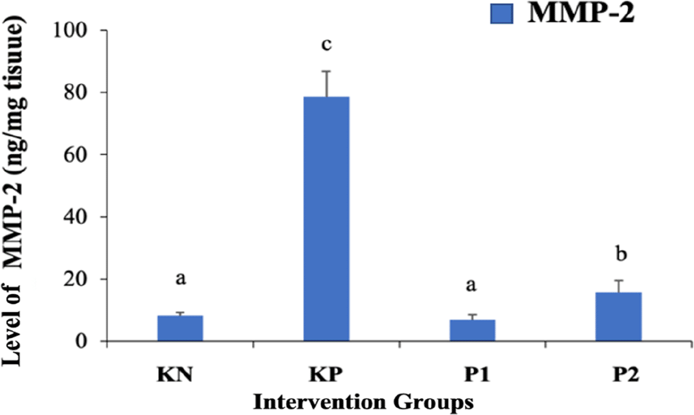
Different superscripts a,b,c above the histogram indicate significant differences (P<0.05).
In this research as shown in Table 1 and Figure 2 both in the AVF group, those given placebo therapy had a higher level of HIF-1α compared to those given in situ MSC intervention and intra-venous MSC. The latter groups had HIF-1α levels closer to those of healthy rabbits. UC-MSCs may be useful in reducing HIF-1α levels in AVF-related hypoxia by modulating HIF-1α expression and activity through hypoxic preconditioning, which can enhance their angiogenic properties and contribute to the reduction of HIF-1α levels.9
Venous neointimal hyperplasia, which causes stenosis in arteriovenous grafts and late AV fistulas, has a well-described pathogenesis involving both upstream and downstream events. Initial insults lead to endothelial injury, triggering a cascade of mediators that regulate oxidative stress, endothelial dysfunction, and inflammation. The response to this injury results in the migration of smooth muscle cells from the media to the intima, eventually forming neointimal hyperplasia.10
HIF-1α, also called Hypoxia-Induced Factor 1 Alpha, is a transcription factor that regulates the adaptive response to low oxygen levels (hypoxia) in cells, also play an important transcription factor that responds to a lack of oxygen in tissues. It regulates the expression of genes that are involved in various cellular processes, such as angiogenesis, metabolism, and cell survival. In the context of AVF-related hypoxia, HIF-1α has been found to play a crucial role in vascular remodelling and angiogenesis.11 During hypoxia, HIF-1α is stabilized and translocated to the nucleus, which regulates the expression of genes involved in angiogenesis, cell survival, and inflammation.12 Recent studies have shown that the stabilization of HIF-1α can improve the functions of mesenchymal stem cells, including cell adhesion, migration, and proliferation. Moreover, HIF-1α can enhance the regenerative potential of adipose-derived stem cells under hypoxic conditions by increasing the secretion of vascular endothelial growth factor.13 HIF-1α is a biomarker that has been shown to play a crucial role in preventing intimal hyperplasia in arteriovenous fistula.
One way in which HIF-1α could be used as a biomarker for preventing intimal hyperplasia is by monitoring its expression levels. Studies have shown that increased expression of HIF-1α occurs early during the maturation of arteriovenous fistulas and that this expression is localized to the venous endothelium. By measuring and monitoring HIF-1α expression, clinicians can potentially identify patients who are at a higher risk of developing intimal hyperplasia. This could allow for early intervention and the implementation of preventative measures to minimize the occurrence and severity of intimal hyperplasia.14
The mechanism through which umbilical cord mesenchymal stem cells exert their anti-intimal hyperplasia effects is multi-faceted. Firstly, these stem cells have the ability to differentiate into various cell types involved in vascular remodeling and repair.15 For example, they can differentiate into smooth muscle cells, endothelial cells, and fibroblasts, which are crucial for maintaining the structural integrity of the vessel wall. By introducing umbilical cord mesenchymal stem cells into the site of arteriovenous fistula creation, it is possible to enhance when introduced into the vessel wall, umbilical cord mesenchymal stem cells can differentiate into smooth muscle cells, endothelial cells, and fibroblasts, which can promote tissue repair and inhibit the development of intimal hyperplasia.16 Furthermore, umbilical cord mesenchymal stem cells have been found to possess immunomodulatory properties. These cells can modulate the immune response and reduce inflammation, which are important factors in intimal hyperplasia development,17 and it may promote vascular repair and inhibit the development of intimal hyperplasia.18
The combination of umbilical cord mesenchymal stem cells and HIF-1α can enhance these angiogenic properties and improve the outcomes of arteriovenous fistula creation. Zhang et al. conducted a study that found human umbilical cord mesenchymal stem cells produced exosomes promote angiogenesis through HIF-1α, which is beneficial to fracture healing.19
The role of umbilical cord mesenchymal stem cells in preventing intimal hyperplasia is a promising area of research. Umbilical cord mesenchymal stem cells have shown potential in the prevention of intimal hyperplasia. In animal models, umbilical cord mesenchymal stem cell transplantation has been shown to reduce intimal hyperplasia formation in arterial prosthetic grafts.20
According to the data presented in Table 2 and Figure 3, the placebo group had the highest level of eNOS concentration, which was significantly greater than the healthy rabbit and treatment groups. The concentration of eNOS decreased in the treatment groups and was significantly lower than in the placebo group. Moreover, the concentration of eNOS was much lower in the in situ UC-MSC treatment group than in the intra-venous group. These findings indicate that UC-MSC therapy can effectively decrease the concentration of eNOS in rabbits suffering intimal hyperplasia from AVF. This is somewhat contradictory to existing theories because eNOS is expected to cause vasodilation of blood vessels. When intimal hyperplasia occurs, the vascular endothelium should secrete enough eNOS to prevent stenosis. Therefore, the high eNOS levels in the AVF group with placebo can be compared to the lower levels in the groups that received MSC interventions.
| Intervention | Mean (ng/mg tissue) | Standart Deviation | P value |
|---|---|---|---|
| KN | 3.73 | 0.71 | 0.000** |
| KP | 24.77 | 4.93 | |
| P1 | 9.21 | 4.11 | |
| P2 | 13.79 | 3.36 |
| Intervention | Mean (pg/mg tissue) | Standart Deviation | P value |
|---|---|---|---|
| KN | 8.24 | 1.08 | 0.000** |
| KP | 78.57 | 8.18 | |
| P1 | 6.98 | 1.63 | |
| P2 | 15.84 | 3.74 |
Endothelial Nitric Oxide Synthase (eNOS) plays a crucial role in maintaining vascular homeostasis by producing nitric oxide, a key regulator of vascular function. Nitric oxide is involved in vasodilation, platelet aggregation inhibition, and smooth muscle cell proliferation suppression. Studies have shown that MSC therapy can enhance the expression and activity of eNOS in vascular endothelial cells, thus promoting nitric oxide production and potentially improving vascular function. In the context of AVF therapy, enhancing eNOS activity through MSC therapy may have beneficial effects on preventing venous neointimal hyperplasia and improving AVF patency.21
MSC therapy has been shown to increase the production of nitric oxide synthase in endothelial cells. This is likely due to the paracrine effects of MSCs, which can secrete various growth factors and cytokines that promote endothelial cell function, including the production of nitric oxide synthase. Specifically, MSCs have been shown to secrete factors such as vascular endothelial growth factor and hepatocyte growth factor, which can stimulate the expression of eNOS in endothelial cells.22 Furthermore, MSC therapy has been associated with the recruitment and activation of endogenous endothelial progenitor cells, which also contribute to the production of nitric oxide synthase. The specific mechanisms by which MSC therapy influences nitric oxide synthase production in endothelial cells are still being studied. However, it is believed that MSCs exert their effects through paracrine signalling, direct cell-to-cell contact, and modulation of the immune system.23
To investigate the communication between MSCs and endothelial cells in the context of nitric oxide synthase production, researchers have analyzed the vascular endothelial growth factor release by MSCs in cell culture supernatants. This is because vascular endothelial growth factor is known to stimulate nitric oxide production by eNOS through an increase in cytosolic calcium concentrations, which leads to enhanced vascular permeability and vasodilation.24
In the context of arteriovenous fistula formation and developing intimal hyperplasia, the enhanced production of endothelial nitric oxide synthase (eNOS) through MSC therapy has the potential to play a crucial role. MSC therapy has shown promising potential in improving the outcomes of arteriovenous fistula formation by increasing eNOS activity. Studies have revealed that MSC therapy can increase the ventricular protein expression of eNOS, an enzyme crucial for proper endothelial function.25 This increase in eNOS expression is associated with improved endothelium-dependent vasodilation and enhanced myocardial neovascularization, which are essential for the maturation and success of arteriovenous fistula formation.26
The mechanism by which MSC therapy enhances eNOS activity in the context of arteriovenous fistula formation involves several pathways. One such pathway is the recruitment and activation of endothelial progenitor cells by MSC therapy. This recruitment and activation of endothelial progenitor cells leads to increased production of eNOS and subsequent release of nitric oxide, promoting angiogenesis and vessel formation.27
Our recent test results demonstrate that rabbits with AVF and UC-MSC present similar MMP-2 levels to healthy rabbits when the UC-MSC is in situ. However, when AVF rabbits are given UC-MSC intravenously, their MMP-2 levels are higher than healthy rabbits but lower than AVF rabbits who received a placebo. Additionally, AVF rabbits with UC-MSC in situ exhibit significantly lower MMP-2 levels compared to those who received UC-MSC intravenously. These findings suggest that administering UC-MSC in situ is more effective in reducing MMP-2 levels, resulting in no significant difference from healthy rabbits. Figure 4 illustrates a comparison of the mean MMP-2 levels (±standard deviation) between treatments.
The role of matrix metalloprotease 2 (MMP-2) in developing and maturing arteriovenous fistulae (AVFs) has been studied extensively. Research has shown that increased levels of MMP-2 in the serum of adult human patients are associated with AVF maturation, indicating that MMP-2 plays a crucial role in the remodeling and maturation process of AVFs. Additionally, MMP-2 has been linked to the severity of intimal thickening, a biomarker of vascular remodeling underlying AVF stenosis.28 Recent studies have also examined the correlation between MMP-2 and AVF loss. One study found that MMP-2 levels were associated with AVF loss, suggesting that the protein may play a role in the occurrence of AVF disturbances that lead to AVF loss. However, this study did not consider MMP-2 inhibitors, which have been shown to influence MMP-2 activity. Therefore, more research is needed to understand the relationship between MMP-2 levels and AVF disturbances or surgical trauma of the vessels.29
It has been observed that surgical procedures and shear stress, both of which are characteristics of AVF creation, can upregulate MMP-2. This finding suggests that MMP-2 may be involved in the pathogenesis of AVF failure. MSC therapy has shown potential in treating AVF by modulating the levels of MMP-2. Further studies are necessary to fully understand the relationship between MMP-2 and AVF disturbances and the potential benefits of MSC therapy in treating AVF.30
Regenerative medicine has found a promising approach in MSC therapy, which makes use of mesenchymal stem cells to repair and regenerate damaged tissues and organs.31 One area where MSC therapy shows potential is in treating vascular injuries, particularly arteriovenous fistulae. These fistulae, which enable hemodialysis treatment in patients with end-stage renal disease, often experience complications such as stenosis and neointimal hyperplasia, leading to access failure and repeated interventions.32 Studies have found that matrix metalloprotease 2 (MMP-2) plays a crucial role in the pathogenesis of AVF failure by promoting vascular remodeling, fibrous connective tissue hyperplasia, and lumen stenosis.33 This has led to investigations on the potential correlation between MSC therapy and MMP-2 levels. Research has shown that surgical procedures and shear stress, both characteristics of AVF creation, upregulate MMP-2 expression. Studies conducted in experimental settings have demonstrated that the administration of tissue matrix metalloproteinase inhibitors or the overexpression of such inhibitors can attenuate vascular remodeling and neointimal hyperplasia in arteriovenous grafts.34
One potential approach for preventing intimal hyperplasia is the use of umbilical cord mesenchymal stem cells. Umbilical cord mesenchymal stem cells have shown promise in various regenerative medicine applications, including tissue repair and angiogenesis. Studies have demonstrated that umbilical cord mesenchymal stem cells can effectively reduce intimal hyperplasia in animal models.35
This can enable early intervention and the implementation of preventive measures to reduce the incidence and severity of intimal hyperplasia. For instance, by closely monitoring HIF-1α, eNOS and MMP-2 expression levels in patients undergoing arteriovenous fistula creation, clinicians can identify individuals at higher risk for intimal hyperplasia development and initiate targeted interventions.36
The limitation of this research is the absence of AVF treatment in animal models at the Experimental Animal Lab, Faculty of Veterinary Medicine, Universitas Syiah Kuala was a limitation in this research. To overcome this, researchers created several AVF animal models using rats and local rabbits.
Our studies have shown that the use of umbilical cord mesenchymal stem cells can prevent intimal hyperplasia in an arteriovenous fistula by controlling the conditions and decreasing hypoxia. We can measure the effectiveness by using HIF-1α, eNOS and MMP-2 as a biomarker and develop better ways to prevent intimal hyperplasia and promote angiogenesis in AVF. However, more research is needed in humans to understand how HIF-1α, eNOS and MMP-2 and umbilical cord mesenchymal stem cells work together, and to optimize their use in preventing intimal hyperplasia.
Ethical clearance approval was given by the Research Ethics Committee Faculty of Veterinary Medicine Universitas Syiah Kuala, date 5 December 2022, No.190/KEPH/XII/2022.
EMAIL OF ALL AUTHORS
Yopie Afriandi Habibie: [email protected]
Dessy Rakhmawati Emril: [email protected]
Azharuddin Azharuddin: [email protected]
Dedy Syahrizal: [email protected]
| Views | Downloads | |
|---|---|---|
| F1000Research | - | - |
|
PubMed Central
Data from PMC are received and updated monthly.
|
- | - |
Provide sufficient details of any financial or non-financial competing interests to enable users to assess whether your comments might lead a reasonable person to question your impartiality. Consider the following examples, but note that this is not an exhaustive list:
Sign up for content alerts and receive a weekly or monthly email with all newly published articles
Already registered? Sign in
The email address should be the one you originally registered with F1000.
You registered with F1000 via Google, so we cannot reset your password.
To sign in, please click here.
If you still need help with your Google account password, please click here.
You registered with F1000 via Facebook, so we cannot reset your password.
To sign in, please click here.
If you still need help with your Facebook account password, please click here.
If your email address is registered with us, we will email you instructions to reset your password.
If you think you should have received this email but it has not arrived, please check your spam filters and/or contact for further assistance.
Comments on this article Comments (0)