Keywords
Alveolar proteinosis, Whole lung lavage, Treatment, Prognosis
Alveolar proteinosis, Whole lung lavage, Treatment, Prognosis
Pulmonary alveolar proteinosis (PAP) is an uncommon disorder in which excessive lipoproteins are deposited in the alveoli. Respiratory failure is caused by an increased respiratory effort and disturbance of air circulation.1 In 1958, SH Rosen, Benjamin Castleman, and AA Liebow identified PAP, also known as the Rosen-Castleman-Liebow triad syndrome.2
Cases of PAP are classified into the following three categories. Idiopathic PAP is the most common type of PAP, representing more than 90% of patients, and includes autoimmune PAP, which is determined by the existence of circulating auto-antibodies against granulocyte-macrophage colony stimulating factor (anti-GM-CSF antibodies). Secondary PAP results from high-dust levels (such as silica) or disease or underlying malignancy, and congenital PAP is caused by defects in surfactant production.1
Chest imaging can suggest PAP. However, bronchoalveolar lavage remains the keystone for the diagnosis of PAP, which gives a milky-looking bronchoalveolar reaction indicating extracellular proteinaceous material which leads to the accumulation of amorphous, periodic acid-Schiff (PAS)-positive lipoproteinaceous material in the distal air spaces. PAP is now rarely confirmed by surgical lung biopsy.2 No uniform treatment exists. However, whole lung lavage (WLL) remains the standard therapy of care as it provides long-lasting results in most patients. Inhaled GM-CSF remains as an alternative therapy of choice.3
WLL is considered a safe technique and can provide most patients with durable recovery. However, in some cases of PAP, WLL may be potentially damaging and dangerous with poor results.4
Here we report a rare case of a patient with pulmonary alveolar proteinosis treated with WLL which led to a clinical complication requiring intubation and hospitalization in the intensive care unit.
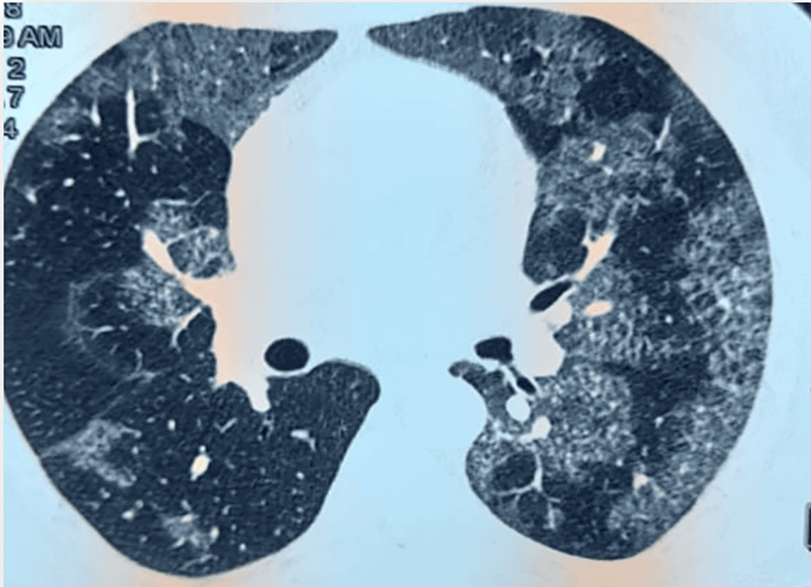
A 33-year-old male patient, policeman, non-smoker, with no reported history of medical problems, complained of dyspnea on exertion and cough for over 5 years for which he had never consulted a doctor before and had never had any treatment. Three months before hospitalization, he reported progressive worsening of dyspnea. An initial physical exam revealed an acute respiratory distress syndrome (polypnea, PaO2=51mmHg) and bibasal crackles. Chest x-ray showed alveoli-interstitial syndrome. CT scan revealed diffuse pulmonary interstitial infiltrates without any particular diagnostic orientation (Figure 1). Bronchoalveolar lavage was milky with cytology of extracellular proteinaceous material with positive periodic acid-Schiff (PAS)-positive. After the stabilization of the respiratory condition and the clear consent of the patient, an abundant lung wash was programmed in the operating room. The procedure began with the administration of curare and sedation and endotracheal intubation with a simple tube. With a bronchial endoscopy placed in the right bronchial tree, we instilled 50mL of saline serum and removed the milky liquid. The procedure was repeated 70 times and a total of 3,5 liters of saline serum was instilled and 2,8 liters of milky liquid was removed. The operation procedure was interrupted because of acute pulmonary edema. The patient was transferred to the intensive care unit. The diagnosis of acute respiratory failure and pneumonia was retained upon admission. After 3 days, under invasive ventilation (FiO2=100%), the patient’s respiratory state worsened (PaO2/FiO2=76, pH=7,30, PaO2=76mmHg, PaCO2=59mmHg). Thus, a second abundant lung wash was performed under general anesthetic with the instillation of 1,5 liters of saline serum and 500mL of milky liquid was removed, with the patient put in a ventral decubitus position. As a result, the evolution was favorable (FiO2=45%, PaO2/FiO2=240) and extubation was possible on day 7 of hospitalization. He received 3 days of noninvasive ventilation and regression of supplemental oxygen. On the 10th day, the patient was eupneic, the physical exam was normal, and SpO2 was 97% in the ambient air.

Faced with the seriousness of the complication, it was decided not to perform a third wash and to monitor the patient functionally and radiologically.
Five years after the lung wash the patient was stable functionally, assessed via spirometry and radiologically with CT scan control (Figure 2).
Whole lung lavage, first reported by Ramirez Rivera, is a well-established approach in the therapy of PAP.5 Nevertheless, there is some debate about the appropriate time to carry out a WLL procedure in patients with PAP. This issue is especially challenging given that some spontaneous remission of PAP has been reported.6 The choice of when to carry out a WLL is still a clinical judgment, although the usual criteria for considering a WLL are the presence of significant respiratory restriction or oxygen desaturation with or without effort.
WLL is an invasive therapy that should be done in the operating room with general anesthesia. The anesthesia and lavage operation risks make it necessary to separate the lungs under general anesthesia and lavage the non-ventilated lung. Patients are intubated with a double-lumen selective intubation tube. One channel of the tube is used for ventilating and the other channel is used for lavage. The supine patient is sedated and curarized and 1-2 L of saline is instilled at 37C. The lavage fluid is discharged by gravity (‘siphoning’).7
After washing lung with 15-20 liters of saline water or even more, the effluent fluid becomes clear and the procedure can be stopped.8,9
In our case, the diagnosis of pulmonary edema induced by the lavage fluid was retained. After the WLL, local hypoxia sets in due to retained fluid which leads to additional edema as a result of activation of inflammatory cytokines and damage to capillaries and alveoli in the corresponding lung fields, as seen in stage 3 of near-drowning pulmonary edema.10
The use of a continuous positive airway pressure (CPAP) valve with a pressure limit of 5 ~ 10 mmHg can increase the removal of accumulated material, as has been reported in previous research.11,12
In a survey by Campo et al. that includes 30 centres, an estimated 1,110 WLL procedures were performed. The most common reported complications were fever in 18% followed by hypoxis (14%) pneumonia (5%) and fluid leakage (4%).13
The decision to perform a WLL remains a clinical decision, although the usual criteria for considering a WLL are the presence of substantial respiratory limitation or oxygen desaturation with or without exercise.3
The particularity of our case report is the favorable evolution of our patient in spite of an interrupted lavage. The lavage remains the treatment of preference but exposes to a risk of complication which can put in stake the vital prognosis.
We reported an acute respiratory distress syndrome caused after a whole lung lavage in a patient with PAP. This uncommon but severe complication allows us to reconsider WLL for each PAP. Considering the evident treatment differences, clinicians must evaluate the patient carefully before suggesting WLL, which is an invasive procedure requiring general anesthesia for a pathology that may evolve spontaneously favorably.
From this case we conclude that:
The authors declare that appropriate written informed consent was obtained for the publication of this manuscript. The patient consented to the publication of their clinical images.
All data underlying the results are available as part of the article and no additional source data are required.
We would like to express our sincere gratitude to Dr. Ines Zendah for her invaluable contributions to the patient’s care. Her dedication played a crucial role in the successful management of this case. We are truly grateful for her exceptional commitment and tireless efforts.
| Views | Downloads | |
|---|---|---|
| F1000Research | - | - |
|
PubMed Central
Data from PMC are received and updated monthly.
|
- | - |
Is the background of the case’s history and progression described in sufficient detail?
No
Are enough details provided of any physical examination and diagnostic tests, treatment given and outcomes?
No
Is sufficient discussion included of the importance of the findings and their relevance to future understanding of disease processes, diagnosis or treatment?
No
Is the case presented with sufficient detail to be useful for other practitioners?
No
Competing Interests: No competing interests were disclosed.
Reviewer Expertise: Rare lung diseases, Pulmonary Alveolar Proteinosis
Is the background of the case’s history and progression described in sufficient detail?
Partly
Are enough details provided of any physical examination and diagnostic tests, treatment given and outcomes?
Partly
Is sufficient discussion included of the importance of the findings and their relevance to future understanding of disease processes, diagnosis or treatment?
Partly
Is the case presented with sufficient detail to be useful for other practitioners?
Partly
References
1. Bonella F, Manali E, Papiris S: Will inhalational GM-CSF replace whole lung lavage as a treatment for autoimmune pulmonary alveolar proteinosis? Many pole positions, not yet the final winner. European Respiratory Journal. 2024; 63 (1). Publisher Full TextCompeting Interests: No competing interests were disclosed.
Reviewer Expertise: PAP, ILD
Alongside their report, reviewers assign a status to the article:
| Invited Reviewers | ||
|---|---|---|
| 1 | 2 | |
|
Version 1 18 Oct 23 |
read | read |
Provide sufficient details of any financial or non-financial competing interests to enable users to assess whether your comments might lead a reasonable person to question your impartiality. Consider the following examples, but note that this is not an exhaustive list:
Sign up for content alerts and receive a weekly or monthly email with all newly published articles
Already registered? Sign in
The email address should be the one you originally registered with F1000.
You registered with F1000 via Google, so we cannot reset your password.
To sign in, please click here.
If you still need help with your Google account password, please click here.
You registered with F1000 via Facebook, so we cannot reset your password.
To sign in, please click here.
If you still need help with your Facebook account password, please click here.
If your email address is registered with us, we will email you instructions to reset your password.
If you think you should have received this email but it has not arrived, please check your spam filters and/or contact for further assistance.
Comments on this article Comments (0)