Keywords
fever of unknown origin, lepra reaction, leprosy,case report,cutaneous
This article is included in the Pathogens gateway.
fever of unknown origin, lepra reaction, leprosy,case report,cutaneous
A fever of unknown origin (FUO) is defined as an unexplained fever of 38.3 °C or higher for at least three weeks’ duration after preliminary investigations.1 In general, infection is the primary cause of FUO, which accounts for approximately one-fourth, followed by neoplasm and noninfectious inflammatory diseases.2
Leprosy, known also as Hansen’s disease, is a chronic granulomatous disease caused by the intracellular bacterium Mycobacterium leprae (M. leprae). M. leprae often spreads by respiratory droplets and close contact and can involve multiple systems throughout the body, especially the nervous, integumentary and musculoskeletal system, whi ch are misdiagnosed as arthritis. Moreover, lepra reaction developed from leprosy responds for a significant morbidity and mortality without relationship to the timing of treatment, despite effective measures counteract the causative M. leprae with antibiotics.3,4 It is regretful that the underlying mechanism of lepra reaction is still poorly understood.5 With development of medical technology and improvement of sanitary conditions, the World Health Organization set a goal of reducing leprosy prevalence to less than 1 per 10,000 inhabitants from 2000 to 2005.6
In this report, a case of fever of unknown origin was eventually identified as type II lepra reaction with the aid of Institute for Dermatologic Diseases Control and Prevention. Due to the prompt intervention, the patient has achieved recovery. The purpose of this case report is to remind clinicians to be familiar with and to detect early for lepra reactions.
A 41-year-old male, who is a minority mainly distributed in Guizhou Province, presented as intermittent fever with body temperature exceeding 39 °C over 16 days, accompanying ring-shaped febrile rash, swelling, pain and deformity of the extremities, was transferred to hospitalization from outpatient. The patient denied a family history or special occupational exposures, with long-term irregular glucocorticoids (prednisone acetate) medication history.
On admission, physical examination revealed facial flush with sparse eyebrows, enlargement of left cervical and inguinal lymph nodes, acromegaly along with eagle talon-shaped deformity of both hands, soft subcutaneous masses, skin keloid scar and concomitant pigmentation (shown in Figure 1). Laboratory examination showed an abnormal haemograms including WBC count 11.58×10⁹/L, neutrophil percentage 79.70%, ESR 38mm/h, Interleukin-6 31.77 pg/ml, PCT 0.26 ng/ml, plasma CRP 165 mg/L. Subcutaneous masses of right elbow joint were detected by ultrasonography, considering to be olecranon bursitis (shown in Figure 2). According to all results at the present stage, a diagnosis of acute infection could not be ruled out, followed by an antimicrobial agent treatment using cefoperazone-sulbactam (2 g, q12h) and levofloxacin (0.5 g, qd) for 3 days and switching to a broad antibacterial spectrum combination of meropenem (1 g, q8h) and vancomycin (15 mg/kg, q12h) for extra 3 days.
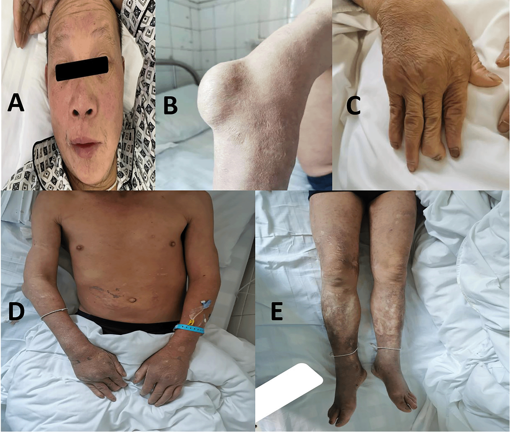
A. Facial flush with spare eyebrows. B. Subcutaneous mass of right elbow joint. C. Aagle talon-shaped deformity of right hand. D-E. Acromegaly along with skin keloid scar and concomitant pigmentation.
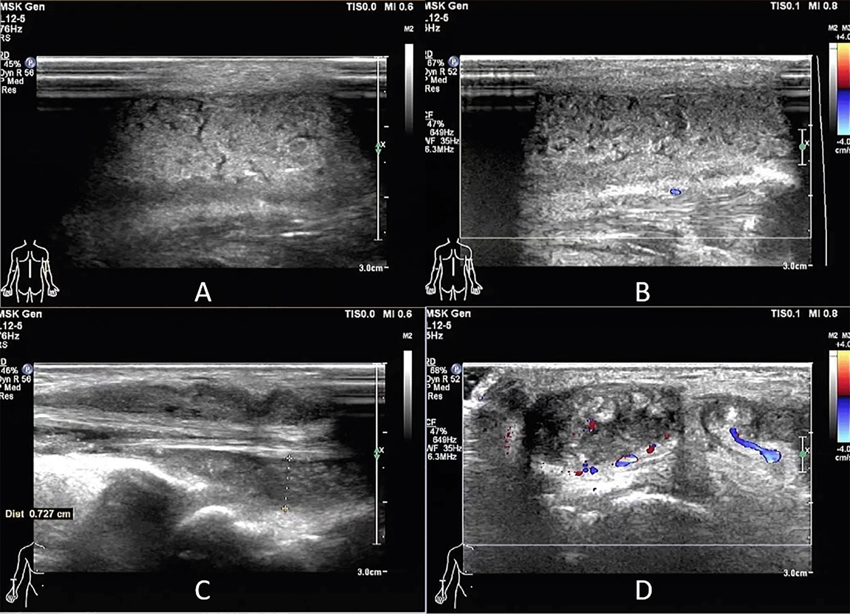
A-B. Hyperechoic mass in the right elbow joint without apparent blood flow signal. C-D. Effusion accumulation at the periphery of right hand tendons.
During anti-infection and supportive treatment, there were no marked improvements in the symptoms. In parallel, further MRI of both ankle and knee joints demonstrated old mechanical injuries shown as cruciate ligament and meniscal damage and soft-tissue edema, excluding suspicious infection foci (shown in Figure 3). No significant abnormalities were found in subsequent comprehensive clinical examinations for FUO, such as cranial and chest-abdomen CT, blood culture, echocardiography and bone marrow biopsy. Note, electromyography investigation suggested peripheral nerve injury, particularly peroneal nerve motor conduction block.
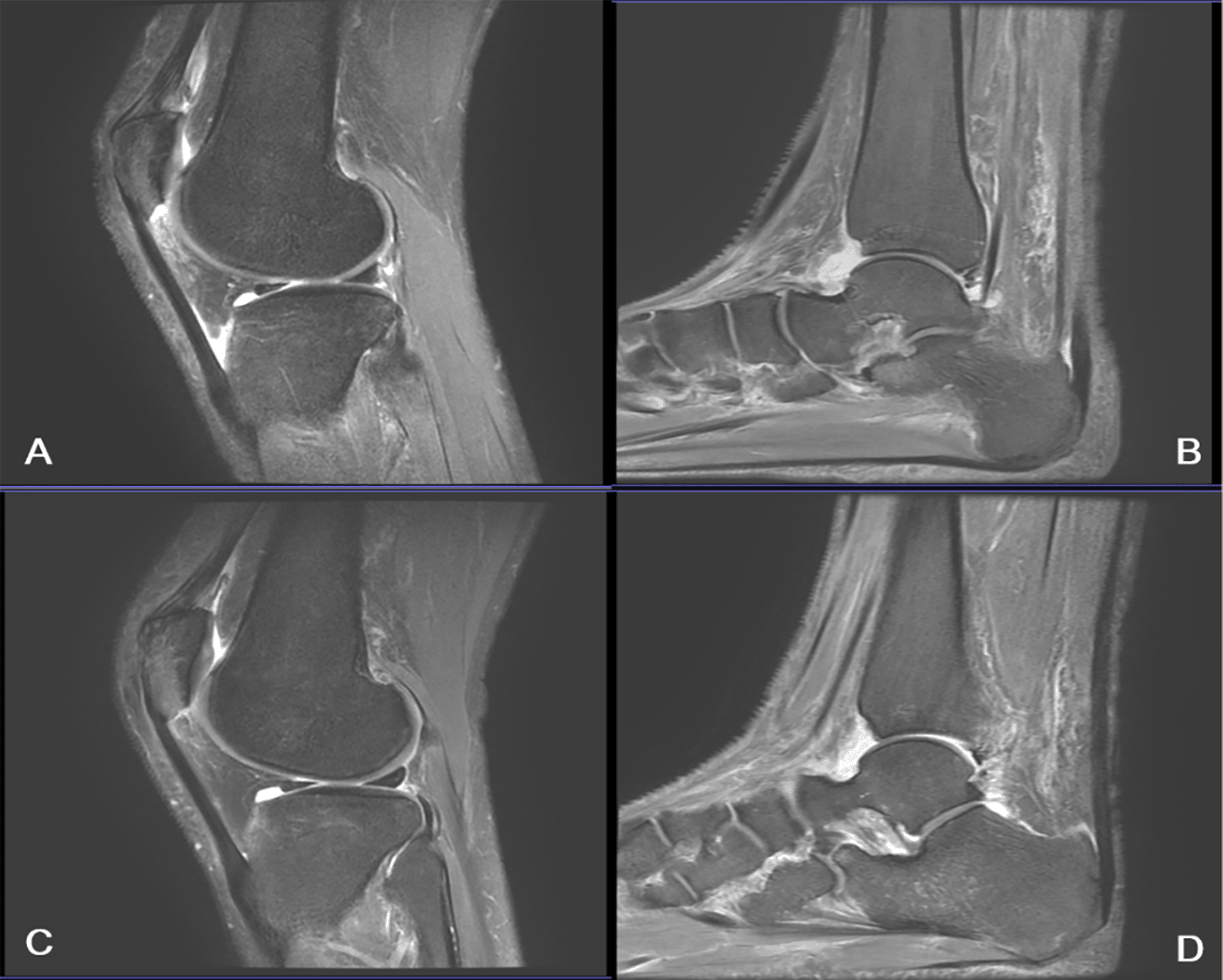
A-D figures represent left knee joint, left ankle joint, right knee joint, and right ankle joint.
In order to specify the reason of FUO, evaluation of a detailed history and physical examination were carefully performed by superior physician again. Some clinical signs and manifestations, for example, spare eyebrows, skin lesions, and peripheral nerve injury, came into view, referring to a leprosy. We immediately contacted the Institute for Dermatologic Diseases Control and Prevention of Guizhou province and applied for a detection of M. leprae in tissue fluid of the patient. Although repeated multiple testing of M. leprae staining were negative, an inspiring diagnostic clue, over 10-year leprosy history of this patient, was probed out from previous archive of the institute. Based on the abovementioned information, fever of unknown origin caused by type II lepra reaction would be regarded as this patient′s final diagnosis. Here, we took the targeted intervention measures, mainly a combination of prednisone acetate (40 mg, qd) and thalidomide (50 mg, qid), in accordance to current treatment guidelines.7 After receiving treatment, the patent′s symptoms were soon relieved without any adverse events and indicators of lepra reaction-related (ESR, Il-6, CRP, ect.) returned to normal level. The patient gradually recovered, and was discharged after nearly a month of admission.
FUO remains an intractable diagnostic and therapeutic problem in clinical daily practice, though medical technologies have undergone rapid development. Data available from published papers, infectious disease is the main reason of FUO in fact. For many pathogenic microorganisms are difficulty to identify, definite diagnose of infection and pathological changes will hardly be verified in the early stage.8
Leprosy is uncommon or rare in China, therefore, current infection and lepra reaction of leprosy are easy to overlook by clinicians. Besides, the pathogenesis of leprosy is not quite clear yet, which poses a huge challenge to physicians. Previous studies have indicated more than one-half had lepra reaction of leprosy, even many years after cessation of the treatment, which is associated with heredity, chronic infections and autoimmune disorders.9 Lepra reaction can be classified into two types. Type I lepra reaction is a delayed, cell-mediated response mainly characterized by cutaneous lesions and neuritis, especially the intense pain in the affected joints, while fever and haematological abnormalities were uncommon.10 On another scale, erythema nodosum leprosum (ENL) also called type II lepra reaction is a severe systemic immune-mediated complication of borderline and lepromatous leprosy, which is a prototypic antibody response to antigens with histopathological alterations of allergic vasculitis.11 An apparent discrepancy of type I lepra reaction patients with skin ulceration, ENL patients usually exhibit erythema nodosum, subcutaneous hypersensitivity reaction, lymphadenitis and interstitial tissue edema, presenting with both fever and abnormal blood index, such as increased neutrophilia, macroglobulin (IgG, IgM), complement 3 (C3) and 2 (C2).12 In addition, cytokine detections may provide important basis for the diagnosis and treatment of patients with type II lepra reaction in terms of the clinical researches.13,14 Taking into account the leprosy and long-term irregular glucocorticoids medication history, existing diagnosis of FUO, excluding of acute infection by negative results of M. leprae staining and MRI, we would consider type II lepra reaction as final diagnosis and started on symptomatic treatment by a combination of prednisone acetate and thalidomide.
The patient’s rapid rehabilitation further confirmed the diagnosis of type II lepra reaction. During the combined drug use procedure, patient developed no adverse reactions with good adherence. The favorable outcome of this patient was consistent with the reported literature,15,16 owing to prompt differential diagnosis and treatment-adjusted. We have been inspired by the present case and draw some insight that clinicians should obtain a comprehensive history, physical examination and laboratory evaluation for each patient suspected of having FUO, as well as enhance diagnostic competency for rare infectious diseases in the absence of clear and unequivocal medical history.
The studies ethics committee approval and fully informed written consent.
Written informed consent was obtained from the patient for publication of this case report and accompanying images. A copy of the written consent is available for review by the Editor-in-Chief of this journal on request.
| Views | Downloads | |
|---|---|---|
| F1000Research | - | - |
|
PubMed Central
Data from PMC are received and updated monthly.
|
- | - |
Provide sufficient details of any financial or non-financial competing interests to enable users to assess whether your comments might lead a reasonable person to question your impartiality. Consider the following examples, but note that this is not an exhaustive list:
Sign up for content alerts and receive a weekly or monthly email with all newly published articles
Already registered? Sign in
The email address should be the one you originally registered with F1000.
You registered with F1000 via Google, so we cannot reset your password.
To sign in, please click here.
If you still need help with your Google account password, please click here.
You registered with F1000 via Facebook, so we cannot reset your password.
To sign in, please click here.
If you still need help with your Facebook account password, please click here.
If your email address is registered with us, we will email you instructions to reset your password.
If you think you should have received this email but it has not arrived, please check your spam filters and/or contact for further assistance.
Comments on this article Comments (0)