Keywords
postoperative optic nerve injury, risk factor, vitrectomy
postoperative optic nerve injury, risk factor, vitrectomy
Vitrectomy is a standard procedure during surgical repair of many posterior eye segment disorders, such as retinal detachment, epiretinal membrane, macular hole, vitreous hemorrhage, and ocular trauma. The procedure of vitrectomy involves removing vitreous gel from the eye and replacing it with saline solution, silicone oil, or gas bubble.1 Recent progress in instrumentation, technology, and surgical technique has resulted in the increasing use of vitreoretinal surgeries to treat posterior eye segment disorders. These developments have created an increase in expectations from both doctors and patients regarding anatomical and functional outcomes.2
While vitrectomy is considered safe and effective, there is a risk of complications, including optic nerve injury, which can result in visual field defects and reduced visual acuity.1 The optic nerve is a component of the visual pathway that is responsible for transmitting visual information from the retina to the brain.3 A review conducted by Taban et al. (2007) reported the outcomes of 1,104 patients who underwent vitrectomy, revealing that 160 cases (14.5%), with a range of less than 1% to 71%, experienced unexplained postoperative visual field defects. Out of these cases, 31 eyes (19.4%) showed indications of optic nerve damage after vitrectomy, such as pallor, relative afferent pupillary defect, and intrapapillary hemorrhage.4
It is important to note that the consequences of this complication can be significant and may lead to permanent vision loss or blindness.1 In this review, we will discuss any risk factors regarding postoperative optic nerve injury after vitrectomy. By understanding the risk factor of this serious complication, it could minimize the risks and achieve the best possible outcomes.
We submitted a protocol for registration before drafting this review and meta-analysis. The protocol was successfully registered in the International prospective register of systematic reviews (PROSPERO) under the code CRD42023453533 on 22nd August 2023.
What is the risk factor of optic nerve injury following vitrectomy for various indications?
We conducted a systematic review through a literature search using electronic databases, including Cochrane, PubMed, Scopus, and EMBASE, as well as a hand search of relevant journals with the keywords “(postoperative optic nerve injury) AND (vitrectomy) AND (risk factor)”. Postoperative optic nerve injury was defined as the damage of the optic nerve component which occurs as a complication of ocular surgery. Postoperative optic nerve damage was measured through imaging techniques, clinical examination, or electrophysiological tests. Vitrectomy refers to a surgical procedure in the eye by removing the vitreous gel in the eye and replacing it with saline, silicone oil, or gas. This study used the Preferred Reporting Items for Systematic Review and Meta-Analysis (PRISMA) guidelines.53
The inclusion criteria of this study are: (1) patients with postoperative optic nerve injury after vitrectomy, (2) clinical trial, case-control, or cohort study, (3) analyzing risk factor of optic nerve injury after vitrectomy, (4) published in the last 15 years and is conducted in English. Exclusion criteria of this study are: (1) studies published >15 years ago, (2) case report or review study, (3) studies with diabetic retinopathies or previous optic neuropathy patients, (4) full-text article is not available, (5) non-English article.
Five investigators independently reviewed titles and abstracts, excluding irrelevant studies. Subsequently, authors AD, AAV, ARY, AKA, and KAS individually assessed each entry using ‘Yes,’ ‘No,’ or ‘Maybe’ based on inclusion and exclusion criteria.
Five researchers independently reviewed extracted data from eligible studies using a standardized data extraction form. The data extracted included study design, patient demographics, length of follow-up, indication for vitrectomy, surgical procedure, risk factor assessed, optic nerve injury complication, and primary outcomes of the study. Discrepancies were resolved through discussion, and the remaining studies were checked for duplicates. Systematic evaluation, following the inclusion and exclusion criteria, was conducted on the final retrieved studies.
The quality of the eligible studies was assessed using the risk of bias (RoB) 2.0 for randomized controlled trials and the ROBINS-E tool for observational studies.
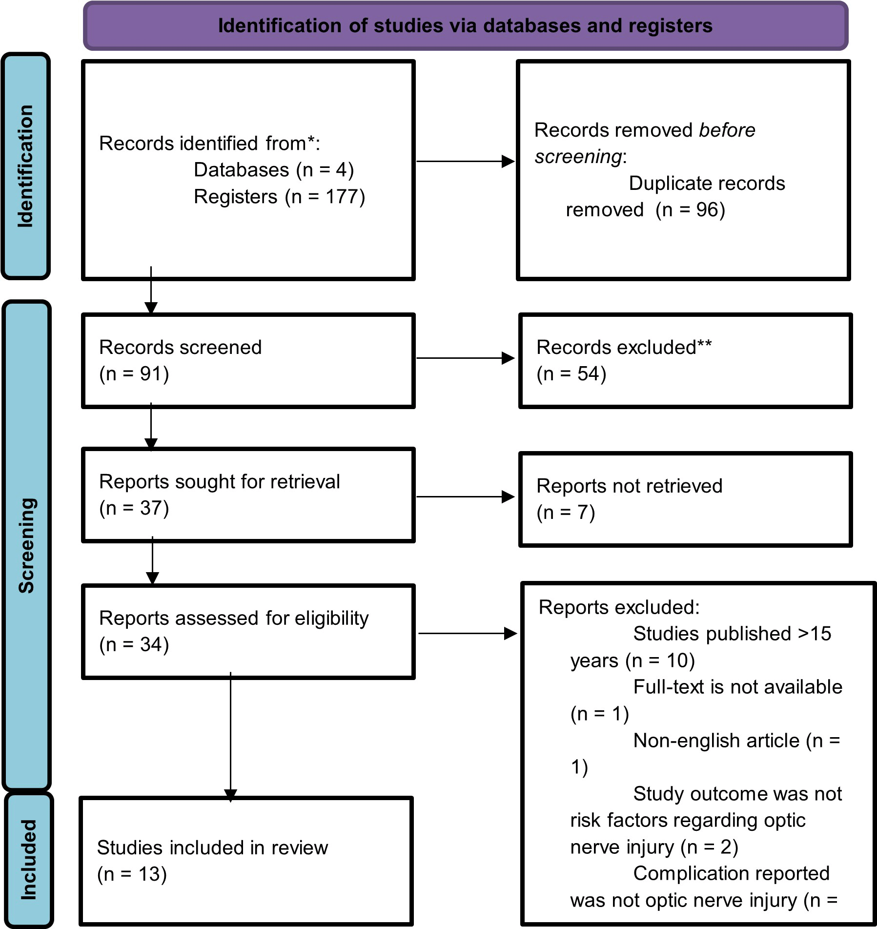
A total of 13 studies were included in this systematic review. The flow chart is shown in Figure 1. Study characteristics are shown in Table 1.
| No | Author | Study Design | Study Population (N) | Age in Years (Median and IQR or mean ± SD) | Gender, % male | Indication | Procedures | Risk Factor Assessed | Optic Nerve Injury Condition | Primary outcomes (as per current review) |
|---|---|---|---|---|---|---|---|---|---|---|
| 1 | Park et al. (2016)5 | Retrospective Cohort | 296 | 63.62 (DONFL group); 64.90 (control group) | 38.5% | Epirerinal Membrane | Vitrectomy | DONFL | ILM peeling (odds ratio, 32.22 [4.33–240.0]; p = 0.001) and gas tamponade (odds ratio, 2.33 [1.03–5.28]; P = 0.042) were significantly associated with DONFL. | |
| 2 | Spaide et al. (2012)6 | Prospective Cohort | 25 | 72.4 (±7.3) | 40% | Macular Hole (40%), Epiretinal Membrane (60%) | Vitrectomy | DONFL | ILM peeling was performed in 13 of 13 eyes (100%) in inner retinal dimpling group and 5 of 12 eyes (42%) in group without inner retinal dimpling. ILM peeling (odds ratio, 36.81 [1.78–761.73]; p = 0.02) was associated with DONFL. | |
| 3 | Bansal et al. (2012)7 | Retrospective Cohort | 49 | 48 (38-72) in optic neuropathy patients; 50 (34-74) in control patients | 43% | Macula-sparing primary RRD | Vitrectomy | Optic Neuropathy | Reduced MOPP of ≤30 mmHg (odds ratio, 15.39 [1.56–774]; p = 0.01) and longer durations during which the MOPP was <30 mmHg (p = 0.02) were significantly associated with post-vitrectomy optic neuropathy. | |
| 4 | Elghawy et al. (2022)8 | Retrospective Cohort | 104 | 55.6 ± 16.8 | 68% | Macula-involving RRD | Group 1: early vitrectomy (<48 hour) Group 2: moderately delayed vitrectomy (3-7 days) Group 3: late vitrectomy (>7 days) | Postoperative Glaucoma | No significant difference regarding postoperative complications among the groups including the development of glaucoma (p = 0.52). Postoperative visual acuity was significantly better in early and moderately delayed repair group compared to late repair group. | |
| 5 | Nikolaenko et al. (2019)9 | Prospective RCT | 59 | 73.32 ± 7.54 | 52.5% | Vitreomacular Traction Syndrome | Vitrectomy | Delayed recovery of retina and optic nerve functional activity | Functional activity of retinal inner layers and the optic nerve in group 1 and group 3 restored twice as actively as that in group 2. The use of gas tamponade, compared to air and BSS tamponade, is a significant negative factor influencing the functional activity of retina and optic nerve inhibition. | |
| 6 | Moharram et al. (2020)10 | Retrospective Cohort | 88 | 39 (12-61) | 54.5% | RD associated with giant retinal tears | Vitrectomy | Postoperative glaucoma | Postoperative glaucoma developed in 6 of 40 (15%) eyes in the SO group and 9 of 48 (18.8%) eyes in the gas group. There was no significant difference between both groups regarding postoperative glaucoma (p = 0.09). | |
| 7 | Lee et al. (2018)11 | Retrospective Cohort | 64 | 46.83±15.00 (SO); 45.01±20.11(C3F8 Gas) | ? | Macula-sparing RD | Vitrectomy | reduction of RNFL thickness | Eyes in the SO group showed a significantly greater reduction of RNFL thickness compared with eyes in the C3F8 gas group at 6 months and 9 months postoperatively (P=0.006 and P=0.005, respectively). | |
| 8 | Inan et al. (2020)12 | Retrospective Cohort | 58 | 60.7±11.2 | 62% | Macula-off RRD | Vitrectomy | reduction of RNFL thickness | There was no significant difference of RNFL thickness between SO and C3F8 gas group. However, a significant thinning in the layers of the ganglion cell layer, outer plexiform layer, and outer nuclear layer of the central subfield was found in the SO group (P = 0.01, p = 0.046, p = 0.024, respectively). | |
| 9 | Zhou et al. (2020)13 | Retrospective Cohort | 21 | 53.86±8.25 | 28% | Macula-on RRD | Vitrectomy | reduction of RNFL thickness | Eyes in the SO group showed a significantly greater reduction of retinal nerve fiber layer thickness compared with eyes in the sterilized air group at 6 weeks postoperatively in fovea section and whole retina (P=0.001 and P=0.003, respectively). | |
| 10 | Picirillo et al. (2021)14 | Prospective RCT | 144 | 67.13 ± 6.71 (DoubledyneTM); 63.96 ± 5.22 (TwinTM) | 61.1% | Macular Hole, Macular Pucker | Group 1: DoubledyneTM (soluble lutein 2%, BBG 0.05 and TB 0.15%.) Group 2: TwinTM (Trypan blue 0.18% and Blulife 0.03%) | No optic nerve injury condition | No toxic dye-related complications or long-term ones affecting the retina were observed in either group. | |
| 11 | Toba et al. (2014)15 | Prospective RCT | 61 | 65.2 ± 7.6 | 37.7% | Macular Hole | Group 1: ICG Group 2: BBG Group 3: TA | reduction of RNFL thickness | The differences in the RNFL thickness among the groups were not significant for at least 12 months postoperatively. | |
| 12 | Rohrig et al. (2019)16 | Retrospective Cohort | 270 | 68.76 | 28.5% | Macular Hole | Group 1: MBB Group 2: AV 17-M | No optic nerve injury condition | There was no significant difference regarding number of IS/OS defects between MBB and AV 17-M group (P = 0.6225). | |
| 13 | Baba et al. (2012)17 | Retrospective Cohort | 63 | 67.1±4.8 (BBG); 65.7±7.3 (ICG) | 41.2% | MH | Group 1: BBG Group 2: ICG | Reduction of retinal sensitivity and CFT thickness | BBG resulted in better mean retinal sensitivity in the central 2° at 3 and 6 months after surgery (p = 0.001 and P = 0.030, respectively), but no significant difference in the central 10°. BBG also had a shorter length of IS/OS junction defect at 3 months (P = 0.048), but not at 6 months. There was no significant difference in the length of ELM defect and GCC thickness between the groups at 3 and 6 months. However, CFT was significantly thinner in the ICG group than in the BBG group at 3 and 6 months. (p = 0.013 and p = 0.001, respectively). |
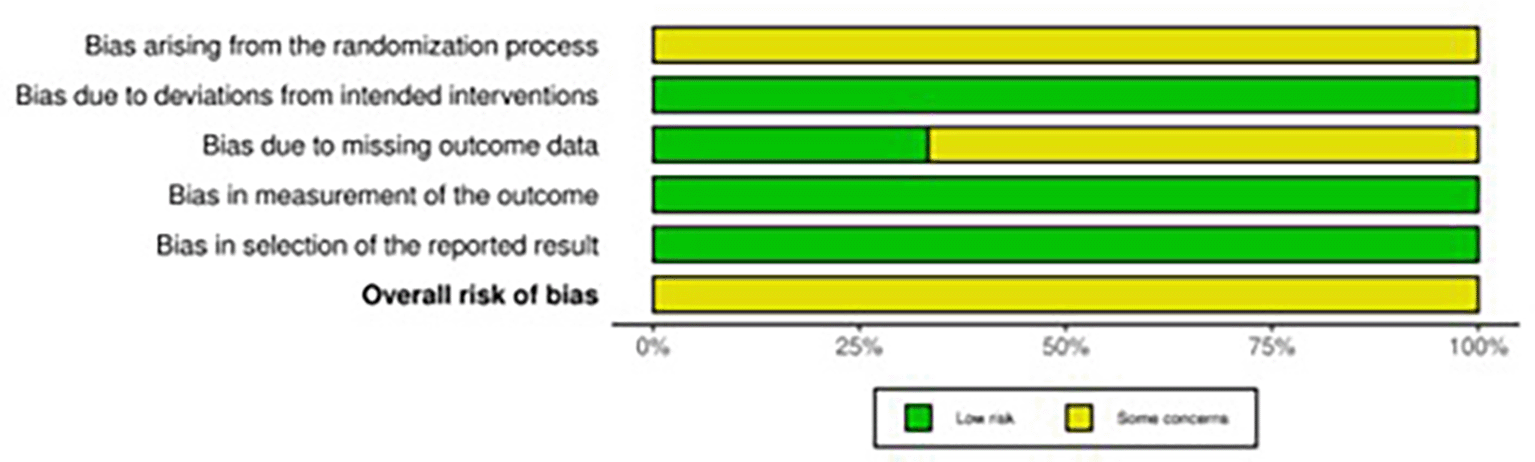
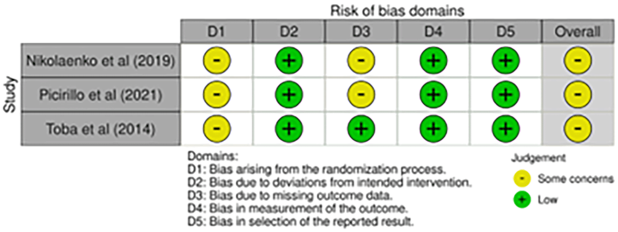
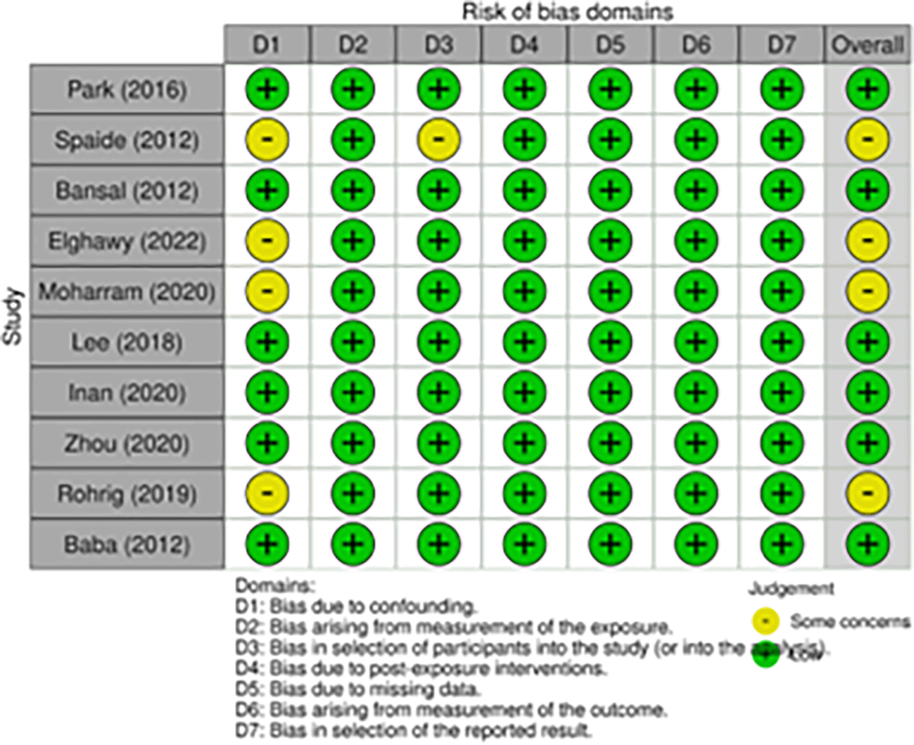
This systematic review included 3 randomized clinical trials (RCT) and 10 observational studies. Quality assessment was conducted using the risk of bias (RoB) 2.0 for RCT and ROBINS-E for observational studies. Bias arising from the randomization process was seen in all RCTs, due to none reported a concealment of allocation sequence. Nikolaenko et al.9 and Picirillo et al.14 also had possible bias due to missing outcome data. The risk of bias in RCT is shown in Figure 2 and Figure 3.
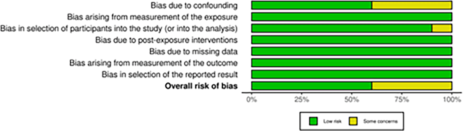
Bias due to confounding was reported in four studies. Demographic data (e.g., age, gender) were different between groups in study conducted by Speide, et al.,6 Elghawy et al.,8 and Moharram et al.10 The confounding factor that may affect study outcome was type of gas used in the study conducted by Rohrig, et al.16 Selection bias was seen in Spaide et al.6 due to no clear explanation regarding eligibility criteria for patient selection. The risk of bias in observational studies is shown in Figure 4 and Figure 5.
Park et al. (2016) investigated the risk factors for a dissociated optic nerve fiber layer (DONFL) after epiretinal membrane surgery. There was no significant difference between the DONFL group and control group in this study regarding mean age (p = 0.745), sex (p = 0.674), mean follow-up time (p = 0.482), mean preoperative best corrected visual acuity (p = 0.967), mean duration of symptoms (p = 0.562), and mean axial length of the eye (p = 0.284). ILM peeling was performed in 29 of 30 eyes (96.7%) in DONFL group and 126 of 266 eyes (47.4%) in control group peeling (odds ratio, 32.22 [4.33–240.0]; p = 0.001).5 Similar result was shown in Spaide et al. (2012), in which ILM peeling was performed in 13 of 13 eyes (100%) in patients with postoperative inner retinal dimpling and 5 of 12 eyes (42%) in patients without postoperative inner retinal dimpling (p = 0.001). In the areas of the dimples, imaging examination showed a thinning of ganglion cell layer with decreased reflectivity from nerve fiber layer.6
Bansal et al. (2012) examined the clinical characteristic and potential risk factors associated with optic neuropathy after vitrectomy for macula-sparing primary RRD. All 7 patients with post-vitrectomy optic neuropathy had visual acuity less than 20/200, RAPD (+), visual field defect, and pallor optic nerve. This study showed that 5 of 7 patients (71%) with post-vitrectomy optic neuropathy experienced a reduced mean ocular perfusion pressure (≤30 mmHg) compared with 7 of 42 patients (17%) in control group (odds ratio, 15.39 [1.56 – 774]; p = 0.01). Furthermore, patients with post-vitrectomy optic neuropathy had longer durations during which the mean ocular perfusion pressure was ≤30 mmHg (70 min) compared with control group (15 min) (p = 0.02).7
According to Elghawy et al. (2022), while postoperative visual acuity between early repair group (<48 h) and moderately delayed repair group (3-7 days) showed no difference (p = 0.163), post-operative visual acuity was significantly better in early repair compared to late repair group (> 7 days) (p = 0.010). However, this study reported no significant difference regarding postoperative complications among the groups including the development of glaucoma (p = 0.52).8
Nikolaenko et al. (2019) reported the effect of tamponade type used in vitrectomy for vitreomacular traction syndrome on retinal and optic nerve functional activity. Electrophysiological testing was performed to assess the functional activity of retina and optic nerve. The functional activity of neurons in inner retinal layers was assessed by electrical sensitivity (ES) threshold and critical frequency of phosphene disappearance (CFPD) values, while the functional activity of optic nerve was evaluated by flash visual evoked potential (FVEP) latency. This study reported that the functional activity of retinal inner layers and the optic nerve in group 1 (vitrectomy + air tamponade) and group 3 (vitrectomy + BSS tamponade) restored twice as actively as that in group 2 (vitrectomy + C3F8 gas tamponade). Park et al. (2016) also reported that the use of intravitreal gas tamponade was significantly associated with DONFL formation (odds ratio, 2.33 [1.03–5.28]; p = 0.042).9 Another study by Lee et al. (2018) reported a greater reduction of RNFL thickness in eyes with silicone oil (SO) tamponade compared to eyes with C3F8 gas group at 6 months and 9 months after surgery in macula-sparing RD (p = 0.006 and p = 0.005, respectively).11 On the contrary, Inan et al. (2020) reported no significant difference of RNFL thickness between SO and C3F8 gas group, even though a thinning in the layers of the ganglion cell layer, outer plexiform layer, and outer nuclear layer of the central subfield was found significantly greater in the SO group.12 Zhou et al. (2020) compared the effect of SO tamponade and sterilized air tamponade on RNFL thickness in macula-on RRD patients. This study reported a greater reduction of RNFL thickness in eyes with SO tamponade compared to sterilized air tamponade at 6 weeks postoperatively.13 Moharram et al. (2020) reported no significant difference between silicone oil tamponade and gas tamponade groups regarding postoperative glaucoma and ERM formation in retinal detachment associated with giant retinal tears. Both groups in this study also achieved similar favorable anatomical outcomes.10
Picirillo et al. (2021) reported no significant difference regarding average RNFL thickness before and after surgery (6 months follow-up) in the use of DoubledyneTM and TwinTM as a dye to perform peeling of epiretinal membrane or internal membrane peeling (p > 0.05). This study also reported no toxic dye-related complications were observed in either group.14 Similar result was shown in Toba et al. (2014), this study compared the changes of RNFL thickness in macular holes after vitrectomy with indocyanine green- (ICG-), brilliant blue G- (BBG-), and triamcinolone acetonide (TA-) assisted ILM peeling. This study reported no significant difference regarding RNFL thickness between those groups for at least 12 months postoperatively.15 Rohrig et al. (2019) also reported no significant difference between the use of MBB and AV 17-M in terms of inner segment/outer segment defect.16 However, Baba et al. (2012) reported faster restoration of inner segment/outer segment junction in brilliant blue G (BBG) group compared to indocyanine green (ICG) group. Also, the central foveal thickness was significantly thinner in ICG used. Therefore, this study suggested that BBG may be a better agent than ICG.17
Vitrectomy is a common procedure used in the surgical treatment of various disorders affecting the back of the eye, such as retinal detachment, epiretinal membrane, macular hole, vitreous hemorrhage, and ocular trauma. Although vitrectomy is considered to be a safe and effective treatment, it does come with the risk of complications, such as optic nerve injury. Optic nerve injury can lead to a reduction in visual acuity and visual field defects.1 In this systematic review, fourteen studies revealed four main risk factors of optic nerve injury following vitrectomy for various indications, including removal of the internal limiting membrane, reduced mean ocular perfusion pressure, the use of particular tamponade agent, and the use of ICG as ILM staining agent.
After a standard surgical procedure of pars plana vitrectomy (PPV), the ILM peeling can be performed by carefully detaching and removing the posterior hyaloid to prevent the growth of cells and the subsequent traction of the retina.18 In our review, two studies reported that the removal of the internal limiting membrane was significantly associated with dissociated optic nerve fiber layer (DONFL) formation. DONFL was seen postoperatively using fundus photographs and optical coherence tomographic (OCT) scan.5,6 There are various possible explanations for the formation of DONFL after ILM peeling procedure. It may occur as an acute response due to direct surgical damage to the inner layer of the retina during the ILM peeling procedure.19 However, Runkle et al. (2018) reported that the DONFL formation is more likely to be caused by the tractional forces from peeling over a large area, rather than the physical contact between the surgical instruments and the tissue.20 Another potential explanation is that membrane peeling might cause harm to the footplates of Müller cells that are attached to the ILM.6 According to Pan et al. (2014), intraoperative trauma to the retinal nerve fiber layer during membrane peeling led to central scotoma and decreased visual acuity postoperatively.21 More research regarding intraoperative OCT in the development of minimally traumatic ILM peeling procedures is needed to achieve the best possible outcome.
Ocular perfusion pressure (OPP) is determined by calculating the difference between the mean arterial pressure (MAP) and the intraocular pressure (IOP). The corresponding mean ocular perfusion pressure (MOPP) in the supine position was calculated as MOPP = 115/130 x (MAP – IOP).22 Thus, a decrease in systemic blood pressure or an increase in IOP can lead to a decrease in OPP. The body’s natural ability to regulate blood flow to the optic nerve and retina occurs within a broad range of perfusion pressures, which helps to minimize the risk of ischemic damage. However, in this review, Bansal et al. (2012) reported a significant association between reduced mean ocular perfusion pressure (≤30 mmHg) and post-vitrectomy optic neuropathy, as manifested by visual acuity less than 20/200, RAPD (+), visual field defect, and pallor optic nerve.7 This result is consistent with Hayreh et al. (2001), which suggested that when the ocular perfusion pressure (OPP) drops below 30 to 35 mmHg, the autoregulation of blood flow is disrupted, and there is a risk of ischemic damage to the optic nerve.23 This scenario is also similar to cases of ischemic optic neuropathy that have been observed following spinal and cardiothoracic procedures, in which hypotension, blood loss, and increased intraocular pressure (IOP) have been associated with decreased ocular perfusion.24 Moreover, Bansal et al. (2012) also reported that patients with post-vitrectomy optic neuropathy had longer durations during which the mean ocular perfusion pressure was <30 mmHg compared with control group, suggesting that the duration of reduced ocular perfusion also has an influence on the occurrence of post-vitrectomy optic neuropathy.7
In the surgery of retinal detachment, an intraocular tamponade agent is used to create surface tension around retinal breaks, which helps to stop additional fluid from entering the subretinal space until a permanent seal is provided by retinopexy. The two primary types of tamponade agents frequently used are gases and silicone oils.25 Air, SF6, and perfluoropropane (C3F8) are the most frequently used gas tamponade in the USA.26 Air tamponade does not expand, unlike 100% SF6 which expands approximately two times over 1-2 days, and 100% C3F8 which expands around four times over 3-4 days.27 After a complete gas-fluid exchange, gas tamponade agents will be absorbed spontaneously from the vitreous cavity, with air taking about 5-7 days, 20% SF6 approximately 2 weeks, and 14% C3F8 around 8 weeks.28 In contrast with gases, silicone oils are permanent and will stay in the eye until they are removed surgically.29 1.000 and 5.000 centistokes (cSt) silicone oils are the most frequently used silicone oils in the USA.30 Gases possess greater surface tension and buoyancy force compared to silicone oils.31
In this review, we compared the effect of each type of intraocular tamponade agent on the occurrence of optic nerve injury. Nikolaenko et al. (2019) reported that the functional activity of retinal inner layers and the optic nerve in air tamponade group and BSS group restored twice as fast as that in C3F8 gas tamponade group in vitreomacular traction syndrome.9 It could possibly be caused by a rise in intraocular pressure (IOP), which is a common complication that occurs when gas tamponades are used. Thus, increased IOP following vitrectomy can result in damage to the optic nerve, retinal ischemia, and subsequent vision loss. This can occur through an open-angle mechanism, closed-angle mechanism, or both. In the case of an open-angle mechanism, the rise in IOP is due to the expansion of intraocular gas tamponade agent.32
While gas tamponade agent has worse outcome compared to air tamponade regarding retina and optic nerve functional activity, gas tamponade is believed to be the preferred agent if compared with silicone oils in many cases. Lee et al. (2018) showed a significantly greater reduction of RNFL thickness compared with eyes in the C3F8 gas group at 6 months and 9 months postoperatively in macula-sparing RD.11 On the contrary, Inan et al. (2020) reported no significant difference of RNFL thickness between SO and C3F8 gas group in macula-off RRD, even though a thinning in the layers of the ganglion cell layer, outer plexiform layer, and outer nuclear layer of the central subfield was found significantly greater in the SO group.12 Different from Lee and Inan, Zhou et al. (2020) compared the effect of SO tamponade and sterilized air tamponade on RNFL thickness in macula-on RRD patients. However, this study also reported a greater reduction of RNFL thickness in eyes with SO tamponade compared to sterilized air tamponade at 6 weeks postoperatively.13 Thinning of RNFL can indicate a loss of retinal ganglion cells which can lead to decreased vision.33 The number of surviving ganglion cells and the regeneration of axons are crucial for preserving and recovering vision after optic nerve injury. Therefore, the thickness of the RNFL can serve as an indicator of changes in visual function.34 There are several possible explanations regarding the effect of SO on RNFL thickness. First, mechanical stress caused by silicone oil in the fovea may lead to the premature loss of outer nuclear layer (ONL) cell bodies.35 Second, subretinal migration of silicone oil due to its use as a tamponade could result in severe optic neuropathy.36,37 The migration of macrophages that have phagocytosed emulsified oil bubbles could be a potential mechanism for the subretinal migration of silicone oil.38 There have been reports that the small molecules in silicone oil may diffuse from the oil and enter the retinal tissue, which could cause inflammation and toxicity in the retina.39,40 Third, retinal thinning may also occur as a result of SO-related idiopathic reactions or changes in the retinal ionic environment. These changes could be caused by localized alterations in potassium concentration due to the failure of potassium siphoning by Müller cells and apoptosis, leading to neuronal degeneration.41,42 Further research is required to clarify the exact mechanism of the reduction in the thickness of RNFL associated with the use of silicone oil.
ILM staining is a technique used in vitreoretinal surgery to visualize and facilitate the removal of the inner limiting membrane (ILM), a thin and transparent layer that separates the retina from the vitreous humor. ILM staining has become an important tool in the treatment of various retinal diseases, including macular holes and epiretinal membranes. This technique involves the use of dyes, which selectively bind to the ILM and make it easier to identify and remove during surgery.14–16 Indocyanine green (ICG) was the initial dye utilized for macular hole (MH) surgery.43 However, it was subsequently discovered that the concentrations of ICG utilized during vitrectomy were toxic to the retina.44 Both in vitro and in vivo studies revealed that exposure of retinal ganglion cells (RGCs) to ICG resulted in damage to the RGCs.45,46 As an alternative to ICG, Brilliant blue G (BBG) has been introduced and can selectively stain the ILM. Studies conducted on rats and monkeys have suggested that BBG is less toxic to the retina than ICG.47,48 However, it has been found that prolonged exposure and high concentrations of BBG can also cause damage to retinal ganglion cells (RGCs).46 In this review, we compared the effect of each type of ILM staining agent on the occurrence of optic nerve injury. Overall, our review revealed that BBG may be a better agent than ICG even though there was no significant difference regarding optic nerve injury complications. Unlike previous studies that used high concentrations of ICG with prolonged exposure during surgery, Toba et al. (2014) used a low concentration of ICG and removed it quickly after application. This may explain why there was no significant decrease in the thickness of the RNFL in the group of patients who received ICG in this study. However, even though ICG did not cause a significant reduction in the thickness of the RNFL compared to BBG or TA, this study found a significant reduction in the photopic negative response (PhNR) of the electroretinogram (ERG) after macular hole (MH) surgery.15 It is in line with Ueno et al., in which also found a reduction in the PhNR of the ERG after ICG-assisted MH surgery.49 These results indicate that reduced function of retinal ganglion cells (RGCs) may occur after MH surgery without loss of RGC axons.
Over the past decade, various new dyes have been developed for the visualization of epiretinal membranes (ERMs) or inner limiting membranes (ILMs).50,51 These dyes are unique in that they possess a high specific weight, which eliminates the need for the potentially unsafe “under air” application that is required for dyes like Trypan Blue (TB) or BBG.52 Picirillo et al. (2021) reported no toxic dye-related complications or long-term ones affecting the retina were observed in DoubledyneTM (soluble lutein 2%, BBG 0.05 and TB 0.15%.) and TwinTM (Trypan blue 0.18% and Blulife 0.03%) group.14 Rohrig et al. (2019) also reported no significant difference between the use of MBB and AV 17-M in terms of inner segment/outer segment defect.16
Our study has a few limitations. First, there are more retrospective studies than prospective studies included, which may hinder data clarification and recoding. Secondly, the patient population was diverse, and the sample sizes varied widely, which could introduce bias. Additionally, there were many risk factors and complications related to optic nerve injury examined in the review. Given these limitations, we need more well-designed prospective studies with lower heterogeneity to further investigate this issue. Limitations in the review process include subjective interpretation, in which how information from individual studies is combined and analyzed can differ between one reviewer and another, potentially affecting the final result. Additionally, limitations in data integration, it can be difficult to merge data from various studies due to differences in original research.
Although vitrectomy is considered to be a safe and effective treatment, it does come with the risk of complications, such as retinal nerve fiber layer damage and optic nerve injury, which can result in visual field defect and reduced visual acuity. After examining the available information, we found that the damage can happen either during or after a vitrectomy procedure. This is linked to four main risk factors: removing the internal limiting membrane, having a lower average ocular perfusion pressure, using silicone oil as a tamponade agent, and using ICG as an ILM staining agent. More research is needed to find out other risk factors that can lead to optic nerve injury. By understanding the risk factor of this serious complication, we can minimize the risks and achieve the best possible outcomes.
| Views | Downloads | |
|---|---|---|
| F1000Research | - | - |
|
PubMed Central
Data from PMC are received and updated monthly.
|
- | - |
Provide sufficient details of any financial or non-financial competing interests to enable users to assess whether your comments might lead a reasonable person to question your impartiality. Consider the following examples, but note that this is not an exhaustive list:
Sign up for content alerts and receive a weekly or monthly email with all newly published articles
Already registered? Sign in
The email address should be the one you originally registered with F1000.
You registered with F1000 via Google, so we cannot reset your password.
To sign in, please click here.
If you still need help with your Google account password, please click here.
You registered with F1000 via Facebook, so we cannot reset your password.
To sign in, please click here.
If you still need help with your Facebook account password, please click here.
If your email address is registered with us, we will email you instructions to reset your password.
If you think you should have received this email but it has not arrived, please check your spam filters and/or contact for further assistance.
Comments on this article Comments (0)