Keywords
Costello syndrome, lung emphysema , giant pulmonary bulla, lung function bullectomy
Costello syndrome (CS) is a rare disease with intellectual disability, characterized by failure to thrive, short stature, joint laxity, loose soft skin, and distinctive facial features. This disease is caused by heterozygous germline mutations in the HRAS proto-oncogene. Cardiac and neurological abnormalities are the most common. Cardiovascular manifestations include valvular pulmonary stenosis, arrhythmia and hypertrophic cardiomyopathy. Neurological manifestations are dominated by hydrocephalus, seizures, and tethered spinal cord. Respiratory system manifestations have been reported in people with CS, but a full description of the different lung involvement and respiratory symptoms in these patients is not available. We report the case of a 19-year-old non-smoking man, followed for a Costello syndrome since the age of 8 months, who complained of exertional dyspnea and chest pain for over 18 months. The chest CT scan showed bullous emphysema of the left lung with a giant bulla in the left upper lobe measured than 20 cm long axis responsible for passive atelectasis. Pulmonary function tests revealed a severe non-reversible obstructive ventilatory defect. Faced with the worsening of his dyspnea despite treatment with bronchodilators and recurrent respiratory infections, it was decided to surgically remove the the giant emphysematous bulla. After bullectomy, a clinical and functional respiratory improvement was noted.
Costello syndrome, lung emphysema , giant pulmonary bulla, lung function bullectomy
Costello syndrome (CS) is a rare genetic disorder that involves delayed physical and mental development. Infants with Costello syndrome are characterized by stunted growth, short stature, joint laxity, loose soft skin, facial dysmorphism, intellectual deficit and heart defects.1 Costello syndrome (CS) estimated number of patients worldwide is 300.2 Cardiac and neurological abnormalities are the most common. Respiratory system manifestations have been reported in people with CS, but a full description of the different lung involvement and respiratory symptoms in these patients is not available.3 We report a case of Costello syndrome with significant bullous emphysema complicated by exertional dyspnea and recurrent respiratory infections.
The patient was a 19-year-old male. He is the second born male of two unrelated Tunisian parents. Prenatal history was remarkable for hypertension and coronary artery disease. The mother has no pathological history. He was delivered at 39 weeks gestation via vaginal vertex delivery. At birth he had he had breathing difficulties that were well controlled by oxygen therapy for 3 days. The patient is unemployed, nonsmoker and never treated for pulmonary tuberculosis. He had been followed for a Costello syndrome since a young age. The patient has a heart defect with a stenosis of the left pulmonary artery, for which he had surgery at the age of 8 months, and neurological impairment leading to moderate mental retardation. The patient had for 18 months before his admission a chest pain and exertional dyspnea.
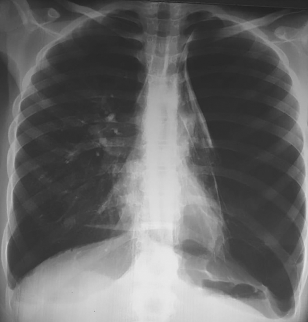
On admission, physical examination revealed the patient had short stature (weight = 54 kg and height = 159 cm) and loose skin (cutis laxa). His facial features were coarses, with a wide forehead, epicanthal folds, low-set ears and thick lips. Examination of the respiratory system revealed absent breath sound in the left hemithorax. Pulse oximetry was 94%. The chest X-ray (Figure 1) revealed a left clearness without a vascular framework evoking a great-abundance spontaneous pneumothorax or a giant emphysema bulla. The chest CT scan (Figures 2 and 3) showed bullous emphysema of the left lung. The giant bulla was in the left upper lobe measured than 20 cm long axis responsible for passive atelectasis. Pulmonary function tests revealed a severe non-reversible obstructive ventilatory defect with a forced expiratory volume in one second (FEV1) = 0,760 L (25% predicted) and a forced vital capacity (FVC) = 0,960 L (26% predicted) and FEV1/FVC = 41.27%. Thus, the patient was maintained under oxygen (oxygen (2 l/min) with a long-acting inhaled bronchodilator (Formoterol: 12 μg twice daily).

Faced with the worsening of his dyspnea despite medical treatment and recurrent respiratory infections one month after hospital discharge, it was decided to surgically remove the giant emphysematous bulla. Bullectomy was performed using the video-assisted thoracoscopic surgery approach. Intraoperatively, we saw multiple bullaes in the upper, middle, and lower lobe. The giant bulla was removed and pleural symphysis was performed. Soon After the operation, the chest pain disappeared with a marked improvement in his dyspnea.
Throughout history, Costello syndrome has been an extremely rare congenital anomaly syndrome that has attracted a lot of interest from doctors and scientists. Since the seminal work of Costello in 1971, scientific research on the Costello syndrome has increased largely.4
This syndrome is characterized by a mental retardation, learning disability, a high birth weight, an absolute or relative macrocephaly, neonatal feeding problems, short stature, curly hair, broad forehead, broad nose, large mouth and thick lips, cutis laxa, papilloma and various defects of internal organs.5,6
Patients with this syndrome have a high incidence of cardiac involvement, including cardiac hypertrophy, congenital heart defect, and arhythmia.7 Complex pulmonary and airway co-morbidities are present in an important proportion of neonates and infants caring CS.7 wheezing and bronchial hypersecretion are frequently observed in children with CS. Involvement of the bronchial tree, particularly bronchomalacia and tracheomalacia, have been reported. These abnormalities can be serious and require a tracheotomy.8 Obstructive sleep apnea syndrome has been shown to be common in these patients, often accompanied by upper airway narrowing.9
Seventy eight percent of CS patients experience respiratory complications as newborns.3 Transient respiratory distress and combined upper and lower airway anomalies are common in neonatal presentations.3 The origin of respiratory airway obstruction is not specifically identified. However, patients with CS generally present common diagnostic described by a combined involvement of the upper and lower respiratory tract with airway malacia being commonly diagnosed.10
Costello syndrome is caused by heterozygous germline mutations of HRAS. These mutations are responsible for the production of an abnormally active H-Ras protein. Among these mutations, the most common is p.Gly12Ser, which is found in approximately 80% of patients.11 The high risk of malignant tumors occurs with patients present this mutation.12 Other rare mutations have been reported, particularly in patients with severe disease phenotypes. These are p.Gly12Cys, p.Gly12Asp, p.Gly12Glu and p.Gly12Val mutations which were found in patients with a severe form of CS progressing early to death.10
Studies related to histopathological aspects of the respiratory tract in CS patients have demonstrated some congenital anomalies, as an example we mention: dysplasia of the pulmonary vasculature, lymphatics, airways, and alveoli. Besides, other histological results have been reported like: fibromuscular dysplasia of arteries, lung fibrosis, and different pulmonary infiltrates. Other histologic findings reported abnormal connective tissue in pleura, septal connective tissue, vessel, and alveolar walls.10 The involvement of elastic fibers in cutis laxa is widely distributed, affecting organs such as the skin, alveoli, aorta, and intestine.13
Other abnormalities of the lung parenchyma have been described in patients with CS such as the deposition of atypical fragmented elastic fibres in the alveolar walls, abnormal collagen fibers in the pleura as well as the development of endogenous lipid pneumonia.14
However, pulmonary emphysema is an exceptional disorder not described in the literature. It can be explained by the deposition of atypical elastic fibers in the alveolar walls. This pulmonary manifestation plays a decisive role in prognosis.
There is no specific treatment for Costello syndrome. The patient may benefit from symptomatic treatment, during the first months of life, such as: enteral or nasogastric tube nutrition, treatment of papilloma, speech therapy, occupational therapy, psychomotricity and physiotherapy for joint and postural anomalies.12
Patients with Costello syndrome share characteristic findings affecting multiple organ systems. Pulmonary emphysema is an exceptional disorder that has not been described in these patients. This type of pulmonary abnormalities can be complicated by pneumothorax, secondary to the rupture of the emphysema bubbles, which can be life-threatening. Thus, the search for pulmonary involvement by performing a chest CT scan and an evaluation of respiratory function should be considered in patients with Costello syndrome.
Written informed consent for publication of their clinical details and clinical images was obtained from the patient.
All data underlying the results are available as part of the article and no additional source data are required.
| Views | Downloads | |
|---|---|---|
| F1000Research | - | - |
|
PubMed Central
Data from PMC are received and updated monthly.
|
- | - |
Is the background of the case’s history and progression described in sufficient detail?
Yes
Are enough details provided of any physical examination and diagnostic tests, treatment given and outcomes?
Yes
Is sufficient discussion included of the importance of the findings and their relevance to future understanding of disease processes, diagnosis or treatment?
Partly
Is the case presented with sufficient detail to be useful for other practitioners?
Yes
References
1. Leoni C, Paradiso FV, Foschi N, Tedesco M, et al.: Prevalence of bladder cancer in Costello syndrome: New insights to drive clinical decision-making.Clin Genet. 2022; 101 (4): 454-458 PubMed Abstract | Publisher Full TextCompeting Interests: No competing interests were disclosed.
Reviewer Expertise: Oncology, Rare Disease, Urogynaecology, Urothelial cancers
Is the background of the case’s history and progression described in sufficient detail?
Yes
Are enough details provided of any physical examination and diagnostic tests, treatment given and outcomes?
Yes
Is sufficient discussion included of the importance of the findings and their relevance to future understanding of disease processes, diagnosis or treatment?
Yes
Is the case presented with sufficient detail to be useful for other practitioners?
Yes
Competing Interests: No competing interests were disclosed.
Reviewer Expertise: children respiratory diseases
Alongside their report, reviewers assign a status to the article:
| Invited Reviewers | ||
|---|---|---|
| 1 | 2 | |
|
Version 1 29 Dec 23 |
read | read |
Provide sufficient details of any financial or non-financial competing interests to enable users to assess whether your comments might lead a reasonable person to question your impartiality. Consider the following examples, but note that this is not an exhaustive list:
Sign up for content alerts and receive a weekly or monthly email with all newly published articles
Already registered? Sign in
The email address should be the one you originally registered with F1000.
You registered with F1000 via Google, so we cannot reset your password.
To sign in, please click here.
If you still need help with your Google account password, please click here.
You registered with F1000 via Facebook, so we cannot reset your password.
To sign in, please click here.
If you still need help with your Facebook account password, please click here.
If your email address is registered with us, we will email you instructions to reset your password.
If you think you should have received this email but it has not arrived, please check your spam filters and/or contact for further assistance.
Comments on this article Comments (0)