Keywords
3D printing, porous strucutre, bone density, CT segmentation, bone model, bone tissue
3D printing, porous strucutre, bone density, CT segmentation, bone model, bone tissue
Aging, rising obesity, and a lack of physical activity have all contributed to a significant increase in joint deterioration and bone abnormalities.1–3 The challenge is focused on making prototypes to replace or support bone parts. It is critical that the implant be functional and remain there without complications or performance deficits. Using traditional two-dimensional radiologic modalities, it is difficult to understand complicated defects, and the evaluation and classification of defects of various kinds are crucial steps to effectively manage clinical conditions. Making three-dimensional models provides both visual and tactile reproduction of bone anatomy, with the potential for better preoperative planning, thus helping to make complex interventions more precise and accurate.4 3D-printing and virtual surgery planning have found significant interest in the field of orthopaedics, leading to considerable advancement in preoperative surgical planning.5–7 Indeed, it is found that 3D models allow surgeons to visualize anatomy three-dimensionally and aid in the planning and execution of complex surgeries.8–10 3D printing of porous structure offers an attractive means to improve the fabrication of bone models and facilitate their understanding for both academic studies and surgical planning.11–13
Here we present the process of 3D-printed porous structure of a femoral bone composed of different infill densities. Among the print parameters of the slicing software, it is possible to change the infill parameters and obtain a 3D print with differentiated densities, effectively replicating the appearance of a bone tissue: a compact and very dense outer part, a trabecular and less dense inner part, and the hollow marrow in the centre. The three-dimensional reconstruction of the anatomical section was performed following the well-established procedure in previous studies.14–16 In brief, by segmentation of medical images from CT scans, different internal zones of the bone are obtained, according to the intensity of the pixels in a grey scale. The study of the distribution of different infill densities as a 3D printing parameter was one of the key points of the research. In particular, the aim was to compare the infill density with the actual bone density obtained by reprocessing medical images. The possibility of obtaining a 3D-printed object that represents a bone both internally and from an external geometric point of view is the main goal of the research. This allows on the one hand a better understanding of the clinical case and on the other hand improves communication in the patient-doctor relationship.17 Moreover, the object studied allows students, professors or doctors to visualise and better understand bone tissue in their studies thanks to an object that faithfully simulates bone tissue. Biomaterials currently used for 3D printing in the medical and orthopaedic fields18,19 include polymers, materials widely used in additive manufacturing because of their ease of structural change due to relatively low melting points;20,21 metals and alloys, including titanium alloy, a compact, lightweight and highly corrosive-resistant material with osteointegrative properties that make it perfect for replacing missing parts or for support during alignment and surgical cutting.22,23 FDM is a manufacturing technology adapted for the fabrication of porous bone tissue at low cost, providing good mechanical properties.24–26 In fact, the 3D printing parameters are set by slicing software, which layers the imported 3D model. The formal accuracy is affected by the G-code setting.27–29 The affordability and capability of reliably reproducing a model show the value of 3D simulations in preoperative planning and implant trajectory prediction to prevent injury and accidental harm to adjacent bone components.30
The aim of this study is to obtain a low-cost model of a 3D-printed femoral bone with a porous structure that is formally equivalent to its real counterpart. with a view to the potential replacement of a diseased piece of bone tissue, the bone in question must be as suitable as possible for the patient, simulating the appearance and weight of the original bone by reproducing its internal density.
The study describes a methodological process by which 3D-printed bone tissue can be obtained with a porous structure infill.
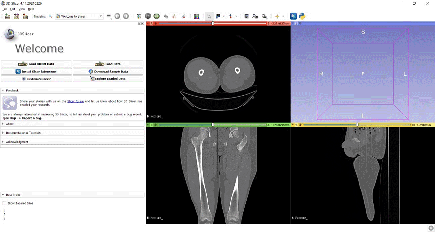
Medical images of a right femur of a 30-year-old man were downloaded from an online database. The procedure involves 3D digital reconstruction of the medical images using 3D Slicer v4.11 software,31 which allows a reading based on the Hounsfield scale, indicating the level of radiation absorption by the bone based on its density32–35 (Figure 1).
According to the Hounsfield scale, intervals are defined for each area of bone:
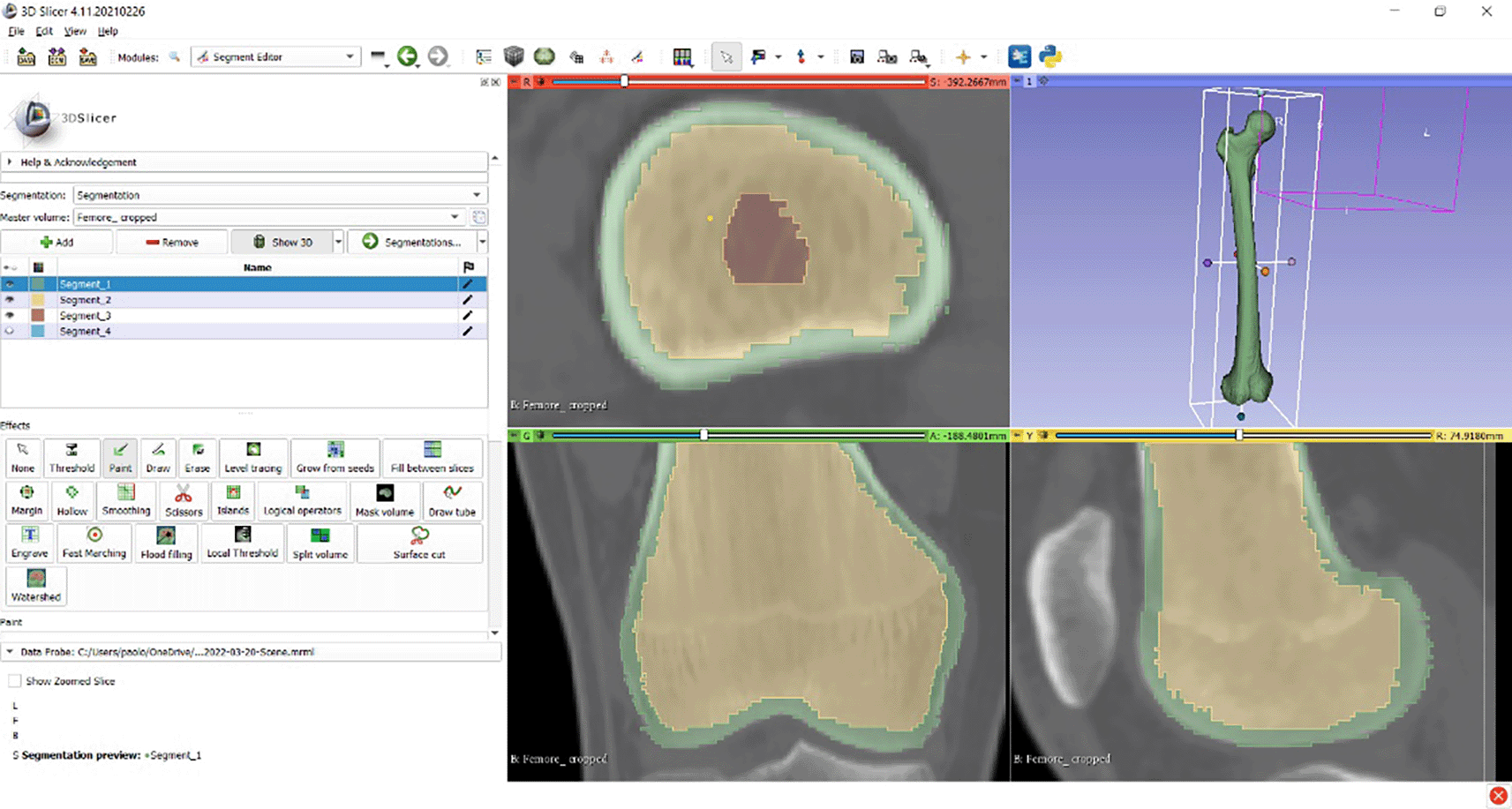
Selecting all the pixels in the region of interest (ROI) produces the three-dimensional anatomical model in which the three zones are placed inside each other. In order to select each of these areas separately, selection masks were defined (Figure 2). Each of this selection masks is identified with a distinct colour. This allows better visualization of the various parts of the 3D model that composed the bone. Once the different areas are identified, only those related to the bone are considered. They can then be exported to an .stl format file.
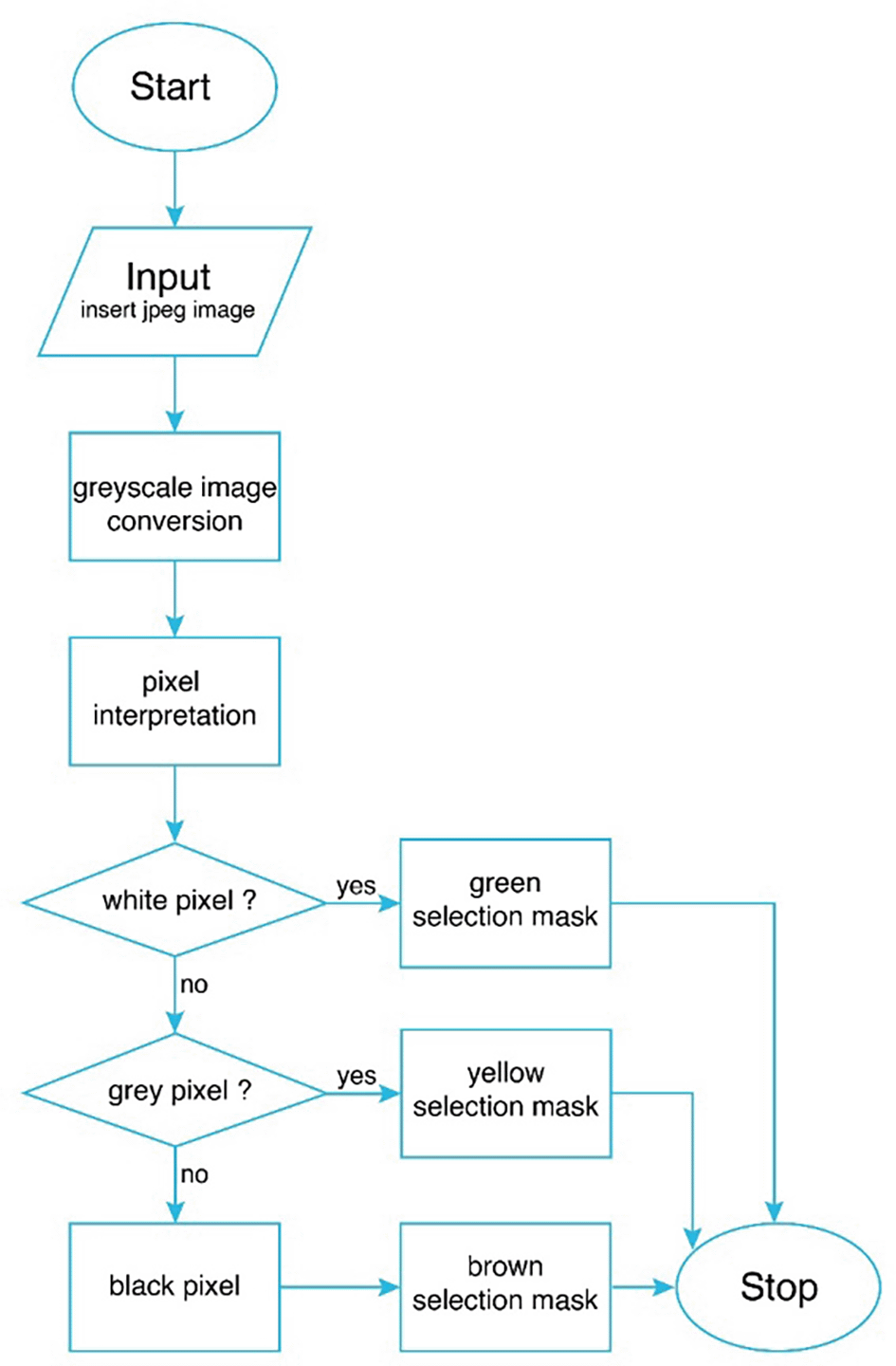
To automate the ROI selection process, a script in the Python language was developed that can reprocess a.jpg image of a CT and automatically identify varied selection masks.
Figure 3 shows the flowchart of the script. By introducing an image packet from CT scan, the script converts and reads each image in grayscale, that is, in pixels in a colour scale of 0 to 256 shades of grey ranging from black to white, respectively. Image manipulation from code is possible through dedicated libraries such as OpenCV.36 The script analyses every single pixel in the images starting from the first pixel in the upper left corner and continuing to the right. Once the first row of an image is finished, it starts again by going down one row of pixels and so on. The script assigns each pixel one of 256 values depending on the gradation in the grayscale. Finally, it converts with a proportion the numbers from 0 to 256 into values from 0 to 100 by sending out a text with all the values. Defined three groups, values are assigned for each selection mask:
• from 0 to 76 in the green mask (white pixel);
• from 77 to 178 in the yellow selection mask (grey pixel);
• from 179 to 255 in the brown selection mask (black pixel).
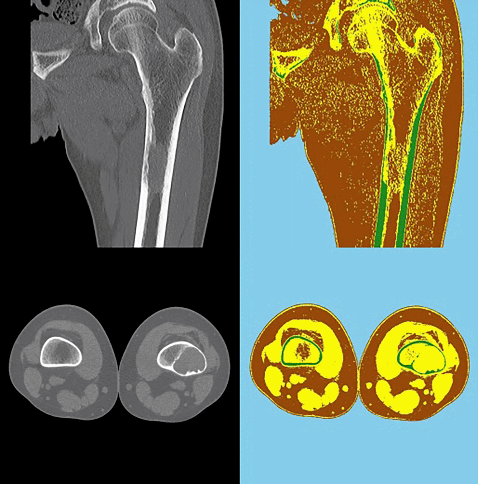
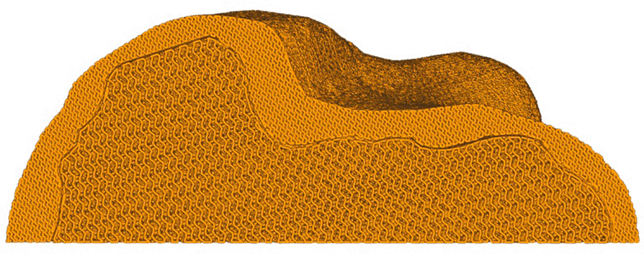
By processing the script, the image is outlined as shown in Figure 4. These three groups identify the three parts that make up the bone. Post-processing was required to exclude areas that are not of interest for selection.
In any case, this automated method needs to be further improved in terms of the pixel selection and categorization process. So, the methodology was carried forward with the manual reconstruction method.
3D printing has been the technology used for making the physical anatomical model. However, a model preparation stage is required for additive technology. First, three meshes in .stl format were exported from 3D Slicer. In the digital environment of Blender v3.3.0,37 a series of simple Boolean operations were performed in order to obtain three distinct final models of compact bone, spongy bone and bone marrow, respectively. In order for the models to maintain their relative positions in space in the slicing environment, they were exported in the .3mf format.
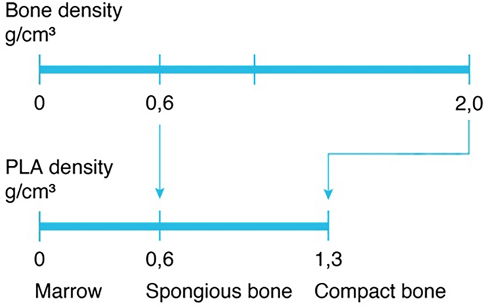
The printer used for this study is a Fused Deposition Modeling (FDM) 3D printer, AnyCubic Predator. FDM technology involves the extrusion of thermoplastic materials using a heated nozzle that melts the material and deposits it, layer by layer, on a printing platform until the part is completed.38 PLA was chosen as the printing material because it is inexpensive, dense, versatile and easy to process.39 To obtain the diversity of the densities of the parts to be printed, a conversion comparing the density of bone with that of PLA filler was performed. Specifically, the density value of compact bone (equal to 2 g/cm3) and that of spongy bone (equal to 0.6 g/cm3)40 were compared with the density of PLA (equal to 1.3 g/cm3) (Figure 5).
Assuming the maximum compact bone density, that is 100% infill, the density of spongy bone will be 46.2%, according to the following proportion:
Ultimaker Cura v5.0.041 is the slicing software used that allows you to manage printing parameters and export a G-code file. Among the various parameters, including layer height, printing temperature, printing speed, are those related to infill of the various layers. An infill density is set as a single, constant value throughout the object. By importing the three patterns in .3mf format to Ultimaker Cura, different densities can be set for each pattern. Gyroid infill was used since it appears to be the one most like the typical trabecular structure of bone. Applying the parameters shown in Table 1, a preview of the compact bone and the spongy bone is visualized (Figure 6). The 3D print for each zone of different density was made without external walls to achieve a more uniform 3D print object with visibly gradual infill.
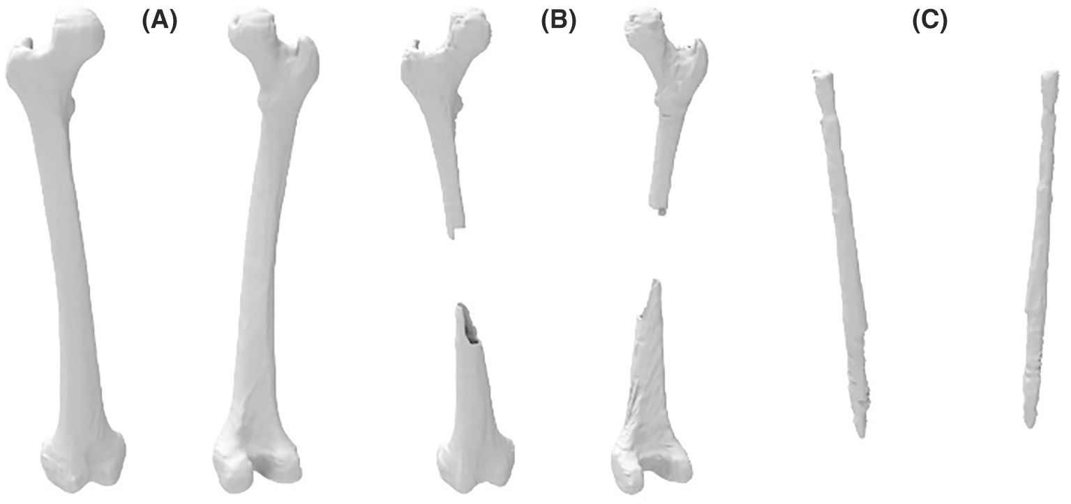
The reconstruction and processing steps resulted in the three distinct three-dimensional parts of the areas of the bone. The areas of the obtained 3D digital models present smooth and compact surfaces in their entirety, maintaining a true-to-life appearance. Figure 7 shows the three obtained digital models of the bone and Figure 8 shows the final 3D printed models.

Nowadays, polymeric materials do not reach a density equal to that of compact bone, but it is possible to study which among them can achieve the same mechanical and structural properties with the right printing settings. An interesting example is the use of PEEK or similar materials that can easily reproduce the structure of bone, but their use involves a complicated and expensive process.42–44 An interesting possibility is offered by bone tissue printing for the fabrication of fractured parts that should be removed or low in bone density through 3D Bioprinting.45,46 Another improvement concerns the use of an optimized infill for printing bone that is more accurate than the one used and more like a trabecular structure so that it is visually increasingly accurate and close to reality. For the future, it also desired to achieve the possibility of making a print that has not only three different zones but that the variation in density is continuous following the bone matrix and not differentiated by n zones.47 Another future development concerns the improvement of automation for the reconstruction phase to have an increasingly accurate as well as fast method.48
By focusing on patient specifics, 3D printing technology in orthopaedics can improve the understanding of clinical cases by creating patient-specific anatomical models. One interesting method to improve the creation of bone models and make them easier to understand for both academic research and surgical planning is variable density 3D printing. 3D printing porous structure allows to obtain an anatomical model that better represents its realistic counterpart in terms of shape, surface area, and weight. By three-dimensional reconstruction of a CT image of a femoral bone, interpreting zones of different densities, it is possible to obtain three zones corresponding to compact bone, spongy bone, and bone marrow, respectively. This process can be done in manual or semi-automatic mode, with a strong potential for automation. This involves writing an articulated computer language code to select and distinguish the different pixels constituting a CT image and categorize them automatically according to their intensity. Finally, through slicing software it is possible to customize the printing parameters, applying different infill densities per part, resulting in a 3D printed object with porous structure. The most interesting application of this process is precisely in making more lifelike bone structures, ensuring eventual comparable replacement. This study is a first approach to obtain a first 3D print model with porous structure. As a future development, it is important to achieve mechanical characteristics comparable to those of real bone tissue. It is first necessary to identify a material with suitable characteristics both structurally and biocompatibility for this application. Finally, it is good to optimize the internal infill geometry that ensures on the one hand the maintenance of mechanical characteristics and on the other hand a good surgical integration.
| Views | Downloads | |
|---|---|---|
| F1000Research | - | - |
|
PubMed Central
Data from PMC are received and updated monthly.
|
- | - |
Provide sufficient details of any financial or non-financial competing interests to enable users to assess whether your comments might lead a reasonable person to question your impartiality. Consider the following examples, but note that this is not an exhaustive list:
Sign up for content alerts and receive a weekly or monthly email with all newly published articles
Already registered? Sign in
The email address should be the one you originally registered with F1000.
You registered with F1000 via Google, so we cannot reset your password.
To sign in, please click here.
If you still need help with your Google account password, please click here.
You registered with F1000 via Facebook, so we cannot reset your password.
To sign in, please click here.
If you still need help with your Facebook account password, please click here.
If your email address is registered with us, we will email you instructions to reset your password.
If you think you should have received this email but it has not arrived, please check your spam filters and/or contact for further assistance.
Comments on this article Comments (0)