Keywords
newborn, tracheal rupture, subcutaneous emphysema, shoulder dystocia, pneumothorax, pneumomediastinum
newborn, tracheal rupture, subcutaneous emphysema, shoulder dystocia, pneumothorax, pneumomediastinum
Tracheal rupture in the newborn represents a rare and severe complication, with a high mortality rate and a difficult diagnosis.1 This complication can be caused by emergency newborn maneuvers such as obstetrical delivery maneuvers in dystocia, endotracheal intubation (ETI), and mechanical ventilation.2,3 For a complete diagnosis, it is necessary to consider a possible preexisting congenital anomaly: tracheoesophageal fistula, tracheal stenosis, or atresia.4
The windpipe wall tear causes air leakage and tissue dissection with subsequent subcutaneous emphysema, pneumomediastinum, pneumothorax, respiratory distress, cyanosis, and dyspnea.5 These can be diagnosed clinically and with a chest radiograph, especially a lateral one, which can reveal a mispositioned endotracheal tube or an accompanying clavicle fracture. A differential diagnosis with the barotrauma following mechanical ventilation should be made.2 Computed tomography (CT) is also useful, but the most accurate diagnosis is made by bronchoscopy (fibroscopy), which can assess the location and aspect of the injury.6
The treatment is either conservatory (tracheal intubation followed by expectative healing) or surgical (suture, anastomosis, or patching of the discontinuity), both accompanied by intensive care.3,7 Whatever the treatment, the mortality is high due to the unexpected complication, the preexisting impaired clinical status, which requires intensive care, and the lack of an immediate, definitive cure. Moreover, the assessment of the severity of the diagnosis and the recommendation for fibroscopy delays the treatment, contributing to an increased risk.5,8
In addition to the few cases found in the literature, that come mostly from clinical specialties (otorhinolaryngology, pediatrics, neonatology), the current case is presented mostly from an autopsy perspective, in which we compared the postmortem with the antemortem diagnosis.9
Here is a full-term macrosomic (birthweight 4780 g) female newborn, vaginally delivered in the morning with shoulder dystocia after a poorly monitored pregnancy, who required manual release maneuvers through rotation and traction. Besides dystocia other risk factors were identified, such as urinary tract infection during pregnancy, green amniotic fluid during birth and single loop nuchal cord.
At birth, the APGAR score was 5 and 6 at 1 and respectively 5 minutes, and postnatally, generalized cyanosis, apnea, anterior cervical subcutaneous emphysema, and limited mobility of the right upper limb (upper right limb paralysis) were noted. In the delivery room, she received oxygen therapy on a balloon mask followed by a cephalic tent in the neonatology department and needle drainage of emphysema at the right subclavicular level. 4 hours post-birth, noting that the subcutaneous emphysema, instead of remitting, extended to the head and upper chest, the transfer to a university hospital with a neonatal intensive care unit was requested for suspected pneumothorax.
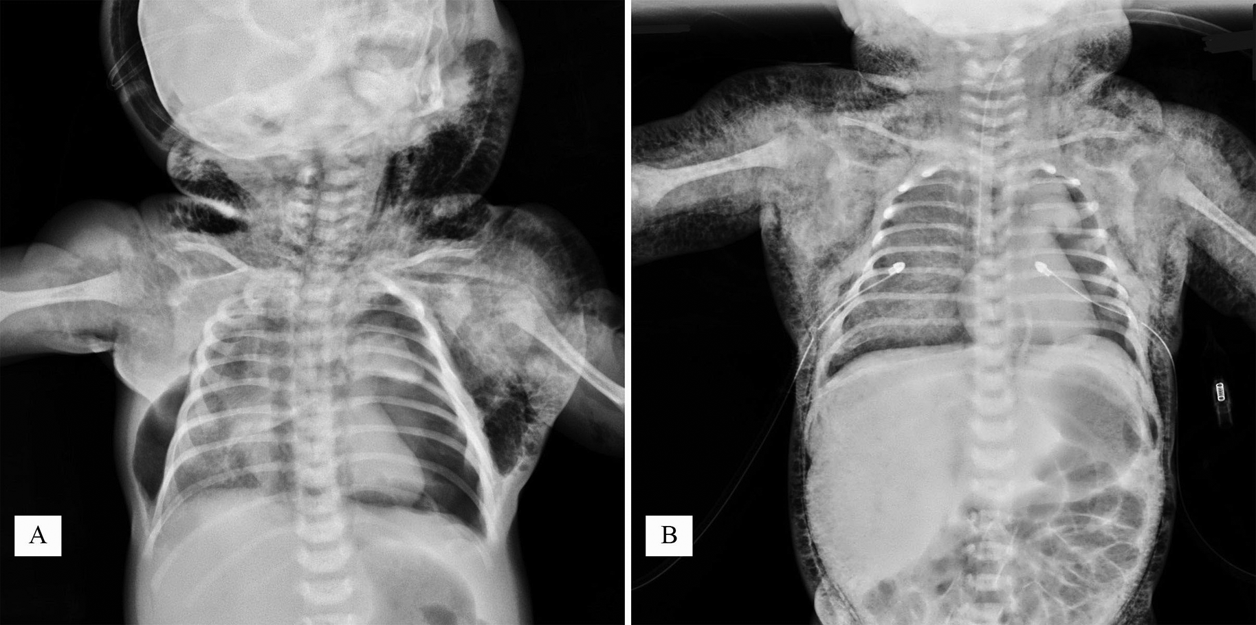
The transfer took less than 1 hour, during which the patient was cardiorespiratory stable while the subcutaneous emphysema persisted. The patient arrived at the next health facility 7 hours after birth and was admitted to the neonatal intensive care unit, where a chest radiograph confirmed emphysema and revealed bilateral pneumothorax with left lung collapse (Figure 1A and B), requiring a bilateral thoracentesis with subsequent oxygen saturation and ventricular allure amelioration.

Along with this, an ETI was performed, and despite this, emphysema was accentuated, and the patient’s condition deteriorated with bradycardia and required resuscitation.
Then, at 8 hours after birth, a fibroscopy through endotracheal tube (ETT) detected a rupture of the tracheal wall, followed by an emergency tracheostomy that confirmed the trachea rupture. During tracheostomy, the patient's evolution was aggravated by cardiac arrest that required resuscitation and by important blood loss (hemoglobin decreased from 21.4 to 6.8 g/dL) that required transfusion.
Under these circumstances, the pneumothorax decreased, but despite this, the general condition remains serious, and the newborn died about 12 hours after birth.
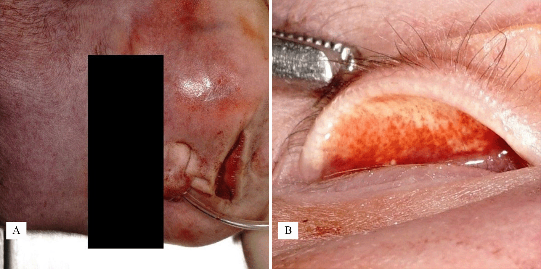
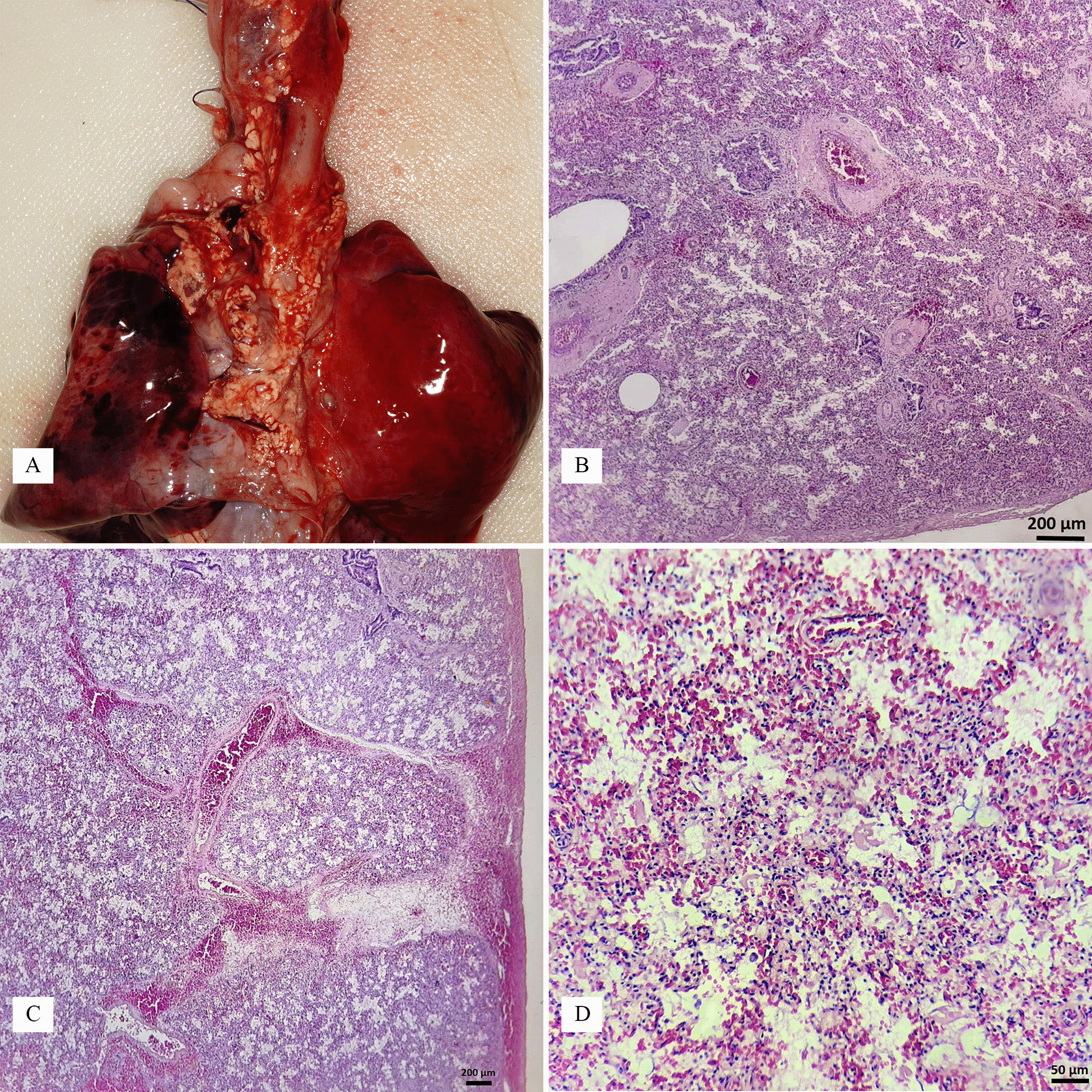
Given the fact that the death occurred in short time after surgical procedure, a medico–legal autopsy was required, that showed as the main features: the emergency tracheostomy with the tracheostomy tube (Figure 3B) and the pleural drain tubes one each side; an intense head cyanosis (clinically misdiagnosed as an ecchymosis due to obstetrical trauma) with subconjunctiva petechiae (Figure 2A and B); subcutaneous emphysema of the thorax, upper limbs, neck, head including the head (Figure 3A); bilateral pneumothorax with pulmonary atelectasis and hemorrhage predominantly on the left lung (Figure 4A, B, C and D). During the dissection of the esophagus, we identified an elliptical rupture (2/0.5) cm in the posterior wall of the trachea, located less than 1 cm above the carina (Figure 5A, B, C and D – in Figure 5A and B the tracheostomy tube can be seen through the rupture); no communication between the esophagus and trachea, congenital or acquired, was found.


Two clinico–pathological patterns of the trachea rupture of the newborn can be summarized as follows6:
For premature/low birth weights infants, it is described the mechanism of tracheal perforation secondary to the ETI; the ruptures can be located anteriorly (beneath the glottis) or posteriorly (above the carina), and even bronchi can be involved; apparently, the cartilaginous rings does not offer protection, as both anterior and posterior wall can rupture.4 The acute clinical sings (subcutaneous emphysema, pneumothorax) occur immediately after the ETI, without being preexistent. This mechanism should be firstly considered if the injury is detected after a cesarean, or after more than one week after birth.1
For high weight births associated with shoulder dystocia, injuries of the brachial plexus, the trachea is mostly injured by a traction mechanism during delivery, and the localization can be anterior subglottic (more frequent) but also inferior, even circumferential; macrosomic neonates require more traction and excessive hyperextension of the neck; maybe there is a weak area; the carina ais fixed—a common injury is within 2.5 cm of the carina; another area is the cricothyroid membrane.5 An endotracheal intubation enlargement of the rupture after a previous undiagnosed rupture cannot be excluded.
In our case, there was a macrosomic newborn with shoulder distocia and possible right brachial plexus paralysis, which was in poor condition immediately after birth and presented extensive subcutaneous emphysema long before any ETI attempt; the most probable mechanism is represented by the releasing traction maneuvers which teared the posterior lower wall of the trachea during delivery, possible enlarged by later intubation attempts.
Noteworthy that between the onset of subcutaneous emphysema (after birth) and the fibroscopy it passed almost 8 hours, maybe due to an apparent preservation of respiratory function. It is also to note the subcutaneous emphysema that was worsened by mechanical ventilation and the difficult emergency tracheostomy was complicated by an extensive local hemorrhage that aggravated the circulation.
The subcutaneous emphysema in newborns can also appear even outside of an identifiable airway tear, due to pneumothorax or pneumomediastinum, as a complication of mechanical ventilation or resuscitation. The pneumothorax itself can be life-threatening, especially if it occurs in an immature lung.10
A few clinical considerations need to be taken into account: an intense blood stasis (cyanosis) can mistake as an ecchymosis, the clinical assessment of severity and the cause of the subcutaneous emphysema is challenging, the emergency surgical and obstetrical procedures involve inherent risks.
Regarding the postmortem examination of the newborn, it is essential to establish the correct sequence of events, in order not to overrate the intubation incidents as the main cause, to differentiate the acute tracheal tear from chronic pressure perforation,4 to carefully dissect the postoperative tissues, and to check for other congenital anomalies: tracheal stenosis, trachea-esophageal fistula, immature lung.
In the presented case, the sequence of events suggests that the traumatic delivery rather than the intubation is more likely the cause of the tracheal rupture. It is possible that the intubation attempts with external ventilation aggravated the rupture and the abnormal passage of air into tissues with subsequent pneumomediastinum, pneumothorax, and subcutaneous emphysema.
A full postmortem investigation was necessary to establish a complete diagnosis, and correctly differentiate the possible congenital anomalies from obstetrical and neonatal intensive care maneuvers.
The study was conducted according to the guidelines of the Belmont Report, Declaration of Helsinki and approved by the Ethics Committee of the National Institute of Legal Medicine Bucharest no. 4/29.12.2022. All the data was obtained from the mandatory forensic autopsy for which legal consent is not necessary. All the identifiable data regarding the patient was concealed and therefore the consent was waived by the Ethics Committee.
| Views | Downloads | |
|---|---|---|
| F1000Research | - | - |
|
PubMed Central
Data from PMC are received and updated monthly.
|
- | - |
Is the background of the case’s history and progression described in sufficient detail?
Partly
Are enough details provided of any physical examination and diagnostic tests, treatment given and outcomes?
Partly
Is sufficient discussion included of the importance of the findings and their relevance to future understanding of disease processes, diagnosis or treatment?
Partly
Is the case presented with sufficient detail to be useful for other practitioners?
Partly
Competing Interests: No competing interests were disclosed.
Reviewer Expertise: Paediatric Intensive Care
Is the background of the case’s history and progression described in sufficient detail?
Yes
Are enough details provided of any physical examination and diagnostic tests, treatment given and outcomes?
Partly
Is sufficient discussion included of the importance of the findings and their relevance to future understanding of disease processes, diagnosis or treatment?
Partly
Is the case presented with sufficient detail to be useful for other practitioners?
Yes
References
1. Romic M, Becejac T, Grbavac D, Romic R, et al.: Conservative treatment of postintubation tracheal laceration with pneumomediastinum, bilateral pneumothorax, and massive subcutaneous emphysema.Lung India. 2021; 38 (1): 77-79 PubMed Abstract | Publisher Full TextCompeting Interests: No competing interests were disclosed.
Reviewer Expertise: Thoracal surgery, pulmology
Alongside their report, reviewers assign a status to the article:
| Invited Reviewers | ||
|---|---|---|
| 1 | 2 | |
|
Version 1 14 Mar 23 |
read | read |
Provide sufficient details of any financial or non-financial competing interests to enable users to assess whether your comments might lead a reasonable person to question your impartiality. Consider the following examples, but note that this is not an exhaustive list:
Sign up for content alerts and receive a weekly or monthly email with all newly published articles
Already registered? Sign in
The email address should be the one you originally registered with F1000.
You registered with F1000 via Google, so we cannot reset your password.
To sign in, please click here.
If you still need help with your Google account password, please click here.
You registered with F1000 via Facebook, so we cannot reset your password.
To sign in, please click here.
If you still need help with your Facebook account password, please click here.
If your email address is registered with us, we will email you instructions to reset your password.
If you think you should have received this email but it has not arrived, please check your spam filters and/or contact for further assistance.
Comments on this article Comments (0)