Keywords
Electrocardiogram, Pulmonary embolism, Prognosis, Mortality
This article is included in the Global Public Health gateway.
Electrocardiogram, Pulmonary embolism, Prognosis, Mortality
Pulmonary embolism (PE) is a common and potentially life-threatening medical condition. It is estimated that thrombo-embolism affects over 1,000,000 people in the United States each year and results in over 25,000 deaths annually.1
Risk stratification is important in determining the likelihood of poor outcomes for patients with acute PE. It dictates the attitude towards patients with PE from early discharge to urgent care in intensive care units. Thus, recent risk stratification algorithms rely on various clinical, laboratory, and imaging factors to estimate the risk of complications and guide treatment decisions.2 A major area of uncertainty in the risk stratification of acute PE is the potential role of electrocardiography (ECG) abnormalities in predicting the risk of complications as several ECG abnormalities have been identified in patients with acute PE, including right ventricular strain and S1Q3T3 type pattern.3–5 These changes are primarily related to right ventricular (RV) overload and reflect right ventricular dysfunction (RVD), injury and enlargement in patients with acute PE.6–9 Furthermore, ECG abnormalities associated with acute PE are more likely to be present in patients with a confirmed diagnosis of PE.2,10 Despite their potential prognostic value, none of these ECG changes are included in the current guidelines for PE risk stratification due to the lack of specificity.2,11 Encouraging evidence has emerged showing the relationship between the right bundle branch block (RBBB) and SIQIII patterns in acute PE events with poor outcomes.10,12,13 In addition, more research is needed to clarify the potential role of ECG abnormalities in the risk stratification of acute PE. By addressing these gaps in the literature, we can improve our ability to manage patients with this common and potentially life-threatening condition.12,14
This study was a retrospective observational cohort study that did not involve human testing and did not challenge human rights. The ethical approval was obtained retrospectively as we did not initially believe that this study needed ethical approval. This issue has been a subject of debate inside our team and that belief has since been revised and updated. Data collection for our study began in January 2011 in two Tunisian tertiary care cardiology departments: Cardiology A Department, Fattouma Bourguiba University Hospital affiliated to the University of Monastir and the Cardiology Department of Kairouan affiliated to the University of Sousse. Our study was retrospectively approved by the Research Ethics Committee of the Faculty of Medicine of Monastir (an independent organization under the aegis of the Tunisian Ministry of Public Health) under the number IORG 0009738 N°114/OMB 0990-0279. We obtained this approval on the 16th of March 2023.
The initial approval to start the study was obtained by consensus from both research teams under the supervision of both heads of the aforementioned departments at the University of Monastir and University of Sousse. The study was performed in compliance with the Declaration of Helsinki. We ensured that participants’ privacy and confidentiality were maintained, and the study results were reported in a way that protected the participants’ identities.
Oral informed consent was obtained from patients, whenever possible. This verbal consent was obtained during initial hospitalization. Each patient was informed that their anonymized data could serve for future research purposes. We did not conceptualize the content of this article at the time of information gathering and we could not obtain this consent in patients initially presenting with critical conditions and fatal outcomes. Verbal consent was approved retrospectively when we received ethical approval and deemed adequate by the ethics committee.
We conducted a retrospective cohort study. We included all patients with confirmed acute PE hospitalized from January 10th, 2008, to December 31st, 2019, in two Tunisian tertiary care cardiology departments: Cardiology A Department, Fattouma Bourguiba University Hospital affiliated to the University of Monastir and the Cardiology Department of Kairouan affiliated to the University of Sousse. The data were gathered by reviewing patients’ hospital records from each hospitalization, supplemented with comprehensive in-hospital assessments and follow-up interviews conducted either in person or via telephone.
PE was diagnosed primarily by either computed tomography (CT) or scintigraphic ventilation-perfusion (V/Q) scans. The CT pulmonary angiogram (CTPA) demonstrated the existence and the extension of a filling defect in the pulmonary artery system. In the cases where V/Q scintigraphic scanning was employed, the results were interpreted by a nuclear medicine specialist. A high-probability V/Q scan was considered sufficient for the diagnosis of acute PE. This examination showed the presence of at least two segmental perfusion defects without ventilatory or radiological abnormalities in the same territories.2,15 PE diagnosis was also assessed by a positive venous doppler ultrasound consistent with deep venous thrombosis (DVT) in patients with high clinical suspicion of PE and positive D-dimer values.2 All CT, scintigraphy and venous doppler ultrasound scans were analyzed and interpreted by experienced specialists.
Complete RBBB was identified according to the Minnesota Code criteria (7-2-1) as a QRS duration must be ≥120 ms in addition to R′ wave>R wave in lead V1 and/or V2 in most beats of leads I, II, III, aVL, aVF; or a QRS complex being predominantly upright with an R-peak duration ≥60 ms in lead V1 and/or V2 or; an S wave duration > to R wave duration in all beats in lead I and/or II.16,17
Incomplete RBBB was identified according to the Minnesota Code criteria (7-1) by a QRS duration in each of leads I, II, III, aVL, and aVF being <120 ms in addition to an R′>R wave in lead V1 and/or V2.16,17
SIQIII-type ECG patterns were defined as a qualitative presence of S wave in lead I and Q wave in lead III.
Right ventricular dysfunction (RVD) was defined as a decreased systolic function of the RV (TAPSE <17 mm) and/or the presence of a paradoxical interventricular septal movement and/or a systolic pulmonary artery pressure ≥40 mmHg.
Major Adverse Cardiovascular Events (MACE) were defined as the presence of at least one of the following: death during hospitalization, cardiogenic shock in initial presentation or the presence of a thrombus in the right heart.
Renal failure was defined as an eGFR < 60 ml/min per 1.73 m2.18
Positive troponin levels were defined as any value above the 99th percentile of the upper reference limit.19
Patients with PE with a systolic blood pressure (SBP) <90 mmHg at admission were classified as high-risk of mortality patients.2
The sex of each participant was defined based on self-report and assigned following external examination of body.
Patients were eligible if: i) The diagnosis of PE was confirmed using one of the three diagnostic tools described above; ii) they were managed in Cardiology A Department, Fattouma Bourguiba University Hospital affiliated to the University of Monastir and the Cardiology Department of Kairouan affiliated to the University of Sousse; and iii) aged 18 years or above.
Exclusion criteria included: i) patients aged under 18 years and ii) if the diagnosis of acute PE was unclear.
A standard 12-lead ECG was assessed in the Emergency Department immediately upon initial contact. This first ECG taken at admission was selected for analysis. Both the ECG and echocardiography exams were performed and interpreted by experienced examiners. The retrospective analysis of ECG parameters focused on the existence of RBBB, either complete or incomplete, and SIQIII-type patterns.
Data analysis was performed using IBM SPSS Statistics (RRID:SCR_016479) version 26.0. The mean ± standard deviation (SD) was used to describe the normally distributed data. The median and interquartile range (IQR) was used to describe the skewed distribution data.
Frequencies and percentages were used to present categorical variables. The comparison between frequencies was performed using the Chi-squared test (χ2 test) or Fisher’s exact test. Means were compared using the Student’s t test for independent samples. A Kaplan–Meier survival analysis was performed to assess the association between the RBBB and/or SIQIII and overall mortality using the log-rank test. In order to identify the independent factors associated with the occurrence of MACEs, univariate and multivariate analyses were performed. First, covariates with a p-value less than or equal to 0.20 were retained in the multivariable model. Then, a binary logistic regression was performed for assessing the independent risk factors for MACEs.
Associations were reported as adjusted Odds Ratios (aOR) with 95% Confidence Interval (95% CI). A p-value≤0.05 was considered as statistically significant.
A total of 255 patients were included in the analysis. All of the medical reports for these patients included a usable ECG from the time of their acute PE episode. Patients were divided into two groups: i) Group I (n=122, 47.8%) included patients with PE without RBBB nor SIQIII patterns; and ii) group II (n=133, 52.2%) included patients with RBBB and/or SIQIII type patterns. Patients in both groups did not differ by age nor sex. The proportion of diabetes mellitus (DM), hypertension (HTN) and dyslipidemia were homogeneous between the two groups (Table 1).25
HTN, hypertension; VTE, venous thromboembolism; SBP, systolic blood pressure; DBP, diastolic blood pressure; TTE, transthoracic echocardiogram; IVS, intact ventricular septum; SPAP, systolic pulmonary artery pressure; RVD, right ventricular dysfunction; TAPSE, tricuspid annular plane systolic excursion; ECG, electrocardiography; RBBB, right bundle branch block; MACE, major cardiovascular event.
Group II patients had an active cancer (17.3% vs. 12.3%) more frequently but this difference did not reach statistical significance (p=0.29). Patients presenting with SIQIII pattern and/or RBBB were also significantly more likely to have history of systolic or diastolic heart failure (13.5% vs. 4.9%, p=0.03).
We analyzed the association between the presence of RBBB and/or SIQIII type pattern with PE severity and patient outcome.
Patients in group II presented more frequently with acute right heart symptoms (45.1 vs. 18%, p<0.001), more often showed cardiogenic shock (31.6 vs. 4.1%, p<0.001) and had lower systolic blood pressure (SBP) (110.3 vs. 122.8 mmHg, p<0.001) and diastolic blood pressure (DBP) (67.5 vs. 73.9 mmHg, p<0.001) at admission. The difference was statistically significant for all these parameters. Syncope occurred more frequently in Group II patients but the difference did not reach statistical significance (14.3 vs. 9%, p=0.24) (Table 1).
Patients in group I had a significantly lower likelihood of positive Cardiac troponin I (cTnI) or high-sensitivity cardiac troponin (hs-cTn) levels (26.9 vs. 51.1%, p<0.001).
Echocardiographic parameters indicating a right heart injury also occurred more frequently in group II patients: SPAP >40 mmHg (63.2 vs. 28.3%, p<0.001), paradoxical interventricular septum (51.6 vs. 26.5%, p<0.001), tricuspid annular plane systolic excursion (TAPSE) <17 mm (55.6 vs. 4.2%, p<0.001), the presence of a thrombus in the right heart (25 vs. 4.2%, p<0.001). Thus, RVD was significantly more frequent in Group II patients by univariate analysis (p<0.001).
A total of 101 patients (39.6%) had RBBB, 66 (26%) had SIQIII type patterns, and 34 patients (13.4%) had both SIQIII pattern and RBBB.
Negative T waves in leads V1-V3 were significantly more frequently associated with Group II patients in univariate analysis (p=0.01) (Table 1).
There were slightly more patients with atrial fibrillation (AF) in Group I patients, but this was not statistically significance (10.6 vs. 7.6%, p=0.4).
Groupe II patients were significantly associated with in-hospital mortality (22.6 vs. 6.1%, p=0.002) and MACEs during hospitalization by univariate analysis (43.3 vs. 13.7%, p<0.001) (Table 1).
Multivariate logistic regression analysis demonstrated five independent predictors of MACE: SIQIII and/or RBBB (OR=2.4; 95% CI: 1.04, 5.9; p=0.039), renal failure (OR=2.5; 95% CI: 1.08, 5.8; p=0.031), positive troponin levels (OR=5.4; 95% CI: 2.34, 12.6; p<0.001), RVD (OR=7.9; 95% CI: 2.16, 28.8; p=0.002) and right heart failure symptoms during initial presentation (OR=2.5; 95% CI: 1.1, 5.8; p=0.025) (Table 2). Using these models, SIQIII pattern (OR=4.8; 95% CI: 1.9, 12.2; p=0.001), RVD (OR=15.2; 95% CI: 1.9, 12.4; p=0.01) and cardiogenic shock at admission (OR=2.7; 95% CI: 1, 6.9; p=0.034) were independently associated with in-hospital mortality, on the other hand, no association was found between in-hospital mortality and group II patients (Table 3).
MACE, major cardiovascular event; RBBB, right bundle branch block; RVD, right ventricular dysfunction; OR, odds ratio; CI, confidence interval.
| Variables | Multivariate | ||
|---|---|---|---|
| OR | 95% CI | p-value | |
| SIQIII and/or RBBB | 2.4 | 1.04, 5.9 | 0.039 |
| Renal failure | 2.5 | 1.08, 5.8 | 0.031 |
| Positive troponin | 5.4 | 2.34, 12.6 | <0.001 |
| RVD | 7.9 | 2.16, 28.8 | 0.002 |
| Right heart failure | 2.5 | 1.1, 5.8 | 0.025 |
RVD, right ventricular dysfunction; OR, odds ratio; CI, confidence interval.
| Variables | Multivariate | ||
|---|---|---|---|
| OR | 95% CI | p-value | |
| SIQIII | 4.8 | 1.9, 12.2 | 0.001 |
| Renal failure | 3.8 | 1.5, 9.6 | 0.003 |
| RVD | 15.2 | 1.9, 12.4 | 0.01 |
| Cardiogenic shock | 2.7 | 1, 6.9 | 0.034 |
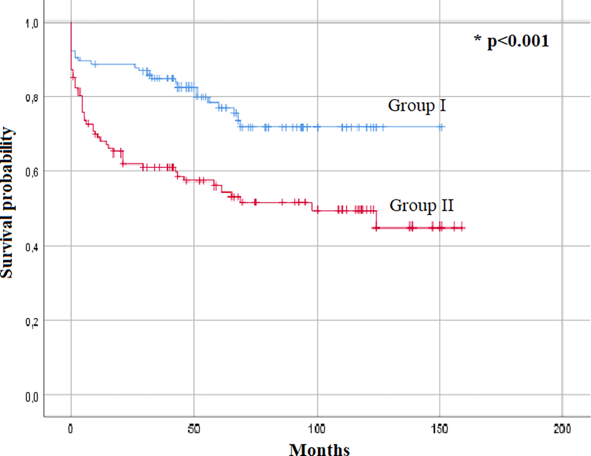
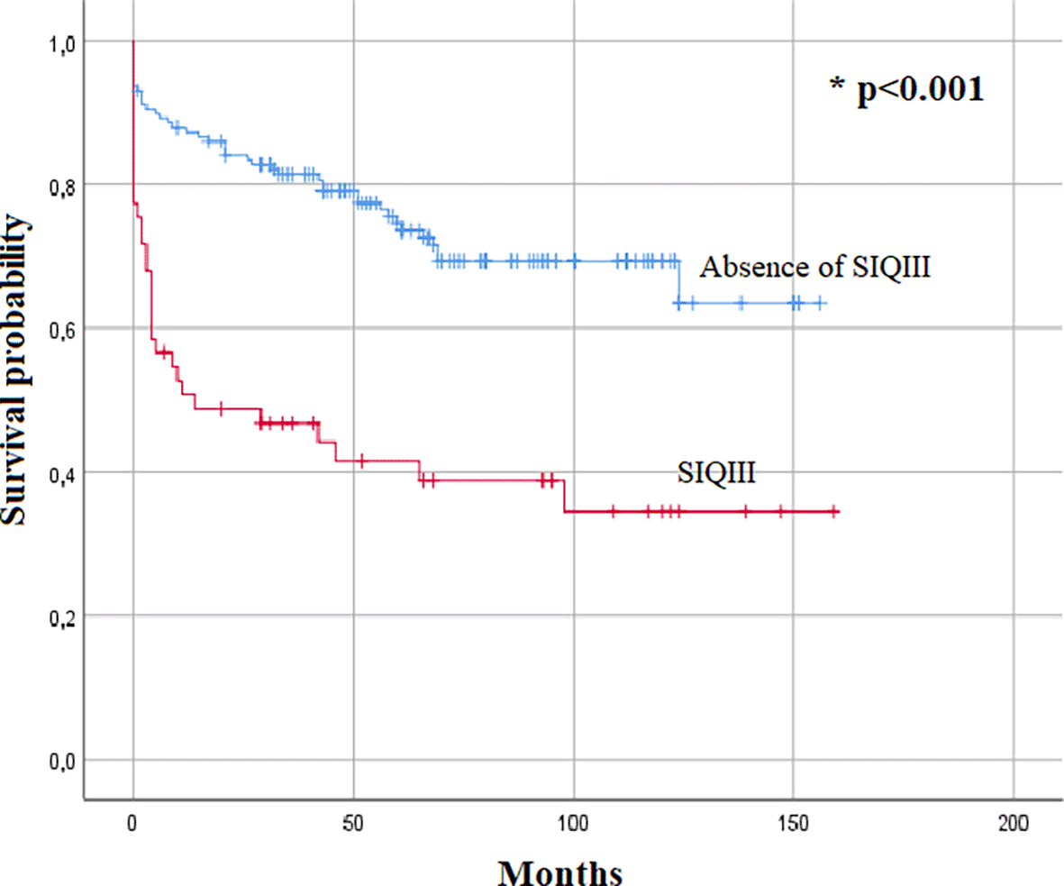
A medium or long term follow-up was done for 83% of the cohort. The Kaplan–Meier survival analysis also demonstrated SIQIII patterns as a predictor of overall mortality (p<0.001) (Figure 1). The Kaplan–Meier analysis also identified the presence of SIQIII and/or RBBB patterns as a predictive factor for overall mortality (p<0.001) (Figure 2).
Early and risk-oriented diagnosis and management of patients with acute PE are key for a better prognosis.2 In our study, RBBB and SIQIII-type patterns at admission were found to be associated with worsening clinical, echographic and biological profiles and with a poor clinical outcome.
Meta-analyses by Shopp et al., reported different ECG patterns, such as inverted T wave in leads V1-V4, a QR pattern in lead V1, SIQIII, complete or incomplete RBBB and ST elevation in lead aVR as being associated with increasing severity and poor outcomes.4
ECG scoring by Daniel et al., was notable for providing an applicable and reliable prognostic tool.5 However, recent studies indicate that there are several ECG abnormalities that can provide valuable prognostic information, but are not currently included in these scores.9 These ECG findings were also associated with early clinical deterioration.20 Despite these findings, ECG abnormalities were not included nor recommended in the latest guidelines for risk stratification of acute PE.2 This is probably due to the lack of specificity as these changes can be encountered in acute and chronic cor pulmonale.21
The SIQIII pattern was found to be a strong independent predictor of in-hospital mortality, contrary to RBBB. Acute onset of SIQIII patterns mirror a longitudinal dextrorotation of the RV and appears to be more associated with poor hemodynamic outcomes than RBBB. RBBB alone was not found to be independently associated with in-hospital mortality The findings concerning RBBB were interpreted as follows: this characteristic can be observed in both acute pulmonary embolism (PE) and different conditions affecting the right ventricle (RV). Consequently, distinguishing between a new onset RBBB and a preexisting chronic RBBB is challenging. Furthermore, this challenge may be attributed to the limited sample size in various studies exploring the correlation between ECG abnormalities and risk assessment in acute PE.
New onset SIQIII and/or RBBB is likely to increase right heart failure, cardiogenic shock and intra-hospital mortality. These ECG changes seem to profoundly impact the overall survival as patients with SIQIII and/or RBBB patterns, and especially those with SIQIII, have significantly lower survival rates with curves diverging in less than a year. Parallel to the Pulmonary Embolism Severity Index (PESI) and to the simplified Pulmonary Embolism Severity Index (SPESI) scores, these findings suggest the importance of the RBBB and SIQIII patterns as important criteria in risk stratification of PE.12 The recent onset of these patterns may improve its specificity especially if other causes of acute cor pulmonale are ruled out.
Given the predictive value of these parameters, it would be beneficial to integrate them into new and more accurate risk stratification scores for future guidelines. This would probably outperform guideline-backed risk scores.22
Furthermore, our study suggests that ECG alone is a useful tool in determining the outcome of PE, particularly in limited resources environments where advanced diagnostic tools are not available.23,24
Risk stratification is key for the management of patients with acute PE. New-onset RBBB and/or SIQIII and especially SIQIII patterns are likely to worsen patient outcomes. Our study suggests important and independent prognostic values of RBBB and SIQIII patterns and their usefulness in determining the outcome of acute PE patients.
| Views | Downloads | |
|---|---|---|
| F1000Research | - | - |
|
PubMed Central
Data from PMC are received and updated monthly.
|
- | - |
Provide sufficient details of any financial or non-financial competing interests to enable users to assess whether your comments might lead a reasonable person to question your impartiality. Consider the following examples, but note that this is not an exhaustive list:
Sign up for content alerts and receive a weekly or monthly email with all newly published articles
Already registered? Sign in
The email address should be the one you originally registered with F1000.
You registered with F1000 via Google, so we cannot reset your password.
To sign in, please click here.
If you still need help with your Google account password, please click here.
You registered with F1000 via Facebook, so we cannot reset your password.
To sign in, please click here.
If you still need help with your Facebook account password, please click here.
If your email address is registered with us, we will email you instructions to reset your password.
If you think you should have received this email but it has not arrived, please check your spam filters and/or contact for further assistance.
Comments on this article Comments (0)