Keywords
Marginal fit, internal fit, wax pattern, CAD CAM
This article is included in the Datta Meghe Institute of Higher Education and Research collection.
Marginal fit, internal fit, wax pattern, CAD CAM
Lost wax casting technique is used widely in dentistry for production of restoration. The technique involves the production of a wax pattern for required extra coronal restoration over the master cast, which is then invested in a heat-proof investment material. The invested pattern is subjected to a high temperature in a furnace. This eliminates the wax pattern and a mould cavity is created within the investment material. This mould cavity is later filled with liquid molten metal (alloy) which takes up the shape of the required restoration. The accuracy of the wax pattern completely depends on the technician’s skill in manual method. It is a laborious process, and the quality of wax pattern depends on the person performing it. Because the wax is glossy, it can be challenging to spot small flaws during the manual production process when the wax pattern is being removed from the die. Dental crowns can be successful or unsuccessful depending on a variety of factors, including how precisely the crown is fitted and seated.1
Clinical acceptability and the success of dental restorations depend heavily on the dental crown prosthesis’s internal fit and small marginal gaps. A dental crown with flawed margins can cause a series of issues, including food build up, gingival inflammation, dental caries and ultimately failure of the crown and tooth.2
According to some studies, the marginal gap must be less than 120 μm, while 150 μm is clinically accepted.3 Marginal fit is made up of two components: absolute marginal discrepancy4 and marginal discrepancy, where absolute marginal discrepancy is the misfit between the restoration and the prepared tooth in both the vertical and horizontal directions. Absolute marginal discrepancy is the difference between the axial wall of the prepared tooth and the internal surface of the casting at the margin. The axial and occlusal cement thickness make up the internal fit,5,6 and the luting cement’s internal distribution determines the dental restoration’s stability and retention.7
The purpose of this study is to evaluate the fit of metal crowns produced from wax patterns that were formed by two different wax pattern fabrication procedures: conventional wax pattern fabrication and wax pattern milled by computer-aided design and manufacture (CAD CAM) method. This study was motivated by the importance of marginal and internal fits and the need for research into the accuracy of new digital technologies in wax pattern production.7,8
To evaluate the marginal fit and internal fit of metal crowns over a prepared tooth made from wax patterns fabricated by conventional technique and CAD CAM technique.
1. To evaluate marginal and internal fit of metal crowns made from wax pattern prepared by conventional carving technique.
2. To evaluate marginal and internal fit of metal crowns made from wax pattern prepared by milling technique.
3. To evaluate the difference in marginal fit and internal fit between metal crowns made from wax patterns prepared using the above-mentioned methods.
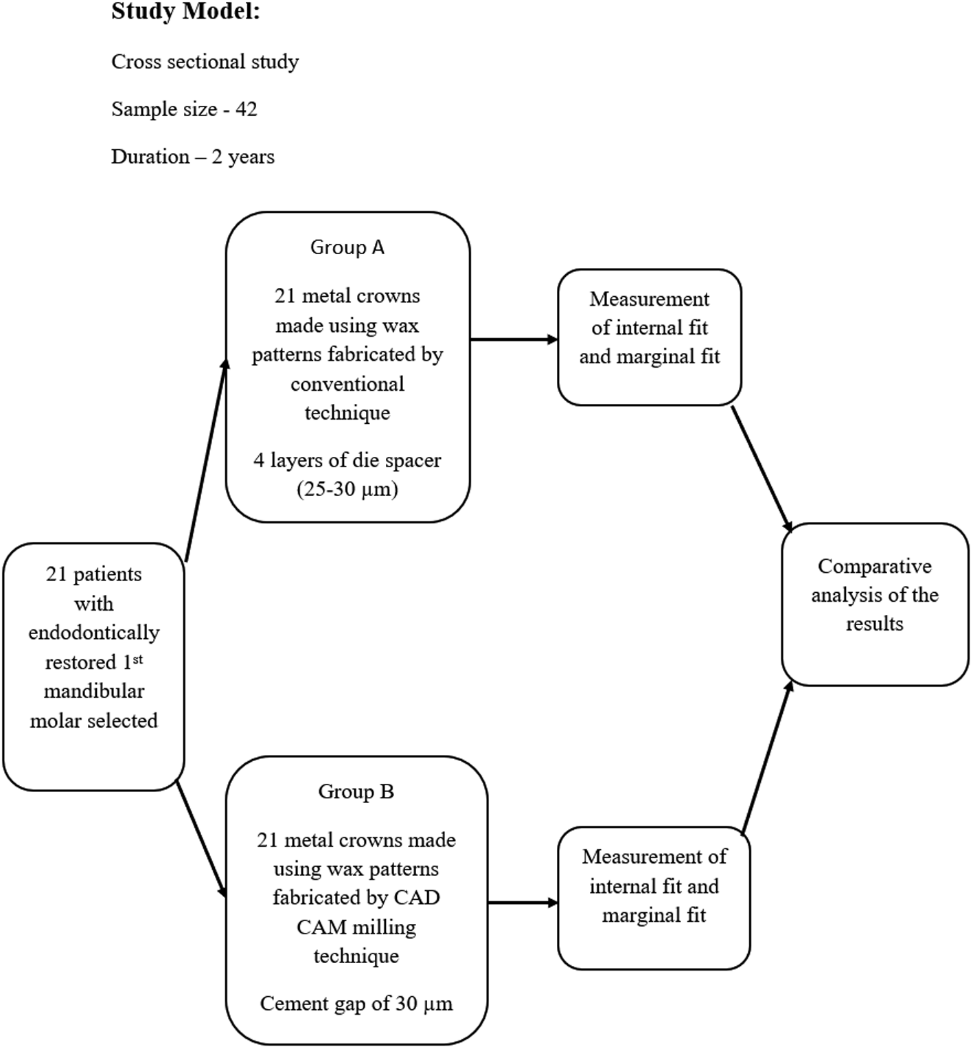
The minimum sample size obtained after calculation from parent article for this research is five per group; so 21 participants will be selected per group, i.e., making 42 the total sample size of this study.
A total of 21 participants with endodontically restored mandibular first molar will be selected.
Two all-metal crowns will be made for each participant, and 42 metal crowns in total will be made. These 42 metal crowns will be divided into two groups based on the method used to create wax patterns: metal crown made using conventional hand carved wax pattern technique (Group I, n=21) and metal crown made using CAD CAM milled wax pattern technique (Group II, n=21).
Preparation of the restored teeth will be performed with chamfer finish line 0.5 mm supragingival following gingival contours with axial walls tapered by 6°-10°, and all the edges will be rounded and smoothened. Gingival retraction will be done with retraction cords. The impressions of the prepared teeth for Group I and Group II will be made using polyvinyl siloxane impression material and will be then poured in Type IV gypsum product to obtain the final master casts. Temporary restoration will be made using a tooth-coloured acrylic (indirect-direct method) and will be cemented using non-eugenol temporary cement at the same appointment.
The study model is shown in Figure 1.
Conventional (hand-formed) production
The 21 master dies are made from the 21 Group I master casts. To prevent layer overlap and make it simpler to distinguish between them, the die spacer will be applied 1 mm from the margin and in two different colours. According to some articles, four layers of “True Fit” die spacer should be used to create a 25–30 mm gap.9
After applying a separator and gently blowing away any excess wax, the dies will be dipped once in molten wax and then covered in medium-hard inlay wax to create the wax patterns.1
Computer-aided design and manufacture (milled)
The 21 master dies will be obtained from 21 master casts for Group II, which will be scanned using Exoscan scanning software version 3. A cement gap of 30 μm will be set to be 1 mm away from the margin. The crown will be designed over this scanned design of die using Exocad. This final designed file in .stl format will be transferred to a milling machine (inLab MCX5) and the wax pattern will be milled out from Ruthinium CAD CAM Wax Disc (98 mm×14 mm).9
The wax patterns will be identically sprued using sprue wax (diameter 2.5 mm). The wax pattern will be sprayed with Debubblizers to lower the surface tension, and the casting rings will be lined with ring liner for investments casting and will be mixed according to the manufacturer’s instructions and with the aid of a vibrator, the investment will be poured into the investment rings and all around the wax pattern, then bench-set for 45 to 60 minutes. The mould will then be placed in an oven that has been preheated to about 480°C, held at that temperature for 20 minutes, and then slowly increased to 700°C and held for 30 minutes. The casting machine will be heated in a melting crucible to a temperature of 1050°C. After that, alloy ingots will be heated in a preheated crucible until they are molten. The casting ring will then be taken out of the furnace and seated onto the induction casting machine where crucible and casting ring will be aligned and the induction casting machine will be started. The casting rings will be removed from the casting apparatus after the casting is finished and set aside to bench-cool to room temperature. On cooling, castings will be retrieved from casting ring. The cast crowns will then be finished and polished using standard burs, stones, rubber wheels, and polishing wheels after being de-vested and separated from their sprue. These procedures of casting will remain the same for each participant’s crown.1
Measurement using silicone replica technique
The inner surface of the crown will be coated with light-body PVS and positioned onto the prepared tooth with finger pressure for 20 s, and then fixed with cotton roll while the patient close their mouth. Excess silicon material will be removed. After 4.30 min, i.e., after setting the silicon layer, the crown will be removed from the prepared tooth. With a regular set putty material of a different colour, the silicon material that adhered to the internal surface of the crown will be stabilised. Both silicon materials will be removed from each crown in one piece after setting. The resulting impression silicon replicas will then be sectioned into six pieces; one mesio-distal cut and two bucco-lingual cuts using a sharp scalpel. The pieces will be examined under the stereomicroscope to measure four points on each piece: occlusal, upper axial, lower axial, and marginal gap to make the measurements under 10× magnification.10
Sample size was calculated using the mean difference formula:
Primary variable (Marginal fit)
Mean ± SD (manual technique) = 160 ± 24
Mean ± SD (milling technique) = 110 ± 11
Difference =160-110 = 50
Standard deviation = 24
As per reference articles.1
Minimum samples size required = 5 per Group.1
The mean and standard error of the mean will be calculated for each group using Microsoft Excel (Office 2021). The statistical package SPSS will be used and student’s unpaired t-test will be employed in the calculation of statistical significance of marginal and internal fit differences between Groups I and II.
Small marginal gaps and good internal fit of dental prosthesis are crucial properties of clinical acceptability and success of dental crown.
1. Some errors can be created in making of die from die stone due to expansion of the gypsum product.
2. The quality of wax pattern made using manual method completely depends on the skill of technicians, and minute cracks on wax patterns cannot be seen with the naked eye because of the glossy surface of wax pattern, which can cause some errors.
In 2012 Paul et al. carried out an in vivo study in which the marginal and internal gap widths of metal ceramic and monolithic zirconia crowns created using conventional and CAD CAM techniques were compared. The study concluded that zirconia crowns made using CAD CAM technology had a better accuracy of fit than metal ceramic crowns made using more traditional methods.1 In 2012, Nawafleh conducted a review and discussed the variables tested and how they affected the results of an investigation in the marginal adaptation of crowns and FPDs. The study came to the conclusion that there is a significant lack of agreement regarding the minimal adaptation of different crown systems because of variations in the testing procedures and experimental design used. The most-used method to reproduce results was direct view technique.2 In 2015 Fathi et al. carried out an in vitro comparison of an internal and marginal fit of crowns made using three different wax production techniques: 3D-printed, milled and conventional, and three different cement gap thicknesses. The conclusion showed that the internal fit of 3D printed wax patterns was far better than the other two fabrication techniques. Regarding internal and marginal fit, wax crown milling is just as precise as traditional hand carving. The most precise internal and marginal fits were produced using the 30 mm offset/die-spacer that the manufacturer recommended.3
| Views | Downloads | |
|---|---|---|
| F1000Research | - | - |
|
PubMed Central
Data from PMC are received and updated monthly.
|
- | - |
Provide sufficient details of any financial or non-financial competing interests to enable users to assess whether your comments might lead a reasonable person to question your impartiality. Consider the following examples, but note that this is not an exhaustive list:
Sign up for content alerts and receive a weekly or monthly email with all newly published articles
Already registered? Sign in
The email address should be the one you originally registered with F1000.
You registered with F1000 via Google, so we cannot reset your password.
To sign in, please click here.
If you still need help with your Google account password, please click here.
You registered with F1000 via Facebook, so we cannot reset your password.
To sign in, please click here.
If you still need help with your Facebook account password, please click here.
If your email address is registered with us, we will email you instructions to reset your password.
If you think you should have received this email but it has not arrived, please check your spam filters and/or contact for further assistance.
Comments on this article Comments (0)