Keywords
Rapidly progressive osteoarthritis hip, RPOH, RPOAH, RPOA , MRI, osteoarthritis, destructive arthritis, Arhtropathy
This article is included in the Datta Meghe Institute of Higher Education and Research collection.
Rapidly progressive osteoarthritis hip, RPOH, RPOAH, RPOA , MRI, osteoarthritis, destructive arthritis, Arhtropathy
Rapidly progressive hip osteoarthritis (RPOH), also known as rapidly destructive arthritis, is a rare illness that can cause joint deterioration as quickly as six months to as long as three years and was first described by Postel and Kerboull in 1970,1 and Lequesne.2 It is defined as chondrolysis >2 mm in 1 year or 50% joint-space narrowing within one year3 with no indication of other forms of rapidly destructive arthropathies, like osteonecrosis or Charcot neuroarthropathy. Osteoarthritis and RPOA (rapidly progressive osteoarthritis) are more common in women and tend to manifest in the sixth decade of life or later, as per all the reported case series.4,5 Although Coste initially identified RPOA in literature in 1959,6,7 the exact etiology of the condition is still unknown. In some case series, infectious etiologies have been ruled out using intraoperative biopsies and/or culture. Based on the histology of the resected tissues, primary osteonecrosis has been ruled out as the cause of the rapid destruction of the femoral or humeral head in numerous studies.8
Some proposed pathologic mechanisms include toxic effects of drugs, B-cell-mediated immunological mechanisms, auto-immune pathologies, or fractures with subchondral insufficiency.9 The rapid progression, rate, and severity of joint destruction, as well as some radiographic features, clearly distinguish RPOH from primary osteoarthritis, despite the fact that the histologically degenerative changes are typically similar to those occurring in primary osteoarthritis of the hip (POH).9
A unique clinical and radiologic presentation supports the diagnosis of RPOA. Therefore, doctors and radiologists must be aware of this phenomenon to avoid needless diagnostic testing and guarantee fast, effective treatment.8 Temporizing surgical management in these cases might lead to considerable difficulties in total hip replacement (THR) due to the potentially severe loss of bone stock that can occur in as little as a few months after diagnosis.10 Unlike in osteoarthritis and osteonecrosis, acetabular defects are expected during surgery for rapidly progressive osteoarthritis.11 Joint preservation is the primary goal in all forms of hip arthropathy, especially with young patients. Villoutreix et al. evaluated corticosteroid injections and activity modification with restricted weight bearing in 28 patients with rapidly progressive osteoarthritis. No difference in the need for definitive treatment was noted, with 20 of 28 patients undergoing THA within one year of symptom onset (mean, 6.2 months; range, 0.3-11 months).12
The patient was a 65-year-old male farmer by occupation since adulthood, residing in rural central India involving physical labour with minimal dependence on machinery. He presented to the hospital with complaints of pain in his left hip for two years. The pain started after the patient fell from a bike two years ago. The patient went to a local hospital and was only prescribed analgesic medications and bed rest. However, the pain was not relieved and continued to be dull in nature, progressive, and aggravated by movements such as walking, squatting, and working. The patient also had complaints of getting up from a sitting position. Then the patient had a slip and fell at home 15 days prior to admission, causing the pain to flare up again. The patient now needed support while walking. The patient had no history of fever, cold, cough, abdominal pain, or burning micturition. The patient was not a known case of diabetes, hypertension, and tuberculosis. The patient had no past surgical history and no history of addictions like tobacco, alcohol, or smoking.
General and systemic examinations of the patient were carried out as follows-
The patient was afebrile with a pulse of 76 beats per minute, and blood pressure was 122/80 mmHg. There was no evidence of pallor, icterus, clubbing, cyanosis, lymphadenopathy, or edema. The cardiorespiratory and abdominal systems were within normal limits.
Local examination of the patient was carried out in supine position with both anterior superior iliac spine (ASIS) at the same level. Inspection showed left adduction deformity, left anterior superior iliac spine appeared at a higher level than the right limb suggesting length discrepancy. The greater trochanter was proximally migrated. No swelling, no scars, or sinus was seen. Moderate muscle wasting around the left hip joint was observed. The patient demonstrated Duck gait or waddling gait on walking. On Palpation, there was no local rise in temperature in the left hip. Tenderness was present over the anterior joint line. The apparent length of left limb was 122.5 cm, and the right limb was 124 cm.
The true length of left limb was 96 cm, and right limb was 97 cm. There was 1.5 cm of apparent limb shortening present on the left side and 1 cm of true shortening.
Hip range of motion examinations (Table 1) presented that there was supra-trochanteric shortening by 4 cm (digital Bryant’s triangle), and telescoping was absent. The patient was asked to move all toes of the foot with and without resistance and active toe movements were present. Distal circulation was assessed by palpating the distal arteries with the index and middle finger. On palpations the distal pulsations were intact, so it was concluded that the distal circulation remained intact, suggesting no evidence of any neurological or vascular deficit.
The clinical diagnosis considered was infective or tuberculous monoarthropathy. To confirm, routine blood and urine investigations were done. These involve evaluating the patient blood for signs of inflammation and infections, which includes increased number of red and white blood cells, presence of pus cells in urine, increased levels of certain biochemicals like CRP (C-reactive protein). The blood or urine sample was collected and evaluated for above signs by microscopic examination and biochemical testing of the given sample. After the above evaluation, there were no signs of infection of inflammation in the blood and urine samples.
Acid-fast staining of sputum sample and cartilage-based nucleic acid amplification test (CB NAAT) were also done to assess tuberculosis infection. Acid-fast staining is a microbiological test which involves use of carbolfuschin to determine presence of ‘acid fast bacilli’ in the given sample. Mycobacterium tuberculosis, which is the causative organisms of tuberculosis is one of the acid-fast bacilli and appears purple under a microscope on acid-fast staining of the given sample. The patients sample tested negative for acid-fast staining. Cartilage-based nucleic acid amplification test (CB NAAT) is a complex and modern test to determine presence of a microorganism, in this case mycobacterium tuberculosis, in the given blood sample. It involves detection of nucleic acid of the organism by a process called polymerase chain reaction. The patient’s blood sample was tested negative for tuberculosis using CB NAAT.
The radiological investigations done revealed the following findings:
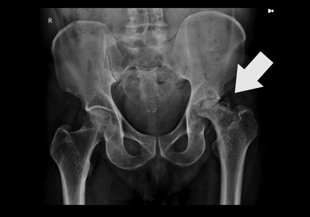
A radiogram of the hip joint in anteroposterior view (Figure 1) revealed complete destruction of joint space, multiple geodes deforming the head of the acetabulum and femur, significant femoral head osteolysis, and ascension of >0.5 cm above the level of the radiological teardrop in the LEFT hip joint. No osteophytes were noted. Minor pelvic tilt to the right can be appreciated.

There was complete loss of joint space.
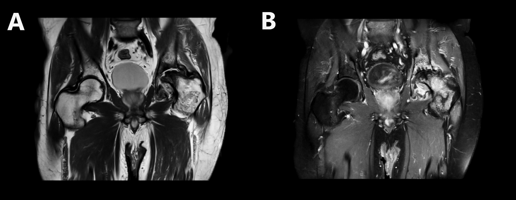
Magnetic resonance imaging (MRI) of bilateral hip joint revealed destruction and thinning of the left acetabulum and loss of normal contour of the left femoral head and neck. There was complete loss of joint space with associated synovitis and joint effusion (Figure 2). There were ill-defined irregular, heterogeneously enhancing areas in the left femoral head, neck, lesser and greater trochanter appearing heterogeneously hypointense on T1WI, heterogeneously hyperintense on T2WI/PDFatSat with few non-enhancing areas (sclerosis) within. There was a low signal intensity in the subchondral area of the femoral head with few subchondral bone cysts (Figure 3). Marrow edema seen from the neck to the upper third of the shaft of the left femur. Bilateral sacroiliac joints and the right hip joint were normal. The above features were suggestive of rapidly destructive osteoarthritis of the left hip joint.

There was a low signal intensity in the subchondral area of the femoral head. There was a complete loss of joint space.

Joint Capsule and Synovium appear thickened and enhancing, suggesting synovitis. There was associated joint effusion and marrow edema present from the neck till upper 1/3rd of shaft left femur. The left gluteus minimus muscle showed strain at its femoral insertion. Right Hip joint and Bilateral Sacroiliac joints appear normal.
July 2021: The patient fell from the bike and went to a local hospital, where he was managed conservatively and was only prescribed analgesic medications and bedrest.
Up to October 2022: Pain was not relieved; however, the patient was mobile and could walk without support. He had complaints of difficulty getting up from sitting position.
October 2022: The patient had a slip and fall at home. The patient had aggravated pain and needed support to walk.
November 2022: Complete physical evaluation and radiological investigations, i.e., X-ray and MRI, were done.
Pathological and microbiological investigations were done.
November 2022: the patient was discharged.
Initially, the patient was planned for a total hip replacement on the left side. However, the patient was not willing to undergo this treatment. Then he was advised skeletal pin traction would be an alternative solution for which the patient was also not willing. So the primary management was by physiotherapy and skin traction.
Although the patient’s acid—fast bacteria staining of sputum and Mantoux test was negative, he has been started on Anti tubercular treatment empirically. The patient was advised to continue skin traction at home and continue ATT treatment as advised. The patient was vitally stable on discharge and was also given the following empirical medications:
As translated from the patient’s mother tongue: I was healthy and fit two years ago when I slipped and fell from my bike. After the accident, I went to a local clinic where the doctor gave me some tablets and advised me to take bed rest. I rested for 2-3 days and again started working as the pain was relatively less but not completely gone. The pain was dull but didn’t affect my work capacity. The pain was only noticeable while squatting or sitting. As I was able to work, I ignored the pain. But I slipped a few days back, and now the pain was unbearable. I couldn’t bear any weight on my left leg, which appeared shorter than my right leg. I came to the hospital, and the doctor examined the leg and told me to get an X-ray and blood investigations done. After looking at the X-ray, the doctor told me to do an MRI. The doctor explained the nature of the disease, advised me to do a total hip replacement, and informed me about various post-operative exercises. However, due to financial constraints, I have denied any surgeries and would like to be treated only medically. After a few days of skin traction, I was discharged from the hospital.
In 1970, a standardised definition of rapidly progressive osteoarthritis was proposed by Lequesne: chondrolysis >2 mm in one year, or 50% joint-space narrowing in one year with no signs of other forms of rapidly destructive arthropathy as mentioned earlier.2,3 However, in 2018 Ancut a Zazgyva et al.10 proposed easily usable clinico-radiological diagnostic criteria and a grading system for practitioners to identify RPOH. These are based on history, clinical aspects, and a single time point radiographic assessment of the hip, without the need for a lengthy observation of the patient, thus hopefully expediting treatment. The cardinal clinical features proposed were: 1) Hip pain starting approximately three years prior, varying in intensity, worsened in the last six to nine months; 2) functional joint mobility, low to moderate limitation in functional joint mobility; 3) Minimal osteophytes; 4) geodes presenting in the femoral head and/or acetabulum.
This case satisfied the above-mentioned criteria as the patient presented with hip pain from two years prior, variable in intensity aggravated only by exercise and after the recent trauma. Moreover, the radiogram did not show evidence of any osteophytes. Donald J. Flemming8 classified RPOH into two categories – with and without trauma. This case has a history of more than one trauma, hence lies in the latter. The grading proposed by Ancut a Zazgyva is as follows – partial joint space narrowing with no femoral head deformation is grade I.10 Complete disappearance of joint space with deformed femoral head with the ascension of femoral head <0.5 cm above the radiologic teardrop is grade II.10 Complete disappearance of joint space with partial osteolysis of the femoral head and ascension of >0.5 cm above the radiologic teardrop is classified as Grade III.10 According to this grading, the left hip joint can be said to be in the final grade, i.e., grade III of RPOH
In a review by Donald J. Flemming,8 femoral head flattening, acetabular remodelling, which parallel femoral head destruction and narrowing of the superior lateral compartment of joint space with sub-chondral sclerosis at the site of bone-to-bone contact were considered as late features.
In the study by Zazgyva A et al.10 Of the 67 patients with RPOH (15 bilateral cases, with a total of 82 hips) Mean age was 66 years females were 67%; males were 32%, and bilateral RPOH was present in only 18% of the cases. With respect to OA, Most authors reported a rather reduced prevalence of RPOH, but it was 9.5% in a cohort of 863 cases of THR performed by the senior author during a period of 10 years.13 As compared to the above study, this case was a 66-year-old male patient with unilateral hip joint involvement.
Many studies suggest that prior subchondral fracture may be involved in the pathogenesis of rapidly progressive arthritis of not only the hip but also that of shoulder.9 However, it has also been noted that in many cases of subchondral fractures, rapidly progressive osteoarthritis does not develop, raising the question of whether subchondral is a sequela of rapidly progressive osteoarthritis or its cause.8
Joint replacement remains the primary treatment for RPOA. Blood loss during total hip arthroplasty is higher than in routine osteoarthritis.4 Survivorship of hip joint reconstruction at five years is greater than 95% in the setting of RPOA.14 Ideally, hip replacement surgery should be performed before the development of acetabular defects, which have occurred in this case.15 A higher degree of vigilance should be maintained in patients suspected of RPOA to reduce operative time and for easier joint reconstruction. Our case was from an economically underprivileged background and was not able to afford and was also not willing for any surgical interventions and preferred conservative management.
The presented case is a classic case of rapidly progressive osteoarthritis of the hip according to newer definitions laid down by Ancut a Zazgyva. With a history of two traumas, a negative history of tuberculosis, and a negative CBNAAT test that rules out tuberculosis as a cause, a preceding subchondral fracture can be considered as one of the major etiological factors. Although other destructive arthropathies are more common, it is essential to consider RPOA as an early differential diagnosis. Appropriate grading and staging of RPOH can help prevent excessive loss of bone tissue and better overall prognosis, i.e., better surgical planning and fewer post-operative complications. As this case progressed to the final grade III (Ancut a Zazgyva), the only effective treatment was total hip replacement. However, the patient was non-affording and preferred the conservative non-invasive approach instead.
Written informed consent for publication of this case report, MRI images and their perspective was obtained from the patient before the patient’s discharge.
Patient management: VN and PD. Data collection: PD and RPD. Manuscript drafting: VN and PD. Manuscript revision: PD, RPD, VN, DN, and PP. All authors approved the final version of the manuscript.
All data underlying the results are available as part of the article and no additional source data are required.
| Views | Downloads | |
|---|---|---|
| F1000Research | - | - |
|
PubMed Central
Data from PMC are received and updated monthly.
|
- | - |
Is the background of the case’s history and progression described in sufficient detail?
No
Are enough details provided of any physical examination and diagnostic tests, treatment given and outcomes?
No
Is sufficient discussion included of the importance of the findings and their relevance to future understanding of disease processes, diagnosis or treatment?
No
Is the case presented with sufficient detail to be useful for other practitioners?
No
Competing Interests: No competing interests were disclosed.
Reviewer Expertise: Rheumatology, Imaging, Arthritis, Biomarkers
Is the background of the case’s history and progression described in sufficient detail?
Yes
Are enough details provided of any physical examination and diagnostic tests, treatment given and outcomes?
Yes
Is sufficient discussion included of the importance of the findings and their relevance to future understanding of disease processes, diagnosis or treatment?
Yes
Is the case presented with sufficient detail to be useful for other practitioners?
No
Competing Interests: No competing interests were disclosed.
Reviewer Expertise: Radiology, Musculoskeletal Disorders, Hip, Sports injuries/imaging, Image guided treatment
Alongside their report, reviewers assign a status to the article:
| Invited Reviewers | ||
|---|---|---|
| 1 | 2 | |
|
Version 1 19 Jul 23 |
read | read |
Provide sufficient details of any financial or non-financial competing interests to enable users to assess whether your comments might lead a reasonable person to question your impartiality. Consider the following examples, but note that this is not an exhaustive list:
Sign up for content alerts and receive a weekly or monthly email with all newly published articles
Already registered? Sign in
The email address should be the one you originally registered with F1000.
You registered with F1000 via Google, so we cannot reset your password.
To sign in, please click here.
If you still need help with your Google account password, please click here.
You registered with F1000 via Facebook, so we cannot reset your password.
To sign in, please click here.
If you still need help with your Facebook account password, please click here.
If your email address is registered with us, we will email you instructions to reset your password.
If you think you should have received this email but it has not arrived, please check your spam filters and/or contact for further assistance.
Comments on this article Comments (0)