Keywords
Antibody, Caries immunotherapy, Freud Adjuvant, Immunoglobulin G, Streptococcus mutans
Antibody, Caries immunotherapy, Freud Adjuvant, Immunoglobulin G, Streptococcus mutans
Streptococcus mutants (S. mutans) is the main cariogenic bacterium that triggers dental caries, which is an infectious disease of the dental tissue characterized by the formation of holes in the teeth. S. mutans is a normal oral flora that belongs to the mutant streptococci group.1 Streptococcus mutans serotype c is the most common serotype isolated from plaque and saliva of caries patients.2,3 S. mutans in the host’s mouth attach to the tooth surface called dental plaque (dental biofilm). The formation of dental plaque is the initial mechanism of the process of dental caries and is an essential stage for bacteremia with the risk of possibly causing infection in systemic organs,4,5 including endocarditis, glomerulonephritis and rheumatic fever.6,7 In addition, S. mutans can spread through blood vessels to various organs such as the heart, kidneys, skin or other parts of the body.4,8–10
Dental caries is an infectious disease of hard tooth tissue due to acids produced by cariogenic bacteria.11,12 This disease is characterized by the dissolution of minerals in enamel and dentin so that a tooth hole is formed. Caries also has an impact on psychological aspects in the form of inferiority due to cavities, black teeth, and bad breath when interacting with the social environment. Caries experienced by children can affect the development of imperfect jaws and mastication so it will have an impact on children’s growth and development. Therefore, various efforts to prevent caries are carried out, one of which is through immunization.8,13,14 Until now, experts continue to conduct various studies to obtain sources of anti-caries immunoglobulin G (IgG) producers that are safe to use in passive immunization to prevent caries. These studies include utilizing antibodies in milk for the production of anticaries-specific antibodies such as goat milk.14–20
Goat’s milk is the second most consumed milk after cow’s milk. In developed countries, such as the Netherlands and Australia, goat and sheep milk are utilized as raw materials for cheese and infant formula.21 The consumption of goat’s milk is preferred by the public because goat’s milk does not cause allergic reactions like cow’s milk. This happens because the amount of protein, namely αS1-casein and β-lactoglobulin in goat milk is very small with a small fat molecule size so that it is easier to digest as a result of which the immune response in the form of an allergic reaction is not triggered.21–25 The use of goat’s milk as a research medium has also experienced rapid development, apart from being hypoallergenic, the frequency of goat milk production is more frequent than cow’s milk due to a shorter gestation period, so that more births occur in goats.22,25
The utilization of goat milk for antibody production in Indonesia has begun, one of which is the production of IgG against Schistosoma japonicum secretory excretory. Immunoglobulin G is part of humoral immunity that functions to provide immune effects against infection.26–28 Sows that experience exposure to bacteria during pregnancy will contain antibodies in their blood serum, namely specific immunoglobulin G, as an effort by the immune system to maintain body homeostasis.29,30 The specific antibodies formed in the mother’s blood serum will be transferred into the colostrum and milk immediately after the mother gives birth as a natural passive immunity for her offspring against infection.31,32
Based on the above description, this study was conducted with the aim of detecting the presence of antibodies, namely anti-Streptococcus mutans immunoglobulin G in goat serum due to injection of Streptococcus mutans antigens originating from caries patients in Banda Aceh City and has been identified in pregnant goats using the Antigen Gel Precipitation (AGTP) method and the serological method, namely Enzyme-Linked Immunosorbent Assay (ELISA) as well as testing the specificity and ability of the specific IgG to react with other serotypes of S. mutans.
This research protocol has been approved by the Ethics Committee of Veterinary Ethics Committee Faculty of Veterinary Medicine Universitas Syiah Kuala, Banda Aceh, Indonesia with number 75/KEPH/XII/2020. The date of approval was 30 December 2020.
Streptococcus mutans from caries patients were serotyped microbiologically and molecularly using Polymerase Chain Reaction (PCR). Streptococcus mutans DNA was extracted using the TIANamp Bacteria DNA Kit according to the instructions (Tiangen Biotech, Beijing). The extracted DNA served as a template for the PCR Master Mix. PCR tubes were filled to a volume of 25 l with PCR master mix (12.5 μl), specific forward and reverse primers (2.5 μl each), template DNA (3.5 μl), and H2O. The primers used were S. mutans serotype primers, which are specific primers that detect the presence of the gtf gene (Table 1).
The PCR tube was then placed in a PCR machine, namely a thermal cycler (BioRad Lab, USA), for DNA amplification. The PCR tube containing 25 μl of PCR master mix (Tiangen Biotech), bacterial DNA, and primers was run through 30 cycles with an initial denaturation temperature of 95°C for 3 minutes, a DNA denaturation cycle of 95°C for 1 minute, annealing 57°C for 30 seconds, and extension 72°C for 30 seconds. The DNA from the amplified sample was separated using an electrophoresis technique with 1.2% agarose (Bio-Rad Laboratories). Samples of PCR running results were taken as much as 6 μl injected into wells on agarose, one well injected marker as a guide. Electrophoresis was run with a current of 100 mA and a voltage of 100 volts for 30 minutes. Furthermore, the agarose was photographed using a gel documentation system for visualization of DNA bands. The identified S. mutans were cultured on liquid media, namely Trypticase Soy Broth (TSB). Streptococus mutans colonies from Trypticase Soy with Sucrose and Bacitracin (TYS20B) media were taken with an osse and homogenized with a vortex. It was then incubated for 72 hours at 37°C under anaerobic conditions.
Pregnant goats used in this study were Etawa breeding goats, which are the result of a cross between Acehnese bean goats (local goats) and Etawa goats, with an ideal body weight of 30 kg and an average age of 2.5 to 3.5 years. The goats were divided into 2 groups: the control group, consisting of 3 heads and the treatment group, consisting of 6 heads. Group K0 is a control group that is not given S. mutans injection, while KP is a treatment group that is injected with pure S. mutans at a dose of 2 ml (108 CFU) per head intravenously. The injection process was carried out three times, namely in weeks 8, 6, and 4 before the goats were expected to give birth. The second injection was done by giving the antigen with Freud Adjuvant Complete subcutaneously, while the third time the antigen was given with Freud Adjuvant Incomplete. The sensitisation dose can be seen in Table 2.
In addition, goats were kept in cages with the same lighting conditions. Goats were fed twice a day, in the morning and evening with forage and concentrate. Drinking water was provided ad libitum.
Blood collection (venesection) was done one week after the third booster. Blood was collected through the Anterior Cephalica Antibrachii Vein located on the distal side of the anterior foreleg of the goat for qualitative examination using the Agar Gel Precipitation Test (AGPT) method. Blood collected as much as 1 ml was collected in a tube and left for 15 minutes so that the blood coagulated. It was then centrifuged to separate the serum from the blood bodies at 3000 rpm for 15 minutes. The resulting serum will be used for the AGPT test.
Agar Gel Precipitation Test (AGPT) consisting of Phosphate Buffer Saline (PBS) with pH 7.4, aquabides, 1% agarose, and 0.001% Na citrate was heated in a microwave until the agar dissolved and boiled. A total of 4 ml of agar solution was poured on the glass slide until the entire surface of the glass slide was covered with agar and left to harden. The hardened agar was perforated using an agar puncher. The hole in the center is filled with antigen (Streptococcus mutans) and six holes around it are filled with antibody serum to be tested, namely goat serum. The agar was kept in a sealed container and allowed to stand for 24-48 hours at room temperature and observed.
Colostrum and goat milk were collected after the goats gave birth and during the peak milk production period at weeks 5, 6 and 7 postpartum.
The ELISA test type used is sandwich ELISA (Cat No. E55-104. Lot No. E55-104-200413) (biothyl laboratories, inc) according to the protocol of the research kit. The test principle is that goat IgG present in the test sample is captured by goat anti-IgG antibodies that have been pre-adsorbed on the surface of microtiter wells. The examination procedure was performed according to the kit’s instructions.
25 ml of 20× Buffer C Dilution was taken and mixed into 475 ml of ultra-pure water and homogenized. The reconstituted 1× Buffer C Dilution was then stored at 2-8°C.
50 ml of 20× Buffer C Dilution was taken and mixed into 950 ml of ultra-pure water and homogenized. The reconstituted 1× Buffer C Dilution was then stored at 2-8°C.
500 ng of goat IgG Standard solution was dissolved in 0.1 ml of 1× Buffer C Dilution, and then homogenized. Next, 7 tubes were prepared that had been marked with 167, 55.6, 18.5, 6.17, 2.06, 0.69, and 0 ng/ml according to the kit’s instructions. Then, 300 μl of 1× Buffer C Dilution was added to each tube. Next, a stratified dilution (1:3) was carried out, in which 150 μl 500-ng Goat IgG Standard solution was added to the first tube (167 ng/ml) and then homogenized. Next, 150 μl 500-ng Goat IgG Standard solution was added to the second tube (55.6 ng/ml) and so on until the sixth tube (0.69 ng/ml). While the seventh tube (0 ng/ml) was not added to the 500-ng Goat IgG Standard solution, it only contained 300 μl 1× Buffer C Dilution because it served as a blank.
Each Goat IgG Standard solution (167, 55.6, 18.5, 6.17, 2.06, 0.69, and 0 ng/ml) of 100 μl was inserted into the well of microtiter plate ELISA column 1 (A-H). The next column was filled with 100 μl of samples that had been diluted (1:50000). The plate was closed and incubated for one hour at room temperature (20-25°C). Next, wash or rinse the antibody that is not attached to the microtiter plate with 1× Wash Buffer 4 times. The plate that was washed added 100 μl anti-IgG Detection Antibody was into each well, then closed and incubated at room temperature for one hour. Washed again four times. Then added 100 μl of HRP Solution C into each well, closed and incubated at room temperature for 30 minutes. Washed and added 100 μl of TMB Substrate Solution and then incubated at room temperature under dark conditions for 30 minutes. In the last step, the reaction was stopped by adding 100 μl Stop Solution and 30 minutes later the optical density (OD) was determined by reading the absorbance using a Microplate Absorbance Reader at a wavelength of 450 nm.
Analysis of cross-reactivity against S. mutans serotypes c, d, e, and f by the immunoblotting method. Colonies of each serotype that had been previously cultured on TYS20B were taken and placed on a nitrocellulose membrane. Then the membrane was blocked with 5% skim milk dissolved in 25 ml PBS for one hour on a shaker. After one hour, the membrane was washed with PBS for 3 times while in a shaker. Then reacted with a primary antibody (antisera against S. mutans), namely IgG anti-S. mutans in goat milk at a dilution of 1:2000 in 5% skim milk and on a shaker. Incubate for one hour or one night at 40°C. Wash the membrane two times with PBS for five minutes each while on a shaker. Add secondary antibody, HRP-Rabbit Antigoat IgG at a dilution of 1:4000 in 5% skimmed milk and shake. This was incubated for one hour. The membrane was washed twice with PBS while on a shaker. It was then developed with 50 ml of develop reagent consisting of 1 ml Chloronaphtol Solution, 10 ml methanol, 30 μl 30% H2O2 and PBS. Furthermore, the membrane was washed with PBS for 5-30 minutes while on a shaker. In the last step, the developing process was stopped by washing the membrane with distilled water and reading the results obtained.
The culture results of Streptococcus mutans (S. mutans) on TSB were successfully inactivated with formaldehyde, but previously the concentration was measured by a spectrophotometer at a wavelength of 625 nm with an absorbance value of 0.08-0.10. The culture results were used as antigens for sensitisation of the immune system of pregnant goats. The dose of bacteria injected into the treatment goats was 2 ml (3×10⁸ CFU/ml) followed by boosters 1 and 2. Boosters were carried out because antibody titers had not been detected in goat serum. sensitization dose can be seen in Table 2.
The resulting serum was used as an antibody in the Antigen Gel Precipitation Test (AGPT) test using the immunodiffusion technique. In the first treatment, namely sensitisation with S. mutans antigen, both control group goats and treatment group goats have not seen the formation of precipitation lines. The same results were also obtained after the first booster using antigen booster emulsified in Freud Adjuvant Complete as shown in Figure 1. The new precipitation line was seen after the second (last) booster using antigen emulsified in Freud Adjuvant Incomplete as shown in Figure 2.


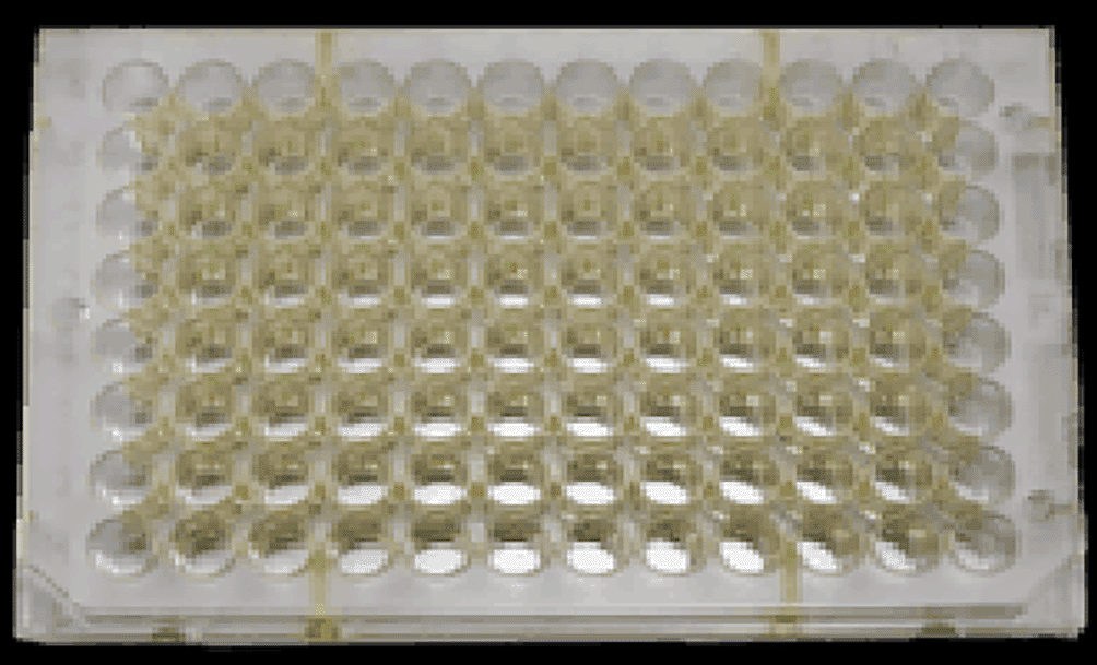
In the study, the colour formed on the ELISA microplate is yellow as shown in Figure 3. The colour formed is then measured absorbance value. The absorbance results of the samples in the form of colostrum and milk, then obtained the concentration as in Table 3.
| No | Sample type | Cut Off values (means±SD) | IgG concentration in goat group | |||
|---|---|---|---|---|---|---|
| Control | Interpretation | Treatment | Interpretation | |||
| 1 | Colostrum | 40.45±0.75* | 39.65 | -a | 43.64 | +b |
| 40.59 | - | 41.58 | + | |||
| 41.13 | - | 42.52 | + | |||
| 44.15 | + | |||||
| 41.93 | + | |||||
| 43.11 | + | |||||
| 2 | Milk, week 5 | 1.46±0.28 | 1.17 | - | 3.21 | + |
| 1.71 | - | 2.92 | + | |||
| 1.51 | - | 3.05 | + | |||
| 3.27 | + | |||||
| 3 | + | |||||
| 3.16 | + | |||||
| 3 | Milk, week 6 | 1.08±0.17 | 0.92 | - | 3.06 | + |
| 1.26 | - | 2.62 | + | |||
| 1.06 | - | 2.8 | + | |||
| 3.02 | + | |||||
| 2.75 | + | |||||
| 2.96 | + | |||||
| 4 | Milk, week 7 | 0.83±0.12 | 0.72 | - | 2.76 | + |
| 0.96 | - | 2.32 | + | |||
| 0.81 | - | 2.55 | + | |||
| 2.72 | + | |||||
| 2.45 | + | |||||
| 2.81 | + | |||||
Streptococcus mutans is a cariogenic bacterium that is commonly isolated from dental caries plaque.33–35 Several studies have been conducted to obtain antibodies from the incidence rate of dental caries in experimental animals, resulting in blood containing specific antibodies that are then injected into other animal models suffering from caries.17,19,36 S. mutans culture results were used as antigens in this study to sensitize the immune system of pregnant goats. The goat’s immune system will produce antibodies to protect its body from the antigen. Furthermore, because the antibody titer was not detected in the goat serum, pregnant goats were given a booster. The booster’s goal is to promote the production of antibodies in the form of immunoglobulin G (IgG) as a form of humoral defense against chronic infections37,38 Boosters were administered by injecting antigen and adjuvant at a 1:1 dose ratio, but the goats miscarried after the second booster, but not after the third repeat dose. As a result, the dose is thought to be safe for sensitizing pregnant goats to produce anti-S. mutans antibody titers. Antibodies or immunoglobulins formed in goat blood, specifically IgG, as a result of S. mutans exposure will be transferred into milk as a natural immune material for the child. IgG is unique in that it can be transferred from blood serum into milk as well as the pregnant goat kid via the placenta.39–41
Blood serum produced after antigen and booster exposure was used for Antigen Gel Precipitation Test (AGPT) using the immunodiffusion technique. AGPT examination aims to prove that anti-S. mutans antibodies have been formed in the goat’s body starting after the first treatment until the last booster.42 Based on the results of the AGPT, a positive reaction is indicated by the presence of white lines or precipitation lines formed between the holes containing antigens and antibodies. If the antibody preparation is not homologous to the antigen, the precipitation line will not form.43
In this study, testing for the presence and concentration of specific antibodies in colostrum and goat milk was carried out using the sandwich method ELISA technique, which detects the presence of the antigen of interest using primary antibodies and reacts with enzyme-labeled secondary antibodies. The primary antibody used was an anti-Goat IgG antibody that would bind to anti-S. mutans IgG present in colostrum and goat milk. While the secondary antibody used was A streptavidin-conjugated horseradish peroxidase (SA-HRP) to detect antibodies that had previously been captured by anti-Goat IgG antibody so that antibody-antigen-antibody complexes were formed. Meanwhile, the substrate used is TMB (3,3′,5,5′-tetramethylbenzidine) which causes a colour change in the liquid in the well with different colour gradations according to the antibody concentration in the sample. The absorbance value obtained expresses the ELISA result which shows the concentration of antibodies detected. The higher absorbance value indicates that the concentration of antibodies contained in the examined sample is also higher.
The normal level of IgG in goat colostrum ranges from 40-60 mg/ml while in milk it is 0.6-7.5 mg/ml. From the observations, it can be seen that IgG against S. mutans is present in colostrum with an average as well as milk collected in the 5th, 6th, and 7th week after birth. Transfer of immunoglobulins from circulating blood to colostrum and milk secretions occurs immediately before, during and after parturition.44 The results of observations on milk samples based on the duration of the week of milk collection decreased the concentration of IgG anti S. mutans in each week, where the largest decrease in concentration occurred in week 7 milk samples, namely 2.601 ± 0.195. However, the decrease was not statistically significant. Meanwhile, based on lactation age, it can be seen that there is a significant decrease in anti-S. mutans IgG concentration in goats with lactation age 5 with an average IgG concentration of 2.618 ± 0.263. Total IgG concentration decreased as the milking time increased. The decrease in total IgG concentration that occurred after the first milking was thought to be due to the reduction or cessation of the colostrogenesis process after the mother goat gave birth.
| Views | Downloads | |
|---|---|---|
| F1000Research | - | - |
|
PubMed Central
Data from PMC are received and updated monthly.
|
- | - |
Provide sufficient details of any financial or non-financial competing interests to enable users to assess whether your comments might lead a reasonable person to question your impartiality. Consider the following examples, but note that this is not an exhaustive list:
Sign up for content alerts and receive a weekly or monthly email with all newly published articles
Already registered? Sign in
The email address should be the one you originally registered with F1000.
You registered with F1000 via Google, so we cannot reset your password.
To sign in, please click here.
If you still need help with your Google account password, please click here.
You registered with F1000 via Facebook, so we cannot reset your password.
To sign in, please click here.
If you still need help with your Facebook account password, please click here.
If your email address is registered with us, we will email you instructions to reset your password.
If you think you should have received this email but it has not arrived, please check your spam filters and/or contact for further assistance.
Comments on this article Comments (0)