Keywords
Surgical Flaps, Ischial tuberosity, Pressure Ulcers
This article is included in the Health Services gateway.
Surgical Flaps, Ischial tuberosity, Pressure Ulcers
Long-term pressure on body tissues, blood circulation problems, necrosis, and skin and soft tissue defects cause pressure ulcers and are most commonly found at bony protrusions. Hip pressure ulcers have the highest clinical incidence. Pressure ulcers in the buttocks are mostly concentrated in three areas: sacrococcygeal, ischial tuberosity, and femoral tuberosity. Because of the presence of a bursa in the Ischial tuberosity, when a pressure sore occurs, the entire bursa is easily affected, resulting in a sore that is larger than the skin necrosis area “the entire bursa is easily affected when a pressure sore occurs”.1 Furthermore, it is close to the perineum, which is easily infected and aggravates the sore, making clinical repair difficult. From January 2016 to August 2018, we successfully used the perigluteal muscle tissue flap to treat 11 cases of grade IV pressure ulcers at the ischial tuberosity.
A spinal cord paralysis caused by a traumatic vertebral fracture affected nine males and two females aged 19 to 52. One patient was bilaterally paralysed, while the others were unilateral paralysed. The average time between recurrent non-healing skin ulcers was 14 months, and all patients had grade IV pressure ulcers of the ischial tuberosity. A wound secretion culture was performed on all patients.
After admission, patients underwent routine examinations and received symptomatic treatment for comorbidities such as anemia, hypoproteinemia, and electrolyte disturbances. We changed the dressing on sore wounds two to three times per day, and the first debridement was performed five to seven days after admission. During the operation, as much necrotic tissue, chronic inflammatory tissue, and infected tissue as possible were removed.
Some of the sores even reached the ischium—bone, causing periosteum and cortical bone degeneration and necrosis. After thorough debridement of the wound, the negative pressure material was placed on it and covered with a film. five to seven days after the operation, the negative pressure material was opened and one or more debridements were performed depending on the condition of the wound base, and VSD treatment was continued. After each debridement, the negative pressure was maintained at -11.97-9.31 kPa, and continuous saline irrigation was administered. The ulcer base was significantly improved after one to three debridement + VSD treatment in 9 patients, and fresh granulation tissue was formed at the bottom of the incubation cavity. The key to surgery is to pack and seal the deep submerged hole because the sore is a deep cavity wound with a narrow opening and a wide base. We opted for a musculocutaneous flap in conjunction with a facial flap or a simple musculocutaneous flap. Four cases were repaired with a gluteus maximus myocutaneous flap combined with a gluteal fasciocutaneous flap, two cases with a biceps femoris myocutaneous flap combined with a gluteal fasciocutaneous flap, two cases with a gracilis myocutaneous flap, one case with a biceps femoris myocutaneous flap, and two cases with a semitendinosus and semimembranosus myocutaneous flap combined with the gluteal fasciocutaneous flap (the size of the flap was 11 cm × 9 cm ~ 28 cm × 18 cm). All 9 patients were sutured directly in the donor area. Anti-infective treatment after surgery was increased and sensitive antibiotics to patients who had precise bacterial culture results were given, for symptomatic treatment.
Example 1
The patient was a 52-year-old Chinese male with paraplegia for eight years and a pressure sore at the left ischial tuberosity caused by a car accident for 13 months. The patient was typically mobile in a wheelchair. The patient’s overall nutritional status was good, and routine examinations revealed no significant abnormalities. The sore on the left ischial tuberosity was 7 cm × 4 cm in diameter and 10 cm deep, with some secretion and a mild odor. On the third day of admission, the first debridement was performed. From shallow to profound, chronic inflammatory tissue and clear necrotic tissue were removed layer by layer. The base was deep up to the ischial cortex, and the sore’s deep cavity was 13 cm × 8 cm. After debridement, negative pressure dressing was applied on the wound surface and the sore was sealed, vacuum sealing drainage (VSD) treatment after the operation was continued; the negative pressure value was kept at -10.64 kPa, and was flushed with normal saline continuously. Every five to seven days, the negative pressure dressing was replaced and the wound was properly debrided. Two such negative pressure treatments were performed. On the 16th day of admission, we performed a repair surgery. The intraoperative design was a 15 cm × 9 cm ipsilateral gluteus maximus muscle flap that was rotated counterclockwise to fill the residual cavity. The flap incision was 15 cm long and extended outward and upward to form a facial flap and fascial skin. To seal the wound, the flap was rotated counterclockwise and covered the muscle flap. The muscle and skin flaps were all alive and well after the operation, and the appearance was good. See Figure 1. During the six- to 12-month follow-up period, the flap remained soft and left a small linear scar.
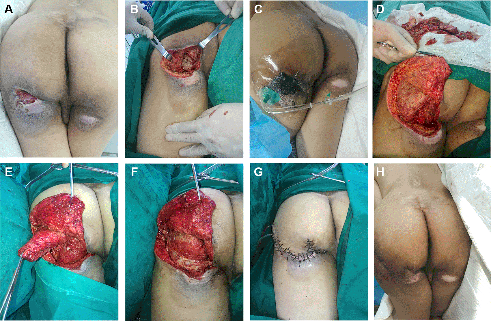
A. Pressure ulcer on the day of admission; B. Ulcer base during the first debridement; C. VSD treatment after the first debridement; D. Ulcer bottom during the second debridement; E. Hip repair during surgery; F. The gluteus Maximus muscle flap rotates counterclockwise to fill the deep residual cavity at the Ischial tuberosity; G. The hip fascia flap rotates counterclockwise to cover and seal the pressure sore; H. The calf fascia flap was rotated counterclockwise to cover and seal the pressure sore. The skin flap was well-shaped three months after surgery, leaving only a small linear scar.
A 36-year-old Chinese man presented with 5 years of paraplegia caused by a traffic accident, as well as 2 months of pressure sores on the right Ischial tubercle. The sores had a typical cystic granulomatous type of grade IV pressure sores, and the skin defect was 5 cm × 4 cm and 3.5 cm deep. On the bottom, there was aging granulation tissue. There was chronic inflammatory skin and soft tissue 1.5-2 cm wide around the wound’s skin, and the injury had little secretion. In the first debridement, the unstable inflammatory tissue surrounding the ulcer was removed. The cyst wall was removed entirely as well. The granulation tissue was removed, the base was enlarged to up to 7 cm × 6 cm, and the bottom was deepened to the superficial muscle layer. Following debridement, the wound was filled with negative pressure material, sealed, and treated with continuous VSD. The negative pressure was -10.64 kPa and was washed continuously with normal saline. One week later, the injury had healed and the medial gracilis muscle flap had been designed. The muscle flap was created within 10 cm of the pubic tubercle-medial semitendinosus muscle connection. The flap was 8 cm × 6 cm in size, and the pedicle was 7 cm × 5 cm in length. The first incision was made at the distal end of the flap to find the gracilis, the myocutaneous flap was separated from the far end and near to 10 cm below the midpoint of the inguinal ligament, and the sore joint was cut to form an open channel; the flap was rotated counterclockwise to cover the wound. We sutured the incision directly on the donor site of the medial thigh. After the operation, all of the flaps survived, and their appearance was satisfactory. Figure 2 depicts this. During the six- to 12-month follow-up, the texture was soft, with only a faint linear pattern.
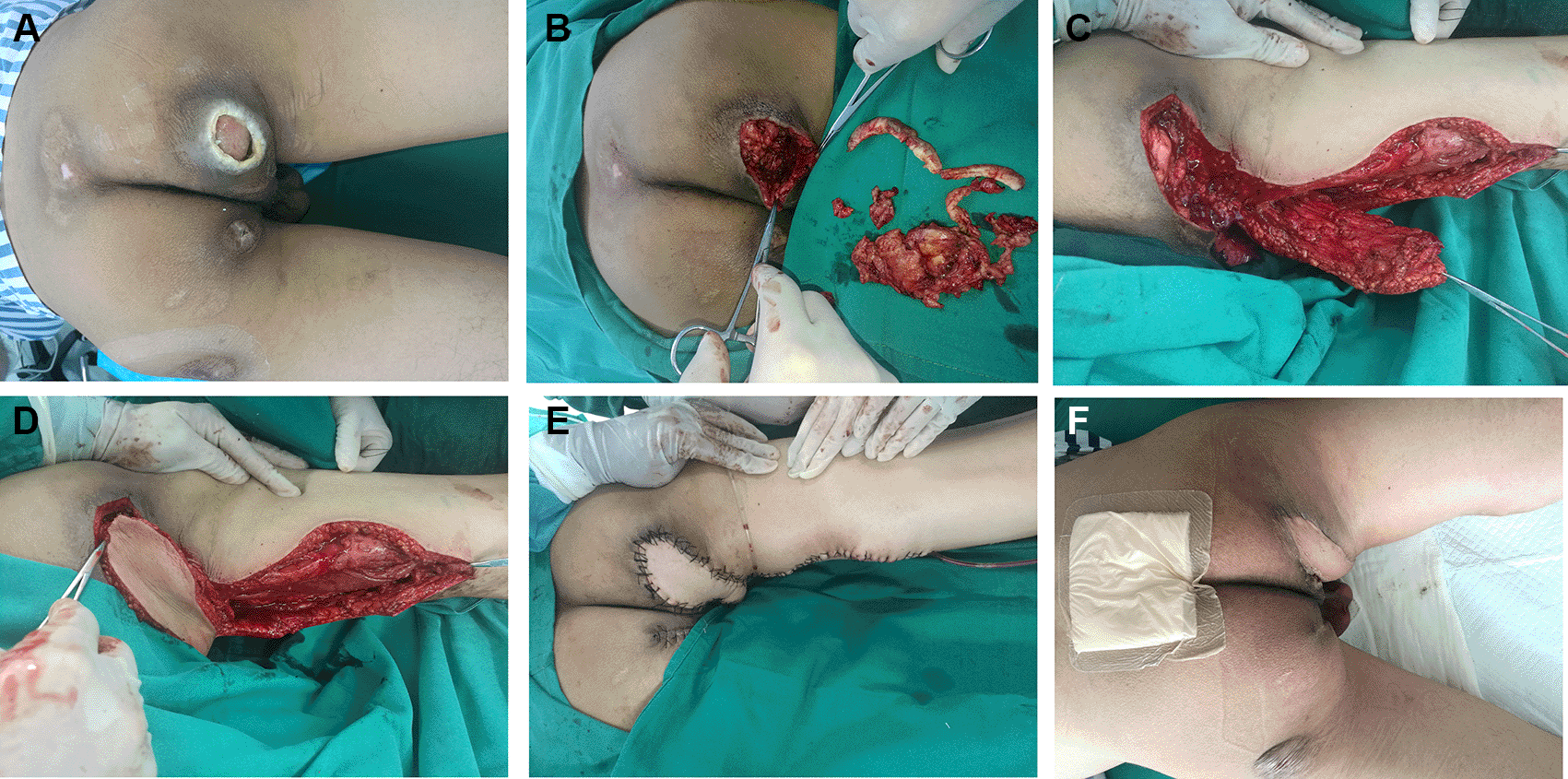
A. Pressure sore situation on admission day; B. During the first debridement, the ulcer base appears; C. The development of a gracilis muscle flap during the repair procedure; D. To cover the wound, the gracilis muscle flap rotated counterclockwise. E. Closed sore, donor area sutured directly; F. Finally, 3 months after the operation, the skin flap appears to be in good condition, with only a small linear scar.
Treatment outcomes
After debridement + VSD + muscle flap combined with fasciocutaneous flap or musculocutaneous flap repair, all patients’ ulcers were healed. Patients in our department were hospitalized for 28 to 81 days. Bacterial cultures were positive in seven of the eleven patients with wound secretions. The appearance of the skin flap was complete after muscular tissue flap repair, with no visible scar hyperplasia. However, after 6 to 12 months of follow-up, the flap was soft and left a small linear scar.
Pressure ulcers are more common in people who have limb dysfunction, are unconscious, or are in forced postures. They were previously known as “bed sores” but they are now more commonly known as pressure ulcers, and they mostly occur in protruding parts of human bones. The buttocks are the most commonly affected area of the body when it comes to pressure ulcers. Pressure ulcers in the buttocks are most common in the sacrococcyx, ischial tuberosity, and femoral tuberosity. The sacrococcygeal region has the highest incidence, which mostly occurs in patients who have been supine for an extended period of time. However, pressure ulcers at the ischial tuberosity are more common in patients who have paralyzed lower limbs and have been in wheelchairs for a long time. Repairing a severe pressure ulcer is complicated, the treatment cycle is lengthy, and the cost is exorbitant. As a result, for people at high risk of pressure ulcers, prevention is preferable to treatment, and the proper family care model is advocated.2 If a pressure ulcer develops, the wound will quickly deepen and expand if not treated properly. Early and effective treatment is critical: 1. Avoid long-term pressure and rotate frequently. 2. Maintain clean and dry skin. 3. Use decompression devices like an air bed and a decompression mat. Early-stage I to II pressure ulcers are mostly treated with protection and conservative dressing changes.
Long-term vertical pressure is the leading cause of pressure ulcers. The normal capillary pressure ranges between 12 and 30 mmHg. When the local pressure exceeds 16 mmHg, capillary tissue perfusion can be blocked, resulting in pressure sores. When the local pressure exceeds 30-35mmHg and lasts for two hours, it can result in a pressure ulcer. Necrotic tissues are classified into ulcer type, full-thickness tissue necrosis type, sinus type, and cyst wall granuloma type based on the depth and clinical manifestations of pressure ulcers.3 Pressure ulcers at the ischial tuberosity have distinct characteristics when compared to the other two parts. The skin of the ischial tubercle is located at the buttock fold, which has a lot of skin and soft tissues. In patients who usually sit for a long time, their soft skin tissues move horizontally repeatedly on the seat. A shearing force is produced by the combination of horizontal friction and vertical pressure. This shearing force causes the adjacent two layers of tissue to slide in the opposite direction, resulting in skin and soft tissue displacement and rupture. Under these conditions, a latent cavity or sinus is formed, which exacerbates the destruction of deep tissues. As a result, ischial tuberosity pressure ulcers have a small-scale skin defect with deep enlarged penetrating ischial tuberosity necrosis.4 Furthermore, the ischial tuberosity is close to the perineum, the surrounding environment is moist, and urine and stool contamination are likely to worsen the infection. In severe secondary infections, there will be a foul smell or a large amount of purulent secretions, which can easily penetrate deep tissues and cause tendons and periosteum to be inflamed, thickened, hardened, and destroying their bones and joints. As a result, it complicates repair treatment. Pressure sores in the ischial tuberosity are difficult to heal in a chronic wound; deep-tissue necrosis is common, and residual bacteria are persistent. As a result, thorough debridement is required before repair, and VSD is a novel type of closed drainage technology. It has been widely used in the treatment of various types of wounds with positive results.5 In addition to the effect of continuous drainage, VSD treatment can hasten necrotic tissue shedding, hasten residual cavity sealing, improve local blood supply, promote vascularization, and prevent microbial invasion and infection in the external environment.6 Pressure sore mouth after VSD treatment can lay the groundwork for future repair. In this group of patients, one or more negative pressure treatments effectively controlled the infection symptoms, and the basal tissue of the latent cavity became fresh. The main goal of surgical pressure sore repair in the ischial tuberosity is to fill the residual cavity, eliminate the sinus tract, and seal the skin and soft tissue defects. It is critical to pack the residual cavity. Muscle tissue has an abundant blood supply and a powerful anti-infection ability. It is ideal for filling a large residual cavity. To repair deep pressure sores at the ischial tuberosity, we used muscle flap packing in conjunction with fasciocutaneous flap covering or a simple muscle flap alone. We chose the lower part of the buttock, the medial thigh, and the posterior thigh as the donor sites for the muscle tissue flap based on the principle of “obtaining materials from nearby.” The gluteus Maximus muscle tissue is densely packed with blood vessels. The gluteus maximus muscle flap is one of the preferred packing tissues for the large residual cavity. The gluteus maximus muscle flap is lifted from the distal end in the direction of the gluteus Maximus muscle bundle. The inferior gluteal artery perforating branches are located in the middle of the gluteal region, 5 cm above the lateral third of the gluteal fold. Depending on the size of the residual cavity, either full-thickness or part-to-section cutting can be performed. We proceeded with caution in order to protect the main inferior gluteal artery during the procedure. The amount of muscle tissue required to fill the ischial tuberosity’s deep residual cavity was obtained. Pressure ulcers frequently consume far more skin and soft tissue than is required to seal the wound. As a result, when selecting the gluteus maximus muscle flap, we usually use only the muscle flap and the fascia flap. Skin soft tissue flaps are typically formed by extending and incising along the buttock folds to inform and transfer. They can be transferred counterclockwise after formation, reducing the fasciocutaneous flap’s tissue volume. The gracilis musculocutaneous flap is located on the inner thigh and is concealed. This muscle is an auxiliary muscle of the inner thigh muscle group, and its removal has little effect on function. The main nutrient vessel is the gracilis branch of the deep femoral artery, and its entry point is about 9 cm below the inguinal ligament midpoint.7 Because the gracilis muscle is thin, musculocutaneous flaps are frequently used to transfer the wound and cover it directly. It is appropriate for wound repair with a shallow wound base. If the amount of tissue filled by the musculocutaneous flap is insufficient, the flap can be appropriately enlarged. To fill the residual cavity, the peripheral part of the tissue is removed from the epidermis to form part of the subcutaneous tissue flap.8 Posterior thigh muscles include the biceps femoris, semitendinosus, and semimembranosus, which all originate from the ischial tubercle. Their blood supply is given to them in stages. The main blood supply to the posterior femoral side comes from the first perforating branch of the deep femoral artery, located 8 cm below the ischial tubercle.9 Clinically, depending on the size and depth of the pressure sore, either a simple muscle flap packing or a muscle tissue pedicle myocutaneous flap directly covering can be used.
The biceps femoris tissue, on the other hand, is extensive and suitable for formation. The large residual cavity is filled with a single muscle flap. Simultaneously, semitendinosus and semimembranosus are small and thin, making them more suitable for the direct formation of myocutaneous flaps for transfer and repair.
The IV-degree pressure sore of the ischium tubercle takes a long time to heal. The blood circulation of the tissue flap chosen above, for example, is extremely reliable. In the cases, there no occurrence of blood supply disturbance of the tissue flap itself, and the factor that affects the healing time is often that the transplanted tissue flap cannot be adhered to the wound base completely. In summary, the main reasons are as follows: 1. Anemia, hypoproteinemia, and electrolyte imbalances are examples of systemic nutritional status deviations. 2. Bacteria that remain in deep tissues can easily infect. 3. The expansion is insufficiently thorough, as is the basement’s remaining ecological organization. 4. Patients with lower-limb paralysis have poor neutrophil function. 5. Long-term postoperative prone position, patients with more difficult body posture. 6. Turning over activities that can easily cause the ischial tuberosity joint traction. In response to the aforementioned preoperative and postoperative issues, it is recommended to perform multiple expansions + VSD to improve the foundation of the wound before using muscle tissue flap transplantation, in addition to the symptomatic adjustment of systemic nutrition, the use of sensitive antibiotics to fight infection, and strengthening of postural care. As soon as possible after transplantation, it should be ensured that the muscle tissue flap heals and closes the injury.
| Views | Downloads | |
|---|---|---|
| F1000Research | - | - |
|
PubMed Central
Data from PMC are received and updated monthly.
|
- | - |
Provide sufficient details of any financial or non-financial competing interests to enable users to assess whether your comments might lead a reasonable person to question your impartiality. Consider the following examples, but note that this is not an exhaustive list:
Sign up for content alerts and receive a weekly or monthly email with all newly published articles
Already registered? Sign in
The email address should be the one you originally registered with F1000.
You registered with F1000 via Google, so we cannot reset your password.
To sign in, please click here.
If you still need help with your Google account password, please click here.
You registered with F1000 via Facebook, so we cannot reset your password.
To sign in, please click here.
If you still need help with your Facebook account password, please click here.
If your email address is registered with us, we will email you instructions to reset your password.
If you think you should have received this email but it has not arrived, please check your spam filters and/or contact for further assistance.
Comments on this article Comments (0)