Keywords
Ascites, Eosinophilic Ascites, Eosinophilic Gastroenteritis, Eosinophilia
Eosinophilic gastroenteritis is a rare disease characterized by gastrointestinal symptoms and peripheral eosinophilia. It can affect any layer of the gastrointestinal tract. Mucosal, muscular, and sub-serosal subtypes of eosinophilic infiltration are distinguished based on their infiltration depth. Ascites is a characteristic feature of sub-serosal eosinophilic gastroenteritis. However, isolated ascites is a very rare presentation of the disease, making diagnosis challenging.
In this case report, we present a 25-year-old female eosinophilic gastroenteritis patient who initially presented with symptoms and signs of ascites. She also reported seasonal rhinitis, which has been associated with eosinophilic gastroenteritis. She responded effectively to treatment with prednisolone.
This case report highlights the importance of considering rare diseases like eosinophilic gastroenteritis in the differential diagnosis of ascites. Treatment typically involves dietary modifications and corticosteroids. This case also contributes to our understanding of this rare initial presentation of the disease.
Ascites, Eosinophilic Ascites, Eosinophilic Gastroenteritis, Eosinophilia
Eosinophilic gastroenteritis (EGE) is a rare inflammatory gastrointestinal disease affecting children and adults. It is characterized by the infiltration of the gastrointestinal tract by eosinophils. Although it commonly affects the stomach and the duodenum, the colon and the esophagus can also be affected.1
Herein, we present a 25-year-old female who presented with ascites, weight loss, loss of appetite, and fever for six months. A complete blood count revealed an elevated eosinophil count; ascetic fluid analysis was also significant for eosinophilia. EGE was confirmed with an endoscopically taken biopsy from the stomach and duodenum. The patient was started on prednisolone 30mg p.o/day, with tapering after two weeks, and for a total of 6 weeks. All her symptoms subsided on subsequent follow-ups, and she regained her initial weight.
Prednisolone 20-40mg in divided doses is the first line of treatment in EGE. It achieves symptomatic remission in 80% of cases within a week and normalization of eosinophilia within two weeks.2 Additional therapeutic options include antihistamines, sodium cromoglycate, montelukast, and ketotifen.3–6 Surgical intervention is reserved for patients with intestinal obstruction.3,7
The clinical course often alternates between periods of remission and relapse, especially when steroid treatment is stopped in approximately half of patients. To minimize steroid-related side effects, steroid-sparing therapies like antihistamines, mast cell inhibitors, leukotriene receptor antagonists, and anti-interleukin drugs, including ketotifen, can be valuable in managing relapses.4,6 In rare instances where steroid therapy is ineffective, total parenteral nutrition or immunosuppressive agents like oral azathioprine or cyclophosphamide may be added to the steroid regimen for patients with widespread mucosal disease.8
A 25-year-old female presented with progressive abdominal distention for six months. She has also had significant weight loss (5 kg/6 months), loss of appetite, and a low-grade intermittent fever. She had watery diarrhea, alternating with intermittent constipation and crampy abdominal pain. There was no history of jaundice, mental status change, leg edema, skin rash, or joint pain. She denied any history of alcohol or intravenous drug use. The patient has had allergic rhinitis since adolescence. She has no history of hepatitis B or C infection nor any personal or family history of liver disease, hypertension, or diabetes. A physical examination was pertinent for a distended abdomen with signs of intraabdominal fluid collection; other systemic examinations were unremarkable.
A complete blood count revealed white blood cells of 5000/μL, with neutrophils and eosinophils accounting for 57% and 11%, respectively. Other parameters, like hematocrit and platelets, were normal. Hepatitis B surface antigen and hepatitis C antibody were negative. Liver and renal function tests were also in the normal ranges. Serum albumin was 3.56 g/dl. Fluid analysis from the ascites showed a total protein of 4.2 g/dl, albumin of 2.73 g/dl, and a total WBC of 1995 cells/μL with polymorphonuclear cells of 10%. SAAG was 0.83. Cytology from peritoneal fluid revealed eosinophilic effusion, with eosinophils contributing to 12% of the inflammatory cells. Stool examinations were normal three times.
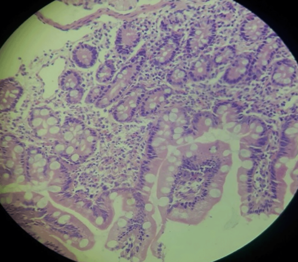
The endoscopy examination found small ulcers in the esophagus and a large ulcer in the first part of the duodenum. The lining of the upper stomach appeared inflamed, but there were no other abnormalities. Multiple biopsy samples were taken from the esophagus, stomach, and duodenum. The upper GI endoscopy showed a Forest class III duodenal ulcer with deformity and narrowing. Biopsies from the stomach showed inflamed cystic cell fragments with dense eosinophil infiltration (35/HPF) in the epithelium and lamina propria (Figure 1). The duodenal biopsy revealed fragmented tissue within the villi surrounded by lymphoplasmacytes and edematous lamina propria with eosinophilic infiltrate (20/HPF) (Figure 2). The biopsy taken from the esophagus was inadequate. These findings were suggestive of EGE.


The patient was initiated on prednisolone at a dose of 30mg per day. After two weeks of treatment, the prednisolone was tapered by decreasing 5mg per/week (cumulative dose 945mg) and discontinued after a total of seven weeks. Her ascites and abdominal pain significantly improved within two weeks of starting prednisolone therapy. Following the completion of treatment, she regained her weight, and all her symptoms resolved completely.
EGE is a rare gastrointestinal disorder that can affect any part of the gastrointestinal tract, and eosinophilic infiltration is the main feature of the disease. Eosinophilic infiltration can affect any layer of the GI tract, with symptoms varying depending on the affected layer.9 Data on the prevalence of EGE is limited due to the rarity of the disease. The prevalence of EGE in the United States is around 22 to 28 per 100,000 people, based on prior survey data.10 Even though the peak onset of the disease is in the third decade of life, it can occur in any age group.11
The prevalence of sub-serosal EGE varies in different studies. According to Klein et al., out of 40 patients with EGE, 57% have mucosal disease, 30% have muscular disease, and only 13% have subserosa diseases.12 A study by Pinte L et al. found that among 171 patients with eosinophilic ascites, 74% had eosinophilic gastroenteritis. The remaining patients had other causes, such as parasitic infections and malignancies.13
Ascites is the most common manifestation of the sub-serosal subtype of eosinophilic gastroenteritis.14 Furthermore, patients with this variant have abdominal bloating, eosinophilia, and a rapid response to corticosteroid therapy.12 Since patients have nonspecific gastrointestinal manifestations, a high index of suspicion is mandatory.
Half of the individuals with EGE have a history of allergy or atopy; a few of them may have IgE specific for food allergens, and they may also have a positive skin test for a specific antigen.15 Our patient has seasonal allergic rhinitis.
Peripheral eosinophilia is present in two-thirds of patients.16 Marked ascitic fluid eosinophilia is common in sub-serosal variants of EGE (45–90%).12,15,17 This patient’s ascitic fluid analysis only shows 12% eosinophils. Eosinophilic infiltration of the GI tract on biopsy is confirmatory after excluding other causes of eosinophilia, such as malignancy, parasitic infestation, and drugs.11
Treatment of EGE is individualized based on patient characteristics. Patients with malabsorption can try an elimination diet. An elimination diet should be tried for at least 4 to 6 weeks. Those who are not responding should be tried on prednisolone (20–40 mg/day); symptoms usually improve within 2 weeks. Prednisolone should be tapered over the next 2 weeks.18
Eosinophilic gastroenteritis should be suspected in a patient with non-specific abdominal symptoms and ascites of unclear etiology. Therapy with corticosteroids reduces the severity and duration of symptoms.
Written informed consent was obtained from the patient for publication of this case report and any accompanying images. A copy of the written consent is available for review by the Editor-in-Chief of this journal.
All the images used in this manuscript are obtained from the patient, and the authors have informed written consent to publish the images along with the manuscript
All data underlying the results are available as a part of the article, and no additional source data are required.
| Views | Downloads | |
|---|---|---|
| F1000Research | - | - |
|
PubMed Central
Data from PMC are received and updated monthly.
|
- | - |
Is the background of the case’s history and progression described in sufficient detail?
Partly
Are enough details provided of any physical examination and diagnostic tests, treatment given and outcomes?
Partly
Is sufficient discussion included of the importance of the findings and their relevance to future understanding of disease processes, diagnosis or treatment?
Partly
Is the case presented with sufficient detail to be useful for other practitioners?
Partly
References
1. Yalçın E, Döngelli H, Dolu S, Akarsu M: Eosinophilic jejunitis presenting as acute abdomen with eosinophilic ascites.BMJ Case Rep. 2024; 17 (9). PubMed Abstract | Publisher Full TextCompeting Interests: No competing interests were disclosed.
Reviewer Expertise: Gastroenterology, hematology and internal medicine.
Is the background of the case’s history and progression described in sufficient detail?
Yes
Are enough details provided of any physical examination and diagnostic tests, treatment given and outcomes?
Yes
Is sufficient discussion included of the importance of the findings and their relevance to future understanding of disease processes, diagnosis or treatment?
Yes
Is the case presented with sufficient detail to be useful for other practitioners?
Yes
Competing Interests: No competing interests were disclosed.
Reviewer Expertise: Eosinophilic GI disease; disorders of gut brain interactions; microbiome
Is the background of the case’s history and progression described in sufficient detail?
Yes
Are enough details provided of any physical examination and diagnostic tests, treatment given and outcomes?
Partly
Is sufficient discussion included of the importance of the findings and their relevance to future understanding of disease processes, diagnosis or treatment?
Partly
Is the case presented with sufficient detail to be useful for other practitioners?
Yes
Competing Interests: No competing interests were disclosed.
Reviewer Expertise: Gastroenterology
Alongside their report, reviewers assign a status to the article:
| Invited Reviewers | |||
|---|---|---|---|
| 1 | 2 | 3 | |
|
Version 1 10 Sep 24 |
read | read | read |
Provide sufficient details of any financial or non-financial competing interests to enable users to assess whether your comments might lead a reasonable person to question your impartiality. Consider the following examples, but note that this is not an exhaustive list:
Sign up for content alerts and receive a weekly or monthly email with all newly published articles
Already registered? Sign in
The email address should be the one you originally registered with F1000.
You registered with F1000 via Google, so we cannot reset your password.
To sign in, please click here.
If you still need help with your Google account password, please click here.
You registered with F1000 via Facebook, so we cannot reset your password.
To sign in, please click here.
If you still need help with your Facebook account password, please click here.
If your email address is registered with us, we will email you instructions to reset your password.
If you think you should have received this email but it has not arrived, please check your spam filters and/or contact for further assistance.
Comments on this article Comments (0)