Keywords
Corneal tear, infected corneal tear, endophthalmitis, secondary IOL, contact lens, BSS (BostonSight Scleral) contact lens
Corneal tear, infected corneal tear, endophthalmitis, secondary IOL, contact lens, BSS (BostonSight Scleral) contact lens
Ocular trauma is an important cause of unilateral vision loss worldwide, especially in young people, and surgical repair is almost always challenging. The successful surgical repair of open globe injury and the subsequent visual rehabilitation of the patient – a topic of great significance and challenge to the practicing ophthalmologists.1,2 Vats et al reported the prevalence of ocular trauma to be 2.4% of the urban population in India. It is the second most common cause of corneal blindness in children.2 Corneal laceration and corneal perforation are common ocular traumas with potentially devastating sequelae, including corneal scarring, astigmatism, and endophthalmitis.3 There is very little literature on management of an infected corneal laceration.
The reported incidence of endophthalmitis in the absence of an intraocular foreign body following an open globe trauma range from 3.1% to 11.9%, of which the paediatric population constitutes 22% to 34.5% cases. The visual prognosis in traumatized eyes with endophthalmitis is extremely poor and worse than that of post operative endophthalmitis.4 Thus ophthalmologists should maintain a high index of suspicion for infection in open globe trauma. They should ensure a prompt management strategy to achieve an optimal anatomic and visual outcome.
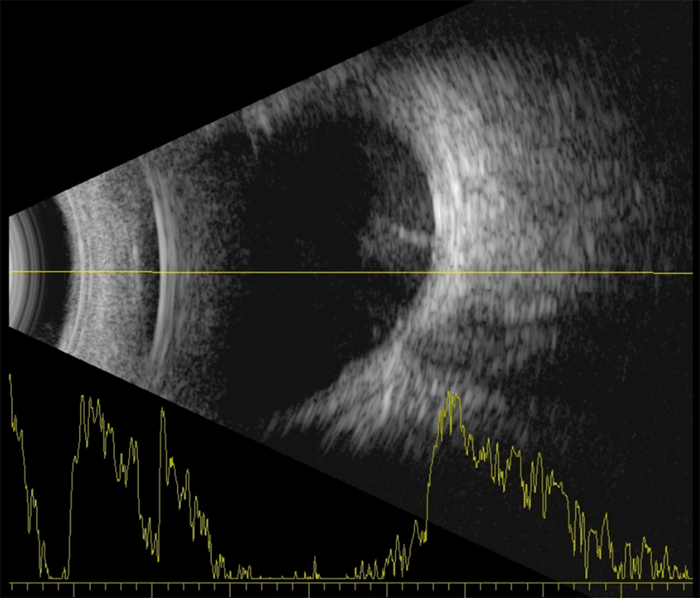
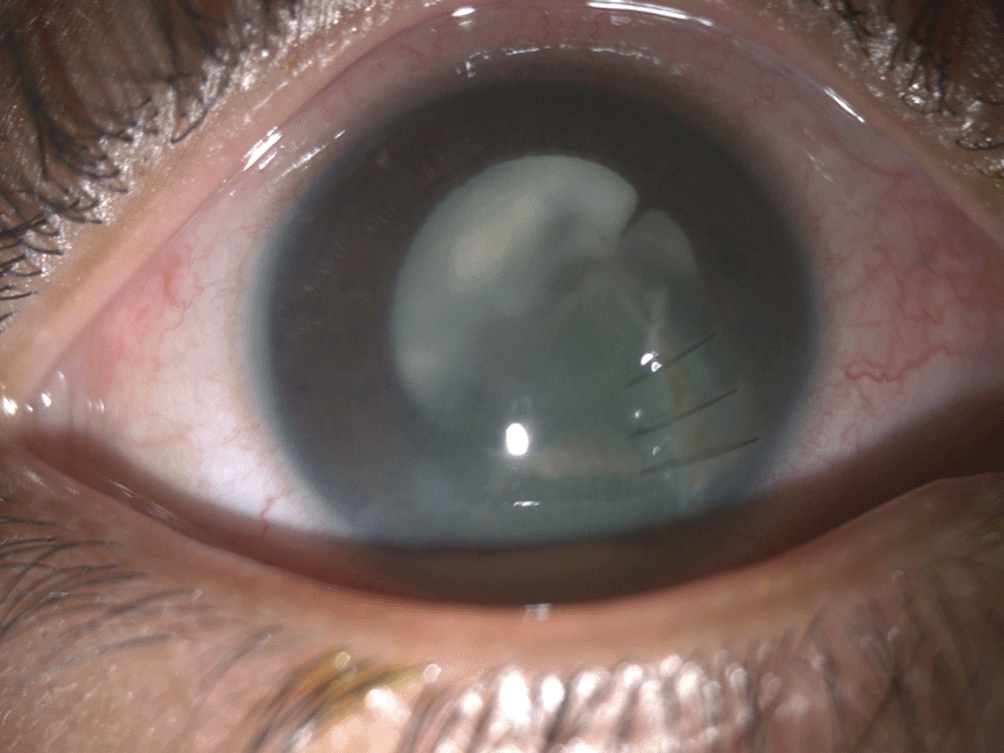
In this case, a boy in his middle childhood sustained an injury to his right eye while playing with scissors. He was taken to a local doctor who prescribed topical medications and then consulted with us two days after the injury. At presentation, his visual acuity was perception of light with an inaccurate response to the projection of rays. A detailed slit lamp examination was performed, and a gentle B-scan was conducted. The examination revealed a full-thickness infected corneal laceration with iris tissue incarceration measuring about 6 mm. Additionally, there were hypopyon and exudates in the anterior chamber. A yellowish tinge in the lens suggested an intralenticular abscess (Figure 1). On B-scan ultrasonography mild to moderate reflective echoes were observed in the posterior aspect of the vitreous cavity suggestive of traumatic endophthalmitis (Figure 2). After the initial treatment with oral antibiotics and analgesics (Amoxicillin+ clavulanic Acid 325 mg with ibuprofen + paracetamol as 15 mg/kg/ml dose), the patient underwent corneal scraping, corneal laceration repair, along with vitreous biopsy, pars plana lensectomy was done. The vitreous cavity was filled with whitish membranous exudates and vitrectomy to the extent safely possible was done and intravitreal antibiotics were administered. The microbiology reports later came out to be positive for the obtained vitreous sample and the organism was confirmed to be Staphylococcus aureus on culture. In addition, three doses of intravitreal injections (vancomycin 1 mg/0.1 ml, Ceftazidime 2.25 mg/0.1 ml and Dexamethasone 0.4 mg/0.1 ml) were administered at 48-hour intervals during initial visits. The patient was closely monitored, and intravitreal antibiotics were repeated, followed by vitreous lavage and complete lensectomy. In four months, with topical medications, surgical interventions, and timely follow-up, he recovered completely from microbial keratitis as well as endophthalmitis. Subsequently, the corneal sutures were removed, and he was scheduled for secondary IOL implantation and a three piece hydrophobic acrylic foldable IOL (Acrysof, Alcon laboratories, Inc). His final visual acuity was 20/50 with BSS (BostonSight Scleral) contact lens, which was considered satisfactory (Figure 3, and Figure 4a and b).


The cornea is an optical surface and requires special treatment to preserve visual acuity during open globe injury repairs.5 Although the time lag between injury and surgery adversely affects the final vision outcome, this is not statistically significant. Issac et al. demonstrated a 1.16-fold increase in the chance of a worse visual prognosis with each day of delayed surgery.6 Our case emphasizes the potential consequences of delayed treatment, underscoring the significance of prompt medical attention for optimizing visual outcomes.
Several cases reported by Salman et al.7 and Henry et al. involved lenticular abscess following penetrating injury. Similar to our case, both Henry et al and Salman et al. reported a positive culture for Staphylococcus species.8 In a study by Rajaraman et al., a variety of organisms causing lens abscess were found, with Staphylococcus epidermidis being predominant.9
In paediatric post traumatic endophthalmitis, the factors affecting visual prognosis have not been extensively analysed especially when there is a concurrent intralenticular abscess. Dave et al, underscored the role of an initial intervention of vitrectomy over a limited vitreous biopsy in attaining a good visual outcome in the management of lens abscess with concurrent endophthalmitis.9 A lens abscess presents distinct challenges compared to infections in other parts of the eye, as the infective organisms are trapped within the avascular, protein-rich environment of the lens. In such cases, surgical removal of the entire infected area is essential. This not only eliminates the majority of the infectious agents but also allows intraocular antibiotics to penetrate and act more effectively. It has also been suggested that the relative softness of the crystalline lens in younger individuals, due to the absence of nuclear sclerosis, facilitates the faster spread of infection across the lens and into the vitreous cavity.10
The visual outcomes in our current case parallel those documented in other reports concerning posttraumatic endophthalmitis.5 Prompt visual rehabilitation in these children, combined with effective amblyopia management, can significantly enhance their visual acuity and overall quality of life. Various optical correction options are available, including spectacles, contact lenses, and intraocular lens (IOL) implantation. In cases where children or their parents are reluctant to opt for glasses or contact lenses, secondary IOL implantation may serve as a viable alternative. In our case, we visually rehabilitated the child with a secondary IOL followed by a BSS contact lens to achieve satisfactory visual improvement.11
This case highlights the challenging nature of managing such cases, where successful management necessitates a combination of careful clinical evaluation, advanced surgical techniques, and tailored treatment plans to optimize visual recovery.
| Views | Downloads | |
|---|---|---|
| F1000Research | - | - |
|
PubMed Central
Data from PMC are received and updated monthly.
|
- | - |
Provide sufficient details of any financial or non-financial competing interests to enable users to assess whether your comments might lead a reasonable person to question your impartiality. Consider the following examples, but note that this is not an exhaustive list:
Sign up for content alerts and receive a weekly or monthly email with all newly published articles
Already registered? Sign in
The email address should be the one you originally registered with F1000.
You registered with F1000 via Google, so we cannot reset your password.
To sign in, please click here.
If you still need help with your Google account password, please click here.
You registered with F1000 via Facebook, so we cannot reset your password.
To sign in, please click here.
If you still need help with your Facebook account password, please click here.
If your email address is registered with us, we will email you instructions to reset your password.
If you think you should have received this email but it has not arrived, please check your spam filters and/or contact for further assistance.
Comments on this article Comments (0)