Keywords
Septic pulmonary embolism (SPE), Infectious foci, Computed Tomography (CT), Soft tissue infection
This article is included in the Health Services gateway.
This is a case of septic pulmonary embolism in a patient presented with a right foot ulcer as foci of infection. A Septic Pulmonary embolism is a thromboembolic event in which case an infected thrombus from an infectious focus; distal to the lung vasculature travels through the lung and results in occlusive, inflammatory and infectious insult to the lung. Clinically it may present as mild respiratory symptoms such as cough, fever or more severe symptoms such as shortness of breath (SOB), hemoptysis, pleuritic chest pain or difficulty breathing1; on computed tomography (CT) imaging, cavitary or non-cavitary peripheral nodules with or without feeding vessel, consolidation and Pleural effusion are prominent findings.2 In this case report, we present a rare case of septic pulmonary emboli from a foci of soft tissue infection.
Septic pulmonary embolism (SPE), Infectious foci, Computed Tomography (CT), Soft tissue infection
Septic Pulmonary embolism (SPE) is characterized by a thromboembolic event wherein an infected thrombus originating from an infectious focus distal to the lung vasculature migrates through the lung, leading to occlusive, inflammatory, and infectious damage to the lung. The clinical manifestation of this condition can range from a gradual onset of symptoms such as fever and respiratory manifestations including cough, dyspnea, hemoptysis, and pleuritic chest pain to a sudden sepsis syndrome.1 Previously, SPE was linked to right-sided infective endocarditis in intravenous drug users,2 postpartum septic thrombophlebitis, and Lemierre’s syndrome.3,4 However, in recent years, there has been a shift in the epidemiology, highlighting the significance of infected vascular catheters, implantable devices,5 and more recently, septic thrombophlebitis due to contiguous deep soft tissue or bone infection of the extremities.6 The diagnostic challenge of pulmonary embolism arises from the non-specific nature of clinical and laboratory data. Presently, computed tomography stands out as the most dependable imaging modality for characterizing chest findings in patients with SPE.7,8 Therefore, clinicians should maintain a high level of suspicion when encountering a patient with an extrapulmonary site of infection who presents with respiratory symptoms. Early clinical identification and prompt administration of broad-spectrum antibiotics significantly enhance the patient’s prognosis.1 Here, we describe a case of septic pulmonary emboli in a patient with a right foot ulcer serving as the focal point of infection.
A female patient aged 55 years presented at Sheno primary hospital, Oromia, Ethiopia, exhibiting symptoms such as high-grade persistent fever, shortness of breath, productive cough with blood-tinged sputum, and right-sided pleuritic chest pain lasting for 2 weeks. She had previously visited a private hospital nearby where she received unspecified oral antibiotics without experiencing any relief from her symptoms. Additional information revealed that she was also dealing with swelling and ulceration on her right foot, accompanied by purulent discharge from the big toe for the past 3 weeks, a condition for which she had not sought medical attention. Her medical history did not indicate any presence of diabetes or hypertension, nor did it include any contact with individuals suffering from chronic cough or tuberculosis. Upon physical examination, the patient displayed signs of respiratory distress, with a respiratory rate of 32 breaths/min, a pulse rate of 104/min, an axillary temperature of 38.9°C, and an oxygen saturation of 82% with room air. Furthermore, edema was observed in her right foot (non-pitting edema) along with an ulcer on the big toe oozing purulent discharge. Laboratory tests conducted on the same day of presentation showed a total white blood cell count of 24,220/μL with 93.8% granulocytes, a platelet count of 487,000 μL, and normal values for other complete blood count (CBC) parameters. Elevated levels of acute inflammatory markers were also detected, with a C-reactive protein (CRP) level of 179.6 mg/L, Highly-sensitive CRP (HsCRP) greater than 5mg/L, and an Erythrocyte Sedimentation Rate (ESR) of 70 mm/hr. Comprehensive metabolic panel and Organ function tests yielded normal results, ruling out the presence of tuberculosis. Additionally, tests for VDRL, hepatitis B and C viral markers, as well as HIV, all returned negative results. The coagulation profile showed no abnormalities, with values of 12.4 sec for PT, 1.02 sec for INR, and 36.7 sec for APTT. Urinalysis results were also normal, indicating no signs of urinary tract infection.
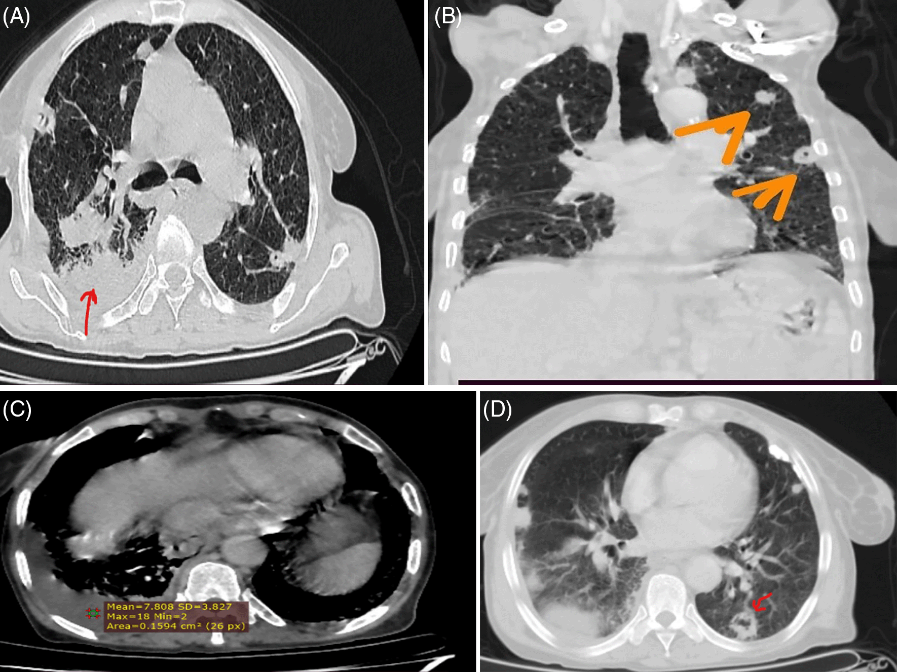
Imaging revealed an unremarkable ultrasound of the abdomen and pelvis, except for right lower lobe consolidation on the lung. Both arterial and venous doppler study of the right extremity exhibited normal findings, with the exception of right leg subcutaneous edema. The Trans-thoracic Echocardiography demonstrated normal results, showing no structural abnormalities or vegetations on the valves. A Chest CT scan with contrast identified multiple peripheral irregular nodules, some with cavitation and a typical feeding vessel sign. Additionally, right-sided pleural effusion and right basal consolidation were observed. Following the comprehensive clinical, laboratory, and imaging assessments, the patient was diagnosed with Septic pulmonary emboli secondary to soft tissue infection. The treatment regimen included Ceftriaxone 1 gm IV BID, Vancomycin 1 gm IV BID, as well as supportive oxygen therapy with 1L of INO2 via nasal prong. Furthermore, wound debridement and twice daily wound care initiation took place, leading to the patient’s admission to the Inpatient department for close monitoring. Subsequent follow-ups indicated a favorable progression in the patient’s condition, ultimately resulting in complete recovery. The patient was discharged from the hospital on the 14th day of admission without any relapses during the follow-up period.
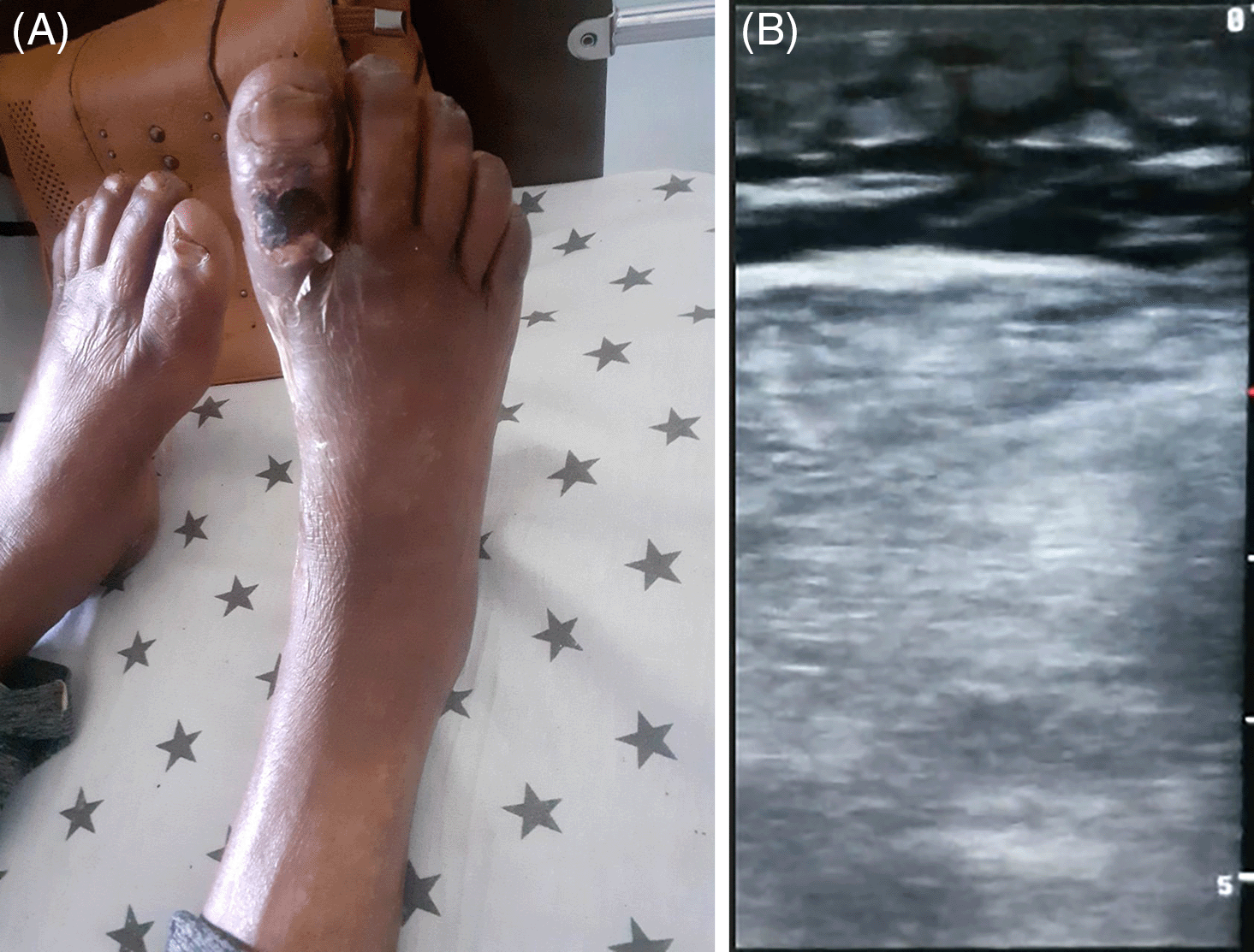
In previous times, Septic Pulmonary emboli were predominantly linked with Lemierre’s syndrome,4 post-partum septic pelvic thrombophlebitis,3and right-sided infective endocarditis in IV drug users.2 However, in the recent decade, there has been a shift in focus towards changes in the epidemiology, notably the significance of infected vascular catheters and implantable devices9 and more recently septic thrombophlebitis arising from contiguous deep soft tissue or bone infection of the extremities.6 Consequently, an escalating number of patients are exhibiting symptoms stemming from an extrapulmonary origin of infection, diverging from the traditional presentation documented in earlier literature. In our case study, we also describe a patient with soft tissue infection of the right foot as the focal point of infection as illustrated in Figure 1A with Sonographic features of cellulitis in Figure 1B indicating the rising occurrence of soft tissue as a primary site of infection.

In a retrospective study conducted from January 2000 to January 2013 at Hennepin County Medical and an urban teaching hospital in Minneapolis, Minnesota, USA, 40 patients were studied with septic pulmonary embolism. The median age of the patients was 46 years, with common presenting symptoms including febrile illness (85%) and pulmonary complaints (66%), such as pleuritic chest pain (22%), cough (19%), and dyspnea (15%). The sources of infection varied, with skin and soft tissue infection accounting for 44%, infective endocarditis for 27%, and infected peripheral deep venous thrombosis for 17%. A majority of cases (85%) were bacteremic with Staphylococcus aureus.1 This study featured a 55-year-old female patient who exhibited high-grade fever, productive cough with blood-tinged sputum, dyspnea, right-sided pleuritic chest pain, and a focus of infection in the right foot and big toe ulcer. As observed by Umesh Goswami et al., skin and soft tissue infection was the most common source of infection (44%), consistent with this case report. While Staphylococcus aureus was the causative organism in the referenced study, blood culture was not performed in the current case due to prior antibiotic exposure and test unavailability. However, many studies identify Staphylococcus aureus as an etiological agent.
In a retrospective analysis of patients with septic pulmonary emboli admitted between January 2007 and June 2018 at the Department of Respiratory and Critical Care Medicine, Hospital of Guangxi Medical University, China, Staphylococcus aureus was the predominant pathogen found in blood cultures (30.3%).10 Similarly, in a recent retrospective study conducted from January 1, 2018 to December 22, 2020 at Mogadishu Somali Turkey Education and Research Hospital, Somalia, Staphylococcus aureus was the most prevalent pathogen, accounting for approximately 52.6% of cases.11
The pathogenesis of septic pulmonary emboli arising from soft tissue infections involves the extravasation or translocation of bacteria from the extrapulmonary site into the systemic venous circulation.9 Subsequently, these bacteria can directly cause damage through toxins12 and indirectly through inflammatory mediators,13 occasionally leading to local thrombosis14 that facilitates bacterial growth. The embolization of these bacteria-containing thrombi to the pulmonary circulation can result in metastatic parenchymal lung infections, characterized by radiologic changes typical of septic pulmonary embolism, including cavitary and non-cavitary nodules, micro lung abscesses, consolidation, and pleural effusion. The diagnosis of septic pulmonary emboli presents challenges to clinicians due to nonspecific clinical and laboratory findings. Suspicions of SPE should arise in febrile bacteremic patients with an identified extrapulmonary source of infection who develop secondary pulmonary symptoms like pleuritic chest pain, dyspnea, and cough.1 With an elevated leukocyte count and increased acute inflammatory markers along with supportive chest findings on physical examination, it is advisable for the patient to undergo chest CT with contrast, which is often the preferred imaging modality in various research studies. Although not pathognomonic, the presence of bilateral wedge-shaped opacity, bilateral cavitary or non-cavitary nodules with a feeding vessel sign is highly indicative of septic pulmonary emboli. A retrospective analysis carried out at Surat Government Medical College in Gujarat, India, between 2018 and 2019 involved the study of chest CT scans from 10 patients. The findings revealed that 9 patients (90%) exhibited the classical feeding vessel sign, 8 patients (80%) displayed bilateral involvement, 7 patients (70%) presented with nodular cavitary and non-cavitary lesions, 2 patients (20%) showed pleural effusion, and 3 patients exhibited wedge opacities, while 6 patients (60%) displayed patchy ground glass opacity and consolidation.8 Another retrospective study conducted at Mogadishu, Somali Turkey Education and Research Hospital in Somalia involved chest CT scans of 19 patients. The results indicated that bilateral opacities were the most prevalent radiographic lesion type, observed in all patients (100%), followed by nodular (73.9%), cavitation (57.9%), consolidation (47.4%), non-nodular (26.3%), pleural effusion (26.3%), and feeding vessel sign (15.8%).11 In our case, the chest CT findings revealed multiple irregular peripheral nodules, some of which exhibited cavitations and demonstrated the typical feeding vessel sign, in addition to right-sided pleural effusion and right basal consolidation, as depicted in Figure 2A to D aligning with findings from numerous studies.

Treatment of septic pulmonary embolism should be commenced promptly in the early stages of the disease course and ought to encompass cause-specific interventions like wound debridement, abscess drainage, valve and catheter replacement, and other similar procedures. Identification of the microbiologic origin through blood, sputum, and pleural fluid cultures, along with deep soft tissue, bone, and explanted device cultures15 when applicable, is crucial for tailoring culture-specific antibiotic therapy. The initiation of empiric antibiotic treatment early on may constrain the microbiologic diagnosis.16 Staphylococcus aureus, notably Methicillin Susceptible Staphylococcus Aureus, remains the predominant pathogen in various research findings, aligning with the frequent occurrence of skin and soft tissue infections as the initial extrapulmonary source. Immediate commencement of empiric antibiotic therapy, starting with Staphylococcus aureus coverage and broad-spectrum antibiotics supplementation, is recommended. Subsequent adjustments to the antibiotic regimen based on culture outcomes should be made, continuing treatment for a minimum duration of 2-6 weeks. Treatment duration should be guided by clinical progress, follow-up culture results, inflammatory markers, and imaging studies. In our case report, blood culture was forgone due to the patient’s prior exposure to antibiotics and the unavailability of the test in our facility. Therapy was initiated with vancomycin 1gm IV BID and Ceftriaxone 1gm IV BID, considering coverage for Staphylococcus and broad-spectrum antibiotic inclusion. This approach yielded favorable outcomes in our patient, as evidenced by clinical amelioration, reduction in white blood cell count and acute inflammatory markers, and resolution of chest abnormalities in subsequent follow-up assessments.
Septic pulmonary embolism is an infrequent clinical entity with a serious prognosis if not promptly identified and managed. A heightened level of suspicion in individuals with infections outside the lungs who present with respiratory symptoms aids in the recognition of this condition. Utilization of chest CT is widely endorsed for imaging in septic pulmonary embolism, revealing distinct features like the feeding vessel sign. The primary treatment involves early administration of intravenous antibiotics and targeted therapies tailored to the specific cause, including procedures like abscess drainage, wound cleansing, and removal of infected medical devices. While it is preferable to tailor antibiotic therapy based on culture results, initiation of empirical antibiotic treatment should not be delayed as numerous studies have identified the predominance of staphylococcus aureus isolation. Therefore, selecting the appropriate antibiotics in light of this particular pathogen is crucial for effectively managing these patients.
According to the review board policy of our institution, ethical approval was obtained from the hospital ethics review committee for this case report and the images that go with it, the Patient’s written informed consent was also obtained for publishing this case as well as on usage of the images that go with it.
We express our gratitude to the patient and her family for supplying all pertinent details, and also to the healthcare providers involved in the management of this patient.
| Views | Downloads | |
|---|---|---|
| F1000Research | - | - |
|
PubMed Central
Data from PMC are received and updated monthly.
|
- | - |
Is the background of the case’s history and progression described in sufficient detail?
Partly
Are enough details provided of any physical examination and diagnostic tests, treatment given and outcomes?
Yes
Is sufficient discussion included of the importance of the findings and their relevance to future understanding of disease processes, diagnosis or treatment?
Partly
Is the case presented with sufficient detail to be useful for other practitioners?
Yes
References
1. Valerio L, Baddour LM: Erratum: Septic Pulmonary Embolism. A Contemporary Profile.Semin Thromb Hemost. 2023; 49 (8): e1 PubMed Abstract | Publisher Full TextCompeting Interests: No competing interests were disclosed.
Reviewer Expertise: Venous thromboembolism, Pulmonary embolism, Deep vein thrombosis, Thrombophlebitis
Is the background of the case’s history and progression described in sufficient detail?
Yes
Are enough details provided of any physical examination and diagnostic tests, treatment given and outcomes?
Partly
Is sufficient discussion included of the importance of the findings and their relevance to future understanding of disease processes, diagnosis or treatment?
Yes
Is the case presented with sufficient detail to be useful for other practitioners?
Yes
References
1. Güleç M, İnanç M, Pazarlı A, İnonu Köseoğlu H, et al.: Septic Pulmonary Embolism Due to Dialysis Catheter. Respiratory Case Reports. 2024; 13 (1): 25-28 Publisher Full TextCompeting Interests: No competing interests were disclosed.
Reviewer Expertise: Pulmonary infections,İntertisiel Lung Diseases,Sleep Apnea
Alongside their report, reviewers assign a status to the article:
| Invited Reviewers | ||
|---|---|---|
| 1 | 2 | |
|
Version 1 07 Oct 24 |
read | read |
Provide sufficient details of any financial or non-financial competing interests to enable users to assess whether your comments might lead a reasonable person to question your impartiality. Consider the following examples, but note that this is not an exhaustive list:
Sign up for content alerts and receive a weekly or monthly email with all newly published articles
Already registered? Sign in
The email address should be the one you originally registered with F1000.
You registered with F1000 via Google, so we cannot reset your password.
To sign in, please click here.
If you still need help with your Google account password, please click here.
You registered with F1000 via Facebook, so we cannot reset your password.
To sign in, please click here.
If you still need help with your Facebook account password, please click here.
If your email address is registered with us, we will email you instructions to reset your password.
If you think you should have received this email but it has not arrived, please check your spam filters and/or contact for further assistance.
Comments on this article Comments (0)