Keywords
Transomental hernia, Internal hernias, Small bowel occlusion, Great omentum
Transomental hernia, Internal hernias, Small bowel occlusion, Great omentum
Internal hernias are protrusions of the viscera through a peritoneal or mesenteric opening, with the herniated viscera remaining within the abdominal cavity.1 Transomental hernias are a rare type, accounting for less than 1% of all internal hernias.2 They occur when abdominal contents protrude through a defect in the omentum, leading to bowel obstruction and potential strangulation.1 Compared to other internal hernias, transomental hernias more frequently present with strangulation of the small bowel.3 We present the case of a strangulated omental hernia to highlight the importance of early diagnosis to reduce the high risk of mortality associated with this condition.
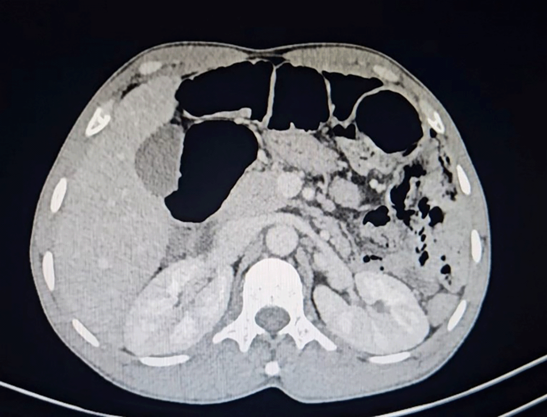
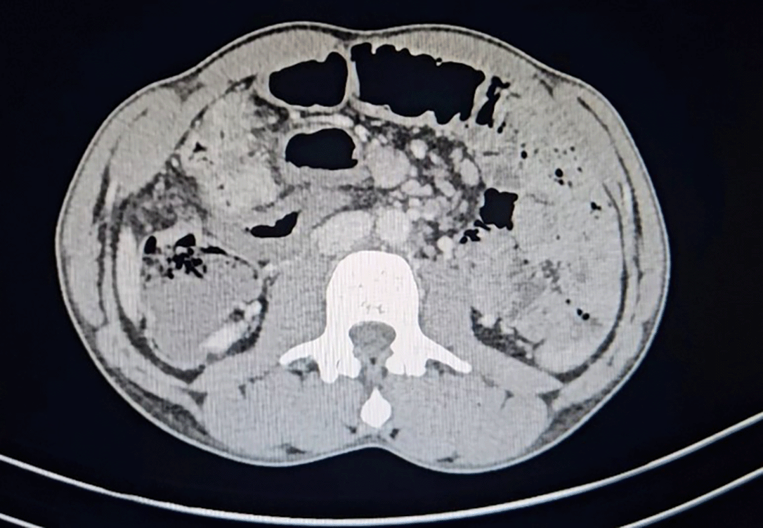
A 38-year-old male patient, with no medical or surgical history, consulted our emergency room for an abdominal pain evolving for 48 hours accompanied by vomiting and a cessation of materials and gas, without a fever context. Clinical assessment revealed a patient who was thin and calm with a deteriorated general condition. Upon physical examination he was apyretic, stable hemodynamically and respiratorily, with a non-scarring abdomen. Notably he has a slight abdominal bloat, a defense of the right iliac fossa, and tympanism. The hernia orifices were free and the digital rectal examination normal. He had leukocytosis and neutrophilia on blood picture at 21000 and a CRP at 144. An emergent computed tomography (CT) scan performed revealed an intestinal distension proximal to a beak-shaped transitional zone located in the right paracolic gutter with decreased enhancement of the small bowel. Suspicion of a congenital band or an internal hernia ( Figures 1 and 2).
Given the clinical and radiological discrepancy, surgical treatment was opted for. Laparoscopy revealed a transomental hernia with part of ileum herniating in the omental defect close to the transverse colon. The obstructed loop of small intestine in hernia was compromised and gangrenous ( Figures 3 and 4). A conversion by the midline incision was requisite.
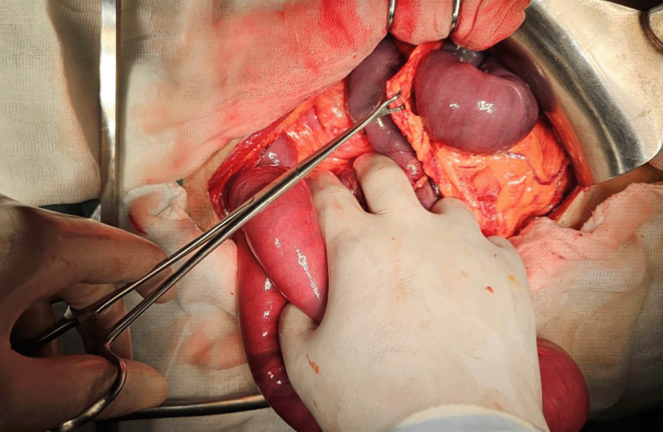
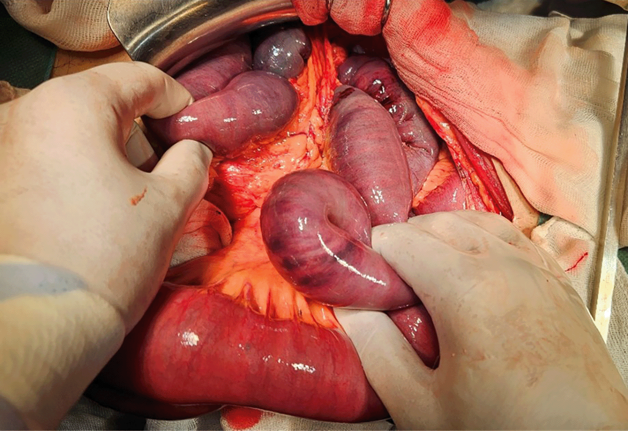
The compromised small intestine was resected leaving 2.5 meters of good vitality downstream of the duodenojejunal angle. Given the perioperative findings of peritonitis contraindicating anastomosis, it was decided to exteriorize the small intestine in a double gun barrel ileostomy. The transomental defect was repaired. The postoperative course was uneventful.
Internal hernias are protrusions of the viscera through a peritoneal or mesenteric opening, with the herniated viscera remaining within the abdominal cavity.1 The apertures for internal hernias can be classified as normal (e.g., foramen of Winslow), paranormal (e.g., paraduodenal, ileocecal, supravesical fossa), or abnormal (e.g., transomental defect) based on the typical anatomy of the abdominal cavity.4 These hernias account for 0.6% to 5.8% of all cases of intestinal obstruction.3 Transomental hernias are a rare type, accounting for less than 1% of all internal hernias.2 The greater omentum extends inferiorly from the greater curvature of the stomach to the transverse colon and beyond, draping into the anterior, inframesocolic abdomen. Omental defects can be congenital or acquired. Bowel can herniate through one to four layers of the omentum, but most commonly penetrates all four layers.5 Yamaguchi classified transomental hernias as type A (peritoneal cavity → greater omentum → peritoneal cavity), B (peritoneal cavity → omental bursa → peritoneal cavity), or C (peritoneal cavity → omental bursa).6 Our case was a type A.
This pathology occurs at all ages. However, the literature notes a predominance between 38 and 48 years. A study of 14 cases of internal abdominal hernias reported a mean age of 71 years.7 Our patient was relatively younger. Regarding gender, several studies have noted a male predominance of this condition.1 The usual way of revealing internal hernia is intestinal obstruction including nausea, vomiting, abdominal pain, distended abdomen, and constipation. Comparing with other types of intern hernias clinical signs of transomental hernia show a severe periumbilical colics accompanied by hyperactive bowel sounds and progressive distension. The surgeon can feel a sensitive abdominal mass, which represents the Gordian knot of the herniated intestine.2 Our patient showed a small bowel occlusive syndrome and a severe colic in the right iliac fossae with a slight distended abdomen.
In 2007, in a series of 14 cases, Armstrong stated that preoperative diagnosis of internal hernias was almost impossible.1 However, nowadays, several authors show the effectiveness and reliability of the abdominal CT not only to confirm the diagnosis of intestinal obstruction, but also to specify the location and type of the internal hernia.8 It can be helpful for diagnosis, revealing findings such as dilated small bowel loops pressed against the abdominal wall without omental fat causing central displacement of the colon segments and a “beak sign” at the transition zone as observed in our case. Because the defect is not visualized, the anomalies of the mesenteric vessels may be seen such as a stretched and agglomerated mesenteric vascular pedicle or a displacement of the main mesenteric.2 However, radiographic findings are often nonspecific and definitive diagnosis is usually made intraoperatively. Hence a skilled radiologist is essential to identify the subtle signs of internal hernias, which can have a significant impact on patient outcomes.
Prompt surgical intervention is critical, as transomental hernias have a high postoperative mortality rate of up to 30% due to the high incidence of strangulation.5 In most cases, a gangrenous bowel is present at the exploratory laparotomy. Treatment involves reduction of the hernia contents, resection of nonviable bowel if present, and repair of the omental defect to prevent recurrence. Mortality is highest when there is a delay in diagnosis and treatment, emphasizing the importance of maintaining a high index of suspicion for this rare cause of bowel obstruction. Our patient consulted our emergency department late, which accentuated digestive ischemia and compromised his bowel loop vitality.
A first laparoscopy is an emerging technic and should be attempted nowadays. A few rare cases of laparoscopic reduction of this type of hernia are described in the literature.7 In our case, it was deemed lucid to proceed with a midline incision conversion due to the presence of intestinal necrosis, in order to preserve the patient’s prognosis and ensure a successful surgical outcome.
Although internal hernias account for a relatively small percentage of cases, they are frequently overlooked as a cause of bowel obstruction. Diagnosis this condition is challenging, both clinically and radiologically. Facilitating prompt surgical intervention to mitigate high mortality is essential. Abdominal CT remains a cornerstone of radiological assessment, and it has already been established that laparoscopic approach is a safe surgical approach.
No generative AI technologies such as Large Language Models (ChatGPT, COPILOT, etc.) and text-to-image generators have been used during the writing or editing of this manuscript.
Written informed consent for publication of their clinical details and/or clinical images was obtained from the patients.
Written informed consent for publication of their clinical details and/or clinical images was obtained from the patients.
Authors and affiliations:
General surgery department, Regional Hospital of Beja, Beja, Tunisia
Ahmed Menif, Souhaib Atri, Mohamed Rached Khelili, Helmi Zebda, Wassim Riahi
Contributions:
WR contributed to work conception, MK and HZ collected data and SA revised the manuscript and AM wrote the manuscript.
Corresponding author:
Correspondence to Ahmed Menif.
| Views | Downloads | |
|---|---|---|
| F1000Research | - | - |
|
PubMed Central
Data from PMC are received and updated monthly.
|
- | - |
Provide sufficient details of any financial or non-financial competing interests to enable users to assess whether your comments might lead a reasonable person to question your impartiality. Consider the following examples, but note that this is not an exhaustive list:
Sign up for content alerts and receive a weekly or monthly email with all newly published articles
Already registered? Sign in
The email address should be the one you originally registered with F1000.
You registered with F1000 via Google, so we cannot reset your password.
To sign in, please click here.
If you still need help with your Google account password, please click here.
You registered with F1000 via Facebook, so we cannot reset your password.
To sign in, please click here.
If you still need help with your Facebook account password, please click here.
If your email address is registered with us, we will email you instructions to reset your password.
If you think you should have received this email but it has not arrived, please check your spam filters and/or contact for further assistance.
Comments on this article Comments (0)