Keywords
dynamic foot posture, biomechanics; motion capture; reliability, Kinovea; gait; 3D motion analyzer.
Dynamic foot posture assessment is essential to determine the pathomechanics of the foot during walking. Currently, there is no affordable method for assessing dynamic foot posture that can be used in a clinical setting. Kinovea, an open-access video analysis-free application, has enabled clinicians to conduct gait analysis at a relatively low cost, but its reliability in assessing dynamic foot posture has not been studied.
32 observations were recorded from various angles throughout the gait cycle, each at varying speeds, and analyzed using Kinovea. Two independent examiners conducted observations to measure the medial longitudinal arch (MLA), first metatarsophalangeal joint (MTP 1), ankle inclination (AI), and calcaneal angle (CA). A third examiner was tasked with comparing the results of intra-class correlation coefficient (ICC) between the two examiners.
The inter-rater reliability showed promising results despite the observations made at 2, 3, and 4 km/h. MLA and MTP 1 observations remained “excellent” at all speeds, while AI and CA had “good” reliability among observers at 2, 3 and 4 kmph, apart from 4 kmph where AI showed “excellent” reliability.
In this study, Kinovea showed good and excellent reliability among observers in measuring the MLA, MTP 1, AI, and CA at various speeds, therefore, being a suitable alternative to a 3D motion analyzer which may not be readily available in certain facilities.
dynamic foot posture, biomechanics; motion capture; reliability, Kinovea; gait; 3D motion analyzer.
The foot is one of the most complex organs within the body, and not only serves as a mode of transportation in our daily lives but also plays a crucial role in maintaining balance. It forms a vital component of a closed-chain system in which the lower limb provides support to the body. Consequently, any deviation in foot posture and stability can affect adjacent segments more proximally.1
Kinematic studies are often required to objectively evaluate human movements during various activities, including gait analysis, clinical research, sports management, footwear, and orthopedics.2 Regardless, the administration of these assessments remains highly dependent on the evaluators’ subjectivity. The goal of kinematic analysis is to obtain quantifiable data to help analyze the patient’s condition and changes during different moments, that is, changes in pre-and/or post-management. Gait analysis is a rigorous examination, requiring advanced medical tools and systems to perform an accurate analysis. Technical challenges with interpretation and setup, accompanied by expensive equipment and software, can restrict their application in both clinical and research settings.3 To mitigate these issues associated with three-dimensional motion capture systems, new low-cost motion analysis applications have been emerging over the past couple of years.4,5
One of these novel applications is Kinovea, which was first created in 2009 by a non-profit collaboration between several researchers, athletes, coaches, and programmers worldwide. Kinovea is an open-access application that allows physicians worldwide to conduct gait analysis with available technology. This application allows examiners to measure angles, distances, coordinates, and spatial-temporal parameters. Measurements can be taken from multiple perspectives, as the software can perform calibration in non-perpendicular planes of the camera-object line.3 Within the realms of medicine, Kinovea has been used in many fields of research, medication or clinical analysis and sports.
Although Kinovea is a user-friendly, portable, and relatively cost-free tool, it is not without its flaws. Prior research has yielded inconclusive findings regarding its reliability, ranging from poor to excellent, with emphasis on the significance of proper configuration before application.6–8
This study aimed to assess the reliability and applicability of a simple, cost-effective, user-friendly, and objective dynamic foot posture evaluation using the Kinovea software. By evaluating the inter-rater reliability of the Kinovea, an assessment of dynamic foot posture, represented by measurements of ankle inclination, medial longitudinal arch, calcaneal angle, and MTP 1 (metatarsophalangeal) angle at each gait phase will be conducted.
The target population for the study was healthy adults without lower extremity abnormalities in Indonesia within the environment of RSUPN Cipto Mangunkusumo, Jakarta outpatient clinic. The subjects were selected according to the following eligibility criteria: older than 18 years of age, able to walk at speeds of up to 4 km/h for at least two minutes, and absence of pathologies that alter gait and/or posture (e.g., deformities, pain, amputees, etc.). Patients were excluded if they had balance and coordination disorders or were unwilling to participate. In this study, 32 observations were conducted for each angle measured at different gait phases and speeds after eligibility criteria was met and written informed consent was obtained. Subjects were selected through consecutive sampling.
Dynamic foot posture videos of participants were collected using a digital camera (GoPro HERO 10) with 1080P resolution at 240 frames per second (fps). The cameras were mounted on a tripod at the appropriate positions to optimize video stabilization 30 cm from the sides of the treadmill at 30 cm from the floor. External lighting with a soft box and contrasting matte sticker marker was used to improve image quality.
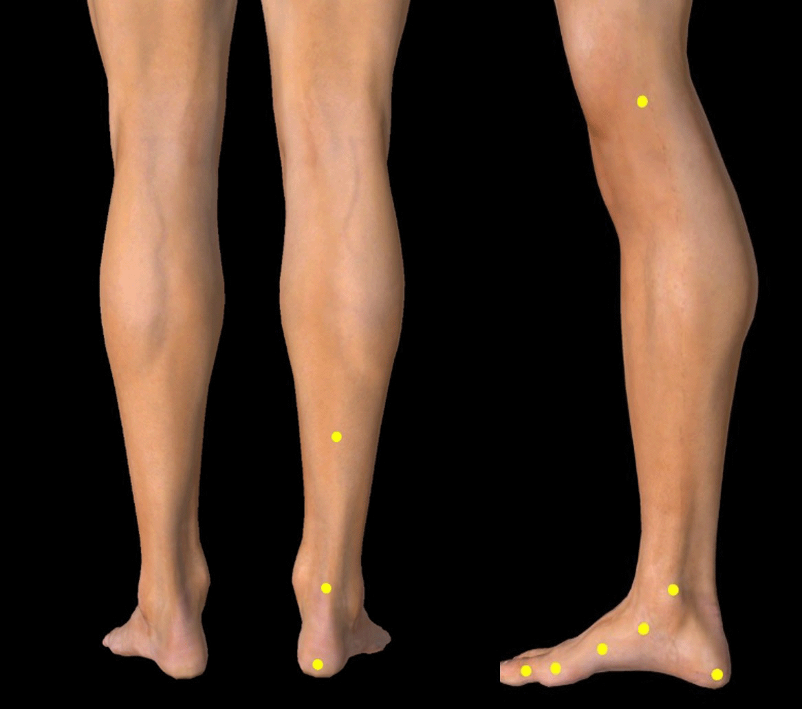
The research was conducted at the Physical Medicine and Rehabilitation outpatient clinic of the Cipto Mangunkusumo Hospital in Jakarta. The participants were asked to take off their shoes, and markers were placed on their feet. Markers were placed at the medial knee joint, first metatarsal tuberosity, navicular tuberosity, medial malleolus, posteromedial calcaneus, midpoint of the first metatarsal, and head of the first proximal phalange in the sagittal portion of the video. For the frontal aspect of the video, markers were placed along the insertion of the Achilles tendon at the posterior calcaneus, midportion of the Achilles tendon at the level of the medial malleolus, and level of the tendon-muscle junction as seen in Figure 1.

Three markers were placed in the posterior aspect at: (1) insertion of the Achilles tendon at the posterior malleolus, (2) midportion of the Achilles tendon at the height of the medial malleolus and (3) tendon-muscle junction of the gastrocnemius muscle.
The cameras were placed 30 cm away from the treadmill at the respective locations to allow simultaneous recording from multiple angles. The height of the camera was adjusted according to the height of the participants to allow perpendicular alignment with the patient’s foot approximately 30 cm above the floor as seen in Figure 2.

Cameras were positioned, 30 cm away from both the lateral and posterior sides of the treadmill at a height of 30cm using a tripod.
Cameras were positioned, 30 cm away from both the lateral and posterior sides of the treadmill at a height of 30 cm using a tripod.
Subjects were then requested to familiarize themselves with walking on the treadmill at a speed of 1 km/h. Once the participants were accustomed to the treadmill, the speed was gradually increased to 4 km/h in increments of 1 km/h every 30 s. All the captured videos were examined individually before analysis to avoid any obvious errors during the recording session. Participants with video defects were asked to repeat the same measurements to improve video quality. Three parameters were measured in the sagittal plane: the medial longitudinal arch (MLA) angle, first metatarsophalangeal (MTP 1) angle, and ankle inclination (AI) angle, while one parameter was measured in the posterior coronal plane: calcaneal angle (CA). The ‘angle’ tool was conducted by each examiner to determine the measured angle at specific moments during the gait cycle. The angle calculation procedure is as follows:
• MLA. The angle is formed by the line between the head of the first metatarsal and the navicular tuberosity and the line between the navicular tuberosity and the posteromedial calcaneus. The following gait phases were analyzed for MLA: initial contact (IC), loading response (LR), midstance (MSt), terminal stance (TSt), preswing (PSw), hallux extension (HE), and initial swing (ISw).
• MTP 1. The angle is formed by the line between the midpoint of the first metatarsal bone and the head of the first metatarsal and the line between the head of the first metatarsal and the head of the first proximal phalange. The following gait phases were analyzed for MTP 1: PSw, HE, and ISw.
• AI. The angle is formed by the vertical line passing through the medial malleolus and the line between the medial knee joint and medial malleolus. The following gait phases were analyzed for AI: LR, MSt, and TSt.
• CA. The angle is formed by the line between the posterior calcaneus and the posterior ankle joint line and the line between the midpoint of the tendon muscle junction and the posterior ankle joint line. The following gait phases were analyzed for CA: IC, LR, MSt, and PSw.
For inter-rater reliability, two independent medical physicians were tasked with analyzing the video independently using the above parameters for each subject separately. Both physicians received training to use the Kinovea application from a senior consultant beforehand to ensure accurate interpretation and analysis. The examination results were then sent to a third physician to analyze and compare the results between the first two physicians.
A univariate analysis was conducted. Numeric data, including ankle inclination, medial longitudinal arch (MLA), calcaneal angle, and MTP 1 angle, will be presented as mean and standard deviation if the data distribution is normal. If the numeric data distribution is non-normal, the data are presented as median, minimum, and maximum values. As the sample size was >50, the Kolmogorov–Smirnov test was used to analyze the data distribution, with a distribution considered normal if p > 0.05.
To evaluate the reliability between the two examiners, the intra-class correlation coefficient (ICC) was used to evaluate the results. The ICC was calculated using SPSS Version 27, and the 95% confidence interval (CI) is displayed. The points estimated for ICC were interpreted as excellent (>0.9), good (0.76-0.9), moderate (0.5-0.75) and poor (<0.50).9 ICC will be calculated from both examiners for each parameter measured at three different speeds.
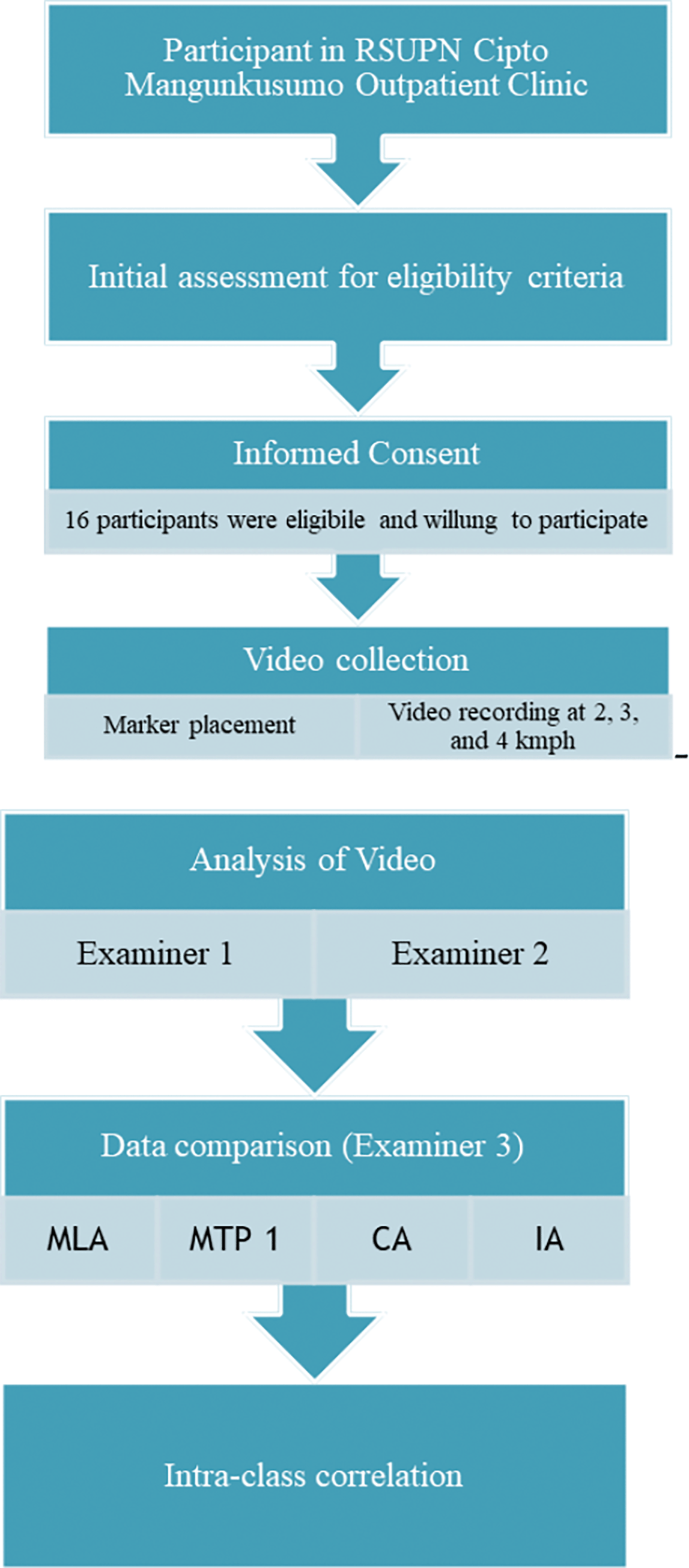
The study recruited 16 participants, consisting of equal numbers of males and females, without any alteration in gait patterns. There were no missing data in the examiners’ results. Video recording of patients were taken for all participants at varying speeds at 2, 3, and 4 kmph. Video analysis was conducted by two different examiners to compare the accuracy and reliability when observing MLA, MTP 1, CA and IA as shown in Figure 3.

Analysis of MLA, MTP 1, CA and IA were conducted by two examiners to analyze ICC.
As mentioned above, the point estimate of ICC will be interpreted as excellent (>0.9), good (0.76-0.9), moderate (0.5-0.75) and poor (<0.50).9 The table below represents the mean angle measured, standard deviation of each examiner as well as the ICC results, 95%CI and p-value.
Table 1 presents the results obtained when the participants were asked to walk on the treadmill at 2 km/h. An inter-rater reliability of >0.9. was observed in both MLA and MTP 1 parameters, indicating excellent correlation, while CA and AI showed good correlation at 0.893 and 0.888 ICC, respectively. As seen in Tables 2 and 3, these observations remained consistent throughout the examination, as the same results were observed at 3 km/h and 4 km/h, with improvements in the AI ICC at 4 km/h, to a value of 0.927 from 0.889 at 3 km/h.
| Parameter (Degrees) | 1st Examiner (M±SD) | 2nd Examiner (M±SD) | ICC | 95% CI | p-Value |
|---|---|---|---|---|---|
| MLA | 148.38 ± 8.00 | 148.93 ± 8.20 | 0.976 | 0.968-0.981 | *<0.001 |
| MTP1 | 143.33 ± 15.95 | 144.11 ± 16.65 | 0.962 | 0.944-0.975 | *<0.001 |
| CA | 174.50 ± 5.45 | 173.82 ± 5.24 | 0.807 | 0.727-0.864 | *<0.001 |
| AI | 171.96 ± 6.13 | 172.67 ± 5.14 | 0.927 | 0.889-0.952 | *<0.001 |
The human foot comprises a complex arrangement of 26 bones, making the assessment of interosseous angles extremely difficult, especially during dynamic processes such as walking. In particular, MTP 1 and MLA angles play essential roles as early indicators of foot pathologies. Restriction of the MTP 1 joint motion, such as in the hallux rigidus, affects propulsion during toe-off. This causes foot inversion and lateral transfer of stress, and can ultimately disrupt stride length. MLA changes are associated with early signs of foot pronation and hallux valgus. Both these pathologies have been shown to increase lumbopelvic muscle activation, causing low back pain and other dysfunctions.10,11
Previous research has been conducted to analyze the inter-reliability of Kinovea to measure its consistency in large joints, such as the knee, ankle, hip, and pelvis.2,3,6,12,13 However, there is a lack of evidence in the reliability of its use in smaller joints such as the foot.2,12,13 The purpose of this study is to examine the consistency among different raters, and reliability of Kinovea as an objective, quantitative, cost-efficient, and straightforward, utilizing a 2D video setup, motion analysis software in analyzing the smaller joints of the human foot. To the best of our knowledge, this study is one of the first to compare the inter-rater reliability of Kinovea to measure dynamic foot posture at varying speeds during a gait cycle.
Kinovea allows for an objective and quantitative dynamic foot posture analysis using free software. The requirement to conduct the examination is relatively simple and allows for repeated measurements, which allows in-depth examination where three-dimensional motion capture systems might not be readily available. However, the primary drawbacks in reliability studies for Kinovea® in the medical literature are the lack of a standardized video protocol and consistent marker placement to allow homogenized and standardized examination.12,14
The primary outcome of this study was to compare the inter-class reliability of the Kinovea to evaluate various dynamic foot posture parameters between examiners. Four parameters (MLA, MTP 1, CA, and AI) were measured during different gait phases by two independent examiners, and the ICC results were analyzed by a third examiner for statistical significance. All measurements had either excellent or good ICC between examiners, indicating that both examiners were able to consistently measure parameters reliably. These findings were especially true for MLA and MTP where ICC values of >0.9 were consistently observed at 2 kmph up to 4 kmph and for AI at 4 kmph, while “good” correlations were seen in when CA and AI were measured throughout 2 kmph to 3 kmph. These findings imply that Kinovea can be used in various settings, including various speeds, as long as the measurements are taken with the same parameters regardless of the examiner.
The high ICCs observed may be ascribed to the effective standardization of marker placement and sufficient training sessions undertaken by the raters, thereby minimizing errors. An accurate, user-friendly, and cost-effective tool is essential in any medical profession. Because Kinovea is free to download applications, it has virtually no cost to use, except for the camera, light source, and sticker marker. 3D motion analysis systems are dependable in medical contexts but pose challenges owing to their inconvenience, high cost, complexity, technical proficiency, and expensive equipment. In 2014, Macleod et al. estimated that the cost of conducting 3D gait studies could reach 2,000 USD, and the cost of building and constructing a movement laboratory could be as high as 300,000 USD. Given the advancements in technology, this value can be even higher.15 As such, dynamic foot posture analysis with Kinovea proved to be a reliable alternative, which is more cost-friendly and can be applied more widely.
In this study, the researcher only included healthy patients; therefore, the results cannot be generalized to other populations or pathologies, and were limited by the small sample size. Another limitation was the lack of intrarater reliability, as the examiners did not perform multiple measurements.
Further studies with larger sample sizes are required to ensure more accurate representation of the general public. Patients with certain pathologies could be recruited to observe and analyze the plausibility of using Kinovea for specific diseases/conditions. A standardized video protocol should be established to reduce any unexpected errors and portray a delineated examination.
This study, conducted on 32 specimens, showed that Kinovea is a reliable and applicable software for examining and analyzing dynamic foot posture parameters at varying speeds. It was observed that reliability was obtained and maintained with “good” result, and even “excellent” in most of the parameters measured. In conclusion, Kinovea as a free software can be used as a reliable alternative for gait evaluation in clinical settings, where 3D motion analyzers might not be readily available.
This study was approved by the Research Ethics Committee of Universitas Indonesia. All methods were carried out in accordance with relevant guidelines and regulations (ethical approval reference number: KET-345/UN2.F1/ETIK/PPM.00.02/2023, March 23, 2023). Written informed consent was obtained from all study participants before the start of the study.
Underlying data: Figshare: Inter-Observer Reliability of Kinovea® Software in Dynamic Foot Posture Analysis in Healthy Population https://doi.org/10.6084/m9.figshare.27366285.
This project contains:
Data are available under the terms of the Creative Commons Zero “No rights reserved” data waiver (CC0 Public domain dedication).
Kinovea is an open-access application available through: https://www.kinovea.org/ https://www.kinovea.org/
The authors would like to thank all parties involved in Cipto Mangunkusumo Hospital and Suyoto Hospital for supporting this research and all the volunteers who participated in this study.
| Views | Downloads | |
|---|---|---|
| F1000Research | - | - |
|
PubMed Central
Data from PMC are received and updated monthly.
|
- | - |
Provide sufficient details of any financial or non-financial competing interests to enable users to assess whether your comments might lead a reasonable person to question your impartiality. Consider the following examples, but note that this is not an exhaustive list:
Sign up for content alerts and receive a weekly or monthly email with all newly published articles
Already registered? Sign in
The email address should be the one you originally registered with F1000.
You registered with F1000 via Google, so we cannot reset your password.
To sign in, please click here.
If you still need help with your Google account password, please click here.
You registered with F1000 via Facebook, so we cannot reset your password.
To sign in, please click here.
If you still need help with your Facebook account password, please click here.
If your email address is registered with us, we will email you instructions to reset your password.
If you think you should have received this email but it has not arrived, please check your spam filters and/or contact for further assistance.
Comments on this article Comments (0)