Keywords
Inflammatory orbital pseudotumor, Behçet’s disease, Inflammatory diseases, Corticosteroids
To provide an original observation of Behçet’s disease, revealed by an inflammatory pseudotumor of the orbit.
We reported an observation of an inflammatory pseudotumor of the orbit that revealed Behçet’s disease.
Twenty-Eight years old patient was admitted to the internal medicine department for painful edema in the right eye with headache and loss of visual acuity.
Ophthalmologic examination revealed eye protrusion with conjunctival hyperemia. Orbital MRI resonance imaging revealed periorbital inflammatory thickening associated with inflammatory myositis. Behçet’s was diagnosed based on a history of recurrent oral aphthous since childhood, pseudofolliculitis, pathergy test positivity, and negativity of the rest of the etiological investigations. The evolution was spectacular with boli methylprednisolone and colchicine prescriptions.
Although the association is rare, Behçet’s disease should be included in the workup of inflammatory pseudotumors of the orbit.
Inflammatory orbital pseudotumor, Behçet’s disease, Inflammatory diseases, Corticosteroids
Behçet’s disease (BD) is a systemic vasculitis that predominantly affects males. Eye involvement is common, occurs in 28-50% of cases, and is dominated by uveitis.1,2
A 28-year-old Tunisian male patient was admitted for right orbital protrusion and edema (Figure 1) with local pain and intense helmet headache lasting for 15 days before admission.
Ophthalmological examination of the right eye revealed a visual acuity of 2/10. A painful non-pulsatile non-reducible exophthalmia with corneal edema, Tyndall to 2 crosses in the anterior chamber with iridocorneal synechia. Examination of the left eye revealed no abnormalities.
Both brain and orbital MRIs were performed using T1, T2, T2*, and Flair diffusion protocols with sagittal axial and coronal cuts with and without gadolinium injection.
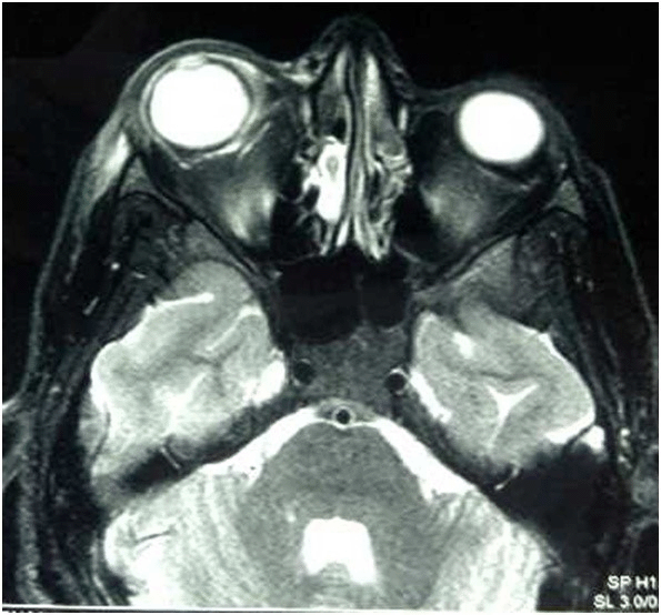
Brain MRI revealed no cerebral venous thrombosis. Orbital MRI revealed thickening of the right anterior orbital soft tissues along with an important inflammatory signal in the extra conal fat, which was moderately enhanced by gadolinium injection, with no intra- or extra-conal soft tissue collection.
MRI also showed a discrete inflammatory signal abnormality with contrast enhancement of the right internal and upper right oblique muscles and periorbital inflammatory thickening associated with inflammatory myositis with no extension to the intra-conal or intracranial fat (Figures 2–5). No abnormalities were observed in the rest of the structures, especially in the optic nerves.


Physical examination revealed a history of recurrent oral aphthous and pseudofolliculitis. The remaining examinations revealed no abnormalities.
B5 HLA typing was negative, and pathergy test results were positive. The diagnosis of Behçet’s disease with inflammatory orbital pseudotumor was confirmed. A workup of the tumor was performed as follows: an ENT examination and bacterial and viral serologies. Thyroid balance, antinuclear antibodies, and tumor markers CEA – PSA, alpha FP, CA 19-9, chest radiographs, sinus, and pelvic abdominal ultrasound showed no abnormalities. Biopsy of the mass was not performed. The patient received three bolus doses of 1 g of methylprednisolone per day, each relayed with corticosteroid (1 mg/kg/day associated with colchicine at a dose of 1 mg/day).
The outcome was favorable after 5 days, with regression of the local inflammatory phenomena (Figure 6). Examination of the control eye showed improvement in the right visual acuity to 9/10, persistent conjunctival injection with normal eye movements, and a quiet anterior segment with a normal eye bottom. The retinal angiography results were normal. Based on both clinical and examination data improvements, we did not opt for subsequent MRI control. The patient did not relapse after 6 months of follow-up.
The idiopathic orbital inflammatory syndrome was first described by Birch and Hirschfield in 1905. It is also known as orbital pseudotumor and represents a nonspecific and nonneoplastic inflammatory process of the orbit.3,4 This condition most commonly involves extraocular muscles. Less commonly, there are inflammatory changes involving the uvea, sclera, lacrimal gland, and retrobulbar soft tissue. The exact etiology is unknown. The main differential diagnosis is malignant tumor progression.5,6
It represents 4.7 to 6.3% (depending on the series) of all orbital conditions.7 We distinguished between localized and diffuse inflammatory pseudotumors of the orbit.
Myositis is the most frequent orbital-localized inflammatory pseudotumor. Orbital myositis is a form of idiopathic orbital inflammation, characterized by inflammation of the external eye muscles. It may lead to proptosis, periocular pain, and diplopia.4–6
The etiologies are dominated by infectious causes (Lyme disease and trichinosis), sarcoidosis, Crohn’s disease, systemic lupus, polyarteritis nodosa, rheumatoid arthritis, Wegner’s granulomatosis, and neoplastic causes.4,5,8
Diagnosis is based on radiological, histological, and clinical data.
The association between Behçet’s disease and inflammatory pseudotumor has rarely been reported, and the first observation was published in 1996, reporting an inflammatory pseudotumor of the terminal ileum in Behçet’s disease.6 Other locations have been described: the heart,9 brain,10 and orbit.11–13
Myositis associated with Behçet’s disease is rare. It mainly affects the skeletal muscles.7
There are few associations between orbital myositis and Behçet’s disease, as mentioned in Table 1.7,8,11,12–14 It is rarely reported as an initial manifestation of Behçet’s disease, as illustrated in our case.7,13,14 To the best of our knowledge, this is the fourth reported case of Behçet’s disease caused by an inflammatory orbital pseudotumor.
| Reference | Year | Age | Gender | Presentation as the initial manifestation | Other manifestations of BD | Treatment | Evolution |
|---|---|---|---|---|---|---|---|
| Bouomrani et al.7 | 2012 | 37 | M | Yes | Recurrent oral and genital ulcers Pseudofolliculitis | Intravenous methylprednisolone Colchicine | Improvement |
| Jee-Hoon Roh et al.8 | 2006 | 36 | F | No | Recurrent oral aphtous Uveitis Erythema nodosum Positive pathergy test | Intravenous methylprednisolone (1 g/day for 5 days) followed by oral prednisolone 50 mg/day | Improvement |
| Hammami et al.11 | 2006 | 35 | F | No | Recurrent oral and genital ulcers Pseudofolliculitis Polyarthralgia Positive pathergy test | Intravenous methylprednisolone (1 g/day) for 3 days followed by oral prednisone (1 mg/kg/day) | Improvement |
| Chebbi et al.12 | 2013 | 45 | M | No | NA | Corticosteroids | Improvement |
| Garrity et al.16 | 2004 | 33 | F | No | NA | Oral corticosteroids, cyclophosphamide, azathioprine, cyclosporine, colchicine, and methotrexate | Improvement then recurrence 6 years later in the controlateral eye |
| Oral corticosteroids Cyclosporine that was stopped because of side effects Orbital radiotherapy Infliximab (4 mg/kg) Methotrexate | Immediate relief then she developed recurrence 9 months later | ||||||
| Methotrexate (10 mg weekly), infliximab (8 mg/kg every two months), and prednisone (20 mg) | NA | ||||||
| Shinya et al.13 | 2022 | 32 | F | Yes | Oral and genital ulcers Folliculitis-like skin rash Presence of ulcers at the ileum | Intravenous methylprednisolone Ciclosporine | Improvement |
| Aiessa Fedrigo et al.14 | 2017 | 26 | F | Yes | Oral and genital ulcers Arthralgias and arthritis | High dose corticosteroids | Absence of improvement |
| Botulinum toxin injection on medial rectus of right eye | Brief improvement | ||||||
| Azathioprine | Absence of improvement | ||||||
| Anti-TNF α | Improvement | ||||||
| Our patient | 2023 | 28 | M | Yes | Oral aphthous Pseudofolliculitis Positive pathergy test | Intravenous methylprednisolon: 1 gram per day for three days Oral corticosteroid: 1 mg/kg/da Colchicine | Improvement |
As orbital biopsies are rarely done, radiological diagnosis is sufficient.7
Treatment is based on corticosteroids,7–9 as is the case in our patient. However, it may be refractory to corticosteroid therapy. Sometimes, immunosuppressive drugs are also required.7,15 The prognosis is favorable with this type of treatment, apart from some recalcitrant forms that require the use of TNF blockers.16
Rituximab has demonstrated effectiveness in cases resistant to glucocorticoids, surgery, or radiation therapy. This finding indicates that rituximab may be a valuable treatment option for managing this condition.17,18
The association between Behçet’s disease and the inflammatory pseudotumor of the orbit is exceptional. Orbital inflammation should be considered as an ophthalmic manifestation of Behçet’s disease and treated precociously to preserve the visual prognosis.
- Dr. Sameh Sayhi: Revising the article, approval of the work, and agreeing to be held accountable.
- Dr. Arij Ezzouhour Yahyaoui: Drafting and revising the article, approval of the work, and agreeing to be held accountable.
- Dr. Rim Dhahri: Drafting, Approval of the work, and agreeing to be held accountable.
- Dr. Nour Elhouda Guediche: Revising the article, approving the work, and agreeing to be held accountable.
- Dr. Bilel Arfaoui: Revising the article, approval of the work, and agreeing to be held accountable.
- Dr. Faida Ajili: Revising the article, approval of the work, and agreeing to be held accountable.
- Dr. Nadia Ben Abdelhafidh: Revising the article, approving the work, and agreeing to be held accountable.
All data underlying the results are available as part of the article and no additional source data are required.
| Views | Downloads | |
|---|---|---|
| F1000Research | - | - |
|
PubMed Central
Data from PMC are received and updated monthly.
|
- | - |
Is the background of the case’s history and progression described in sufficient detail?
Partly
Are enough details provided of any physical examination and diagnostic tests, treatment given and outcomes?
Partly
Is sufficient discussion included of the importance of the findings and their relevance to future understanding of disease processes, diagnosis or treatment?
No
Is the case presented with sufficient detail to be useful for other practitioners?
No
Competing Interests: No competing interests were disclosed.
Reviewer Expertise: Rheumatology, Behcet's syndrome, systemic sclerosis, epidemiology
Alongside their report, reviewers assign a status to the article:
| Invited Reviewers | |
|---|---|
| 1 | |
|
Version 1 20 Mar 24 |
read |
Provide sufficient details of any financial or non-financial competing interests to enable users to assess whether your comments might lead a reasonable person to question your impartiality. Consider the following examples, but note that this is not an exhaustive list:
Sign up for content alerts and receive a weekly or monthly email with all newly published articles
Already registered? Sign in
The email address should be the one you originally registered with F1000.
You registered with F1000 via Google, so we cannot reset your password.
To sign in, please click here.
If you still need help with your Google account password, please click here.
You registered with F1000 via Facebook, so we cannot reset your password.
To sign in, please click here.
If you still need help with your Facebook account password, please click here.
If your email address is registered with us, we will email you instructions to reset your password.
If you think you should have received this email but it has not arrived, please check your spam filters and/or contact for further assistance.
Comments on this article Comments (0)