Keywords
Flabby Ridge; Edentulous Patients; Prosthodontics; Rehabilitation
Flabby Ridge; Edentulous Patients; Prosthodontics; Rehabilitation
The main aim of complete denture prosthodontics is to rehabilitate the patient’s missing teeth and adjacent bone by providing a stable prosthesis, which in turn restores function, esthetics and comfort in the patient. Jacobson and Krol have clearly stated that retention, stability, and support are the paramount factors which determine the success of complete denture prosthesis.1
The restoration of the dento-maxillary system functions by complete denture prosthesis depends on the correct evaluation of the balance between the positive and negative elements of the support areas, the suction areas and the neutral ones in the edentulous prosthetic fields.2
Ideally, the alveolar ridge should be covered by masticatory mucosa of 1.5 – 2 mm thickness for adequate soft tissue support for the denture.3,4
The Glossary of Prosthodontic Terms defines flabby ridge as excessive movable tissue.5 In everyday language, flabby ridge can be described as the mobile soft tissue present over the remaining residual alveolar ridge.6
Massad and Lobel have classified flabby tissues based on their displaceability7
Among the three classes, highly displaceable tissues are difficult to treat.7 Among the various etiological factors proposed by Desjardin and Tolman,4 bone resorption, excessive atrophy of alveolar bone, nutritional deficiencies, improper forces were considered detrimental for flabby ridge development. Lynch and Allen in 20048 and Kelly in 19729 reported that flabby ridges mainly result from clinical situations where the edentulous ridge is opposed by remaining natural teeth. Many studies indicate that the prevalence of flabby ridges occurs up to 24% of edentulous maxilla and in 5% of edentulous mandible.10
Ellsworth Kelly in 19729 reported a peculiar condition where flabby ridge was present in the anterior maxilla due to presence of mandibular anterior teeth. He explained that the remaining teeth generated excessive trauma to maxillary anterior ridge as all the forces of mastication are directed on to this area.
Lammie explained the mechanism behind this by showing that the compressive and rotational forces generated by the remaining mandibular teeth are transmitted to the maxilla causing resorption of underlying alveolar bone.11
Kelly showed that this specific pattern of bone resorption in the anterior maxilla is then followed by hyperplasia of the fibrous tissue overlying the bone.9
Kelly named this condition as Combination Syndrome, as it was associated with other typical features such as extrusion of remaining mandibular anterior teeth, palatal papillary hyperplasia, down growth of maxillary tuberosity and bone loss in the mandibular posterior region.9
Lammie iterated that in clinical conditions of complete maxillary edentulousness opposing mandibular Kennedy Class 1 or 2 situations, combination syndrome is more likely to occur when rehabilitation is done only by a single maxillary complete denture.11
Retention, stability, and support is compromised in complete dentures when flabby tissues are present.3 Constant movement of tissues under the denture causes loss of peripheral seal which results in compromised denture retention.1,6
Support for the complete dentures is jeopardized if the flabby ridge shows more than 2 mm displaceability under stress.12
In the past 45 years, there have not been many advancements and innovations in the materials and techniques used for fabricating acrylic dentures for completely edentulous patients.2
With the current knowledge, materials and techniques available, fabrication of a retentive maxillary acrylic denture for such patients is an onerous challenge. Many techniques have been proposed for management of flabby ridges.8
The commonly used techniques can be divided into:
1. Surgical method – removal of flabby ridge through scalpel surgery or by injecting a sclerosing agent6
2. Surgical ridge augmentation – bone grafts are placed to augment the residual alveolar ridge.1,4,6,9,8
3. Implant-prosthetic management – making of overdentures after bone augmentation and implant placement.2
4. Prosthetic methods such as modifications in impression techniques, balancing the occlusal load are frequently done to manage flabby tissues.6,13,14
5. CAD-CAM technology – with the advent of computerized technique for fabricating complete dentures, the dentist can virtually check the occlusal contacts, select the type of occlusion and also choose the direction of force distribution, making the difficulties faced with flabby mucosa a thing of the past.2
Among the methods described modifications in impression making have captivated the minds of prosthodontists since the 1900s. Two schools of thought have been proposed for recording flabby tissues. Most of the clinicians advocated the compressive technique of impression making. However, after the 1930s, the scenario slowly began to change with many clinicians favoring the static philosophy of impression recording.1
There have been various static methods discussed in literature over the years. These include use of spacers or perforations in the impression trays, use of detachable impression trays, scraping of impression trays.15
The principles of making a good impression which are sought after by prosthodontists include recording the complete area of the maxilla/mandible, recording of peripheries, achieving valve seal without interfering with function, accurate adaptation without causing injurious displacement of underlying tissue.16
There are three philosophies of impression making. They include mucocompressive (displacive, entire denture bearing tissues are displaced), mucostatic (non-displacive, denture bearing tissues are not displaced) and selective pressure impression (denture bearing tissues are selectively displaced).8,17,18
For recording flabby tissues, considerations must be taken for the difference in displaceability shown by the flabby tissue and the rest of the arch. Several modifications have been reported in literature which includes double spacers, multiple relief holes, or a window where the flabby tissue is located.12,19–23
Watson in 1970 proposed a window impression technique. In this technique, a window was created in the custom tray over the flabby tissues. The flabby tissues were recorded using a mucostatic material (impression plaster) and zinc-oxide eugenol impression paste was used for the rest of the healthy denture bearing area.24
In our case report, we wish to demonstrate a modification in the technique of impression making for a patient with flabby ridge in the anterior maxillary region.
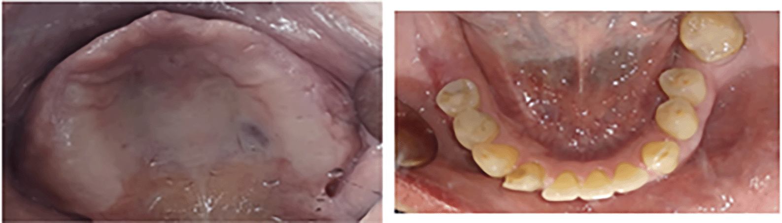
A 65-year-old female patient reported to our clinic at the Faculty of Dentistry, AIMST University with a complaint of ill-fitting maxillary complete denture from a year.
On intra-oral examination, an edentulous maxillary arch with severely displaceable anterior flabby ridge was observed. This was opposed by teeth in anterior region of the mandible. A few posterior teeth were missing. There was a down growth of both maxillary tuberosities. The mandible in the posterior region showed resorption of bone. By the presence of the following factors, it was diagnosed as the starting stage of Kelly’s combination syndrome. A treatment plan was formulated for this patient (Figure 1).
A treatment plan was formulated to provide the patient with a new maxillary conventional complete denture and a mandibular partial denture.
To record the flabby tissues at rest position, a special window impression technique for the definitive impression was designed.
On the first appointment, primary impressions of the maxilla and mandible were made using alginate (Zelgan, Dentsply) material and edentulous stock trays.
Following this, the impressions were poured with dental stone (Dental stone, Type III, Kulzer). In the maxillary cast, the flabby ridge area was marked, followed by fabrication of a specialized custom tray (Figure 2).
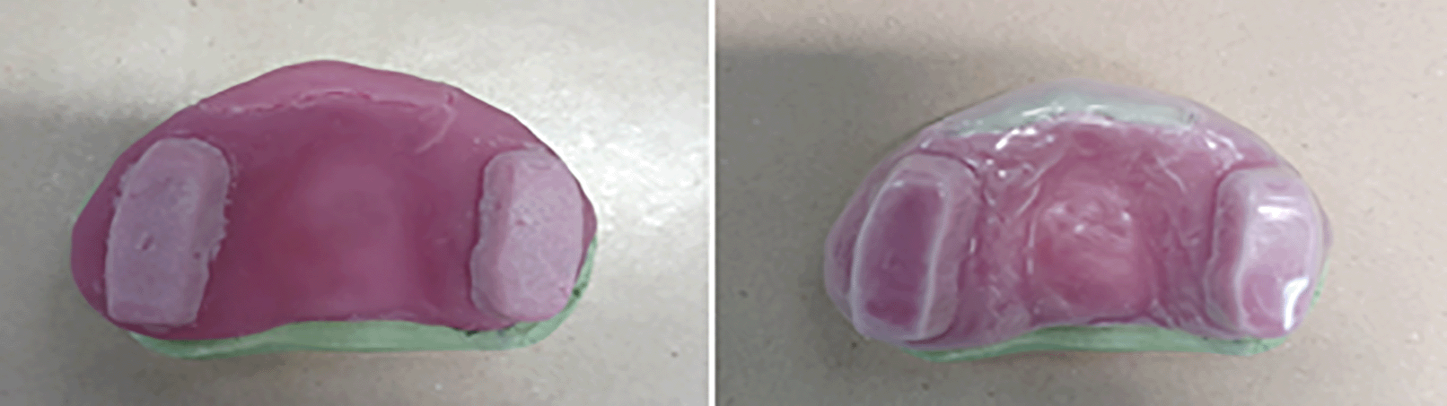
Steps for fabrication of custom tray:
1. A wax spacer (Metrowax, England) was adapted over the maxillary cast. Extra layer of spacer was added over the flabby ridge portion. This will ensure relief over the area.
2. Light cure polymethyl methacrylate sheet (Shandong Huge Dental Material Corp., China) was adapted over the spacer. The extent of the flabby ridge was marked on the cast.
3. Corresponding to that marking, an anterior window was outlined on the tray material using a sharp knife. This was done before curing to facilitate removal of the window at a later stage.
4. Two posterior handles were fabricated over the premolar region on both sides.
5. The light cure resin sheet was cured
6. A thermoplastic vacuum resin sheet of 0.5 mm thickness was adapted over the tray. The excess was cut off with sharp scissors to fit the tray exactly.
5- The light cure anterior window was removed. The resultant tray had only thermoplastic resin sheet covering the anterior flabby region with space in between for passive placement of impression material.
On the secondary impression appointment, the tray was tried in the patient’s mouth. The borders were trimmed to be 2 mm shorter than the depth of sulcus. The extent of the anterior window to the extent of the flabby ridge was confirmed (Figure 3).
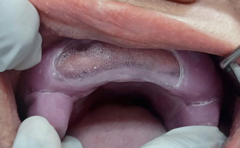
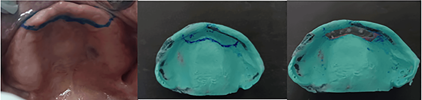
Border molding was performed using green stick compound (Tracing Sticks, Kemdent, England) by the conventional technique. Following the border molding, a secondary impression of the maxilla was made using light body PVS impression material (Panasil Initial Contact Light, Kettenbach GmbH, Germany). The outline of the flabby ridge was marked in the patient’s mouth using an indelible marker. After careful inspection of the impression, the impression was reseated in the mouth to transfer the markings onto the impression surface. This facilitated easy removal of the light body from the area of flabby ridge using a scalpel blade (Figure 4).
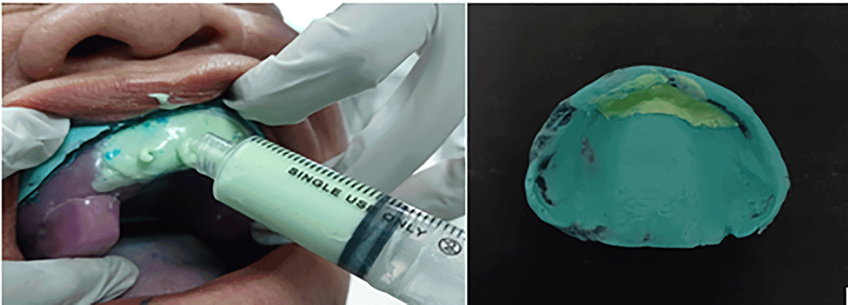
Three holes were made over the anterior region on the thermoplastic resin sheet. The impression was re-seated in the patient mouth. Alginate (Chroma 1, Dentapex, Italy) was mixed in a fluid consistency and loaded into a wide bore syringe. Alginate material was injected through one of the side holes until the entire space was filled with the material and some excess material overflowed through the other holes. Once the material was set, the tray was carefully removed and disinfected (Figure 5).
For the mandibular arch, conventional border molding was done by open tray technique and impression was made using Zinc oxide eugenol impression paste (Impression Paste, SS White, The Netherlands) and a pickup impression was made using a stock tray and alginate impression material.
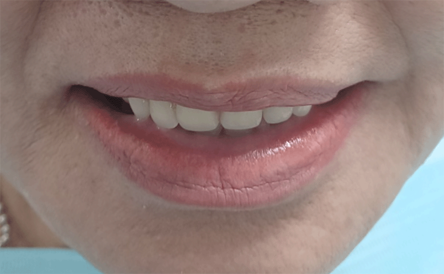
Master casts were poured from the upper and lower impressions. Denture bases and occlusal rims were fabricated for jaw relation procedure. Vertical dimension and centric jaw relation was recorded. The teeth setting was done after articulation of the casts. Following try-in, a maxillary conventional complete denture and a mandibular partial denture was issued to the patient. That patient was comfortable with the denture and satisfied with the esthetics and function (Figure 6).
The key to a successful denture treatment lies in making an accurate impression of the edentulous ridge and functional sulcus. The presence of flabby tissues compromises the retention and stability of the denture. This occurs due to the elastic recoil of the fibrous soft tissues of the flabby ridge during function.6,25 In selective pressure technique, the tissues are compressed during impression making. Using the conventional technique for flabby ridges will lead to compression of the flabby portion also. Fabricating a denture over compressed flabby tissue will lead to compromised retention and stability causing frequent dislodgement of the denture.
Several impression techniques and methods have been described in the literature for recording flabby tissue during impression making. However, there is no evidence to support that one impression technique will provide a stable and retentive denture on flabby ridges as compared to others.17 This report presents an innovative window technique for the impression of anterior maxillary flabby ridge using PVS impression material and alginate.
There have been various studies in literature to make the impression of the entire arch along with the peripheral seal followed by preparation of window and recording the displaceable tissues with a low viscosity impression material.17 On the other side of the coin, some authors have suggested that a custom tray with a window must be made prior to border molding to allow for accurate peripheral tracing of the functional sulcus and improved final impression. The displaceable tissues should be recorded in a static position through the window after final impression.8
The main disadvantage of these techniques is the inability to control the uniform application of the low viscosity impression material. This mainly arises due to difference in the chair positions and the fact that gravity can cause the impression material to flow downwards (maxillary arch).
The various materials that can be used to record the flabby tissues are impression plaster, light body PVS, impression waxes and alginate. These materials provide a mucostatic impression of the displaceable tissue. For the rest of the arch, a multitude of impression materials are available, namely zinc oxide eugenol, heavy body PVS, light body PVS etc. These materials record the tissues in a mucocompressive state.
In the technique described in the article, the use of clear polyethylene sheet helped the clinician to see the flow of the impression material and the adaptation of the impression to the flabby ridge. The clear sheet also acted as a scaffold to hold and prevent the impression material from drooping down, away from the tissue. Furthermore, in this technique, the operator was able to control the amount of material flowing on to the flabby ridge. No pressure was exerted over the thermoplastic resin sheet. Presence of extra vents caused overflow of the excess material, hence there was no shortage of material in the impression.
The initial cutting of the anterior window prior to curing allowed for easy removal of the window. Therefore, the authors recommend clinical application of this window technique for final impression of flabby maxillary ridge in the fabrication of complete dentures.
The modified window technique described in this report demonstrates an easy and effective way to control the amount of pressure applied on the flabby tissues. The tray is easy to fabricate, and the procedure is not cumbersome. This method can also be used with other materials such as impression plaster, which can be poured through the venting holes. Impressions can also be made using a combination of heavy body PVS for the secondary impression and light body PVS to record the flabby tissues.
| Views | Downloads | |
|---|---|---|
| F1000Research | - | - |
|
PubMed Central
Data from PMC are received and updated monthly.
|
- | - |
Provide sufficient details of any financial or non-financial competing interests to enable users to assess whether your comments might lead a reasonable person to question your impartiality. Consider the following examples, but note that this is not an exhaustive list:
Sign up for content alerts and receive a weekly or monthly email with all newly published articles
Already registered? Sign in
The email address should be the one you originally registered with F1000.
You registered with F1000 via Google, so we cannot reset your password.
To sign in, please click here.
If you still need help with your Google account password, please click here.
You registered with F1000 via Facebook, so we cannot reset your password.
To sign in, please click here.
If you still need help with your Facebook account password, please click here.
If your email address is registered with us, we will email you instructions to reset your password.
If you think you should have received this email but it has not arrived, please check your spam filters and/or contact for further assistance.
Comments on this article Comments (0)