Keywords
Adenoid cystic carcinoma, tongue, treatment. Radiotherapy, Chemotherapy
This article is included in the Datta Meghe Institute of Higher Education and Research collection.
Adenoid cystic carcinoma, tongue, treatment. Radiotherapy, Chemotherapy
Adenoid cystic carcinoma (ACC) is a relatively uncommon tumor of the salivary glands that originates from epithelial cells. It represents approximately 5% to 10% of all malignant salivary gland tumors and accounts for 10% to 15% of all tumors in the head and neck region.1 31% of these tumors are found in the minor salivary glands, particularly in the palate. However, they can also occur in the major salivary glands such as the submandibular and parotid glands.2 ACC can also develop in other areas of the head and neck region, including the tracheobronchial tree, esophagus, lacrimal gland, and tongue. The occurrence of ACC in the base of the tongue is relatively rare.
ACC exhibits a wide age distribution, commonly affecting individuals in their fifth and sixth decades of life, with a higher prevalence in females.3 This type of cancer typically progresses slowly and is characterized by the infiltration of nerves, the potential for distant spread, and a significant likelihood of local recurrence.4 Prognostic factors encompass the stage of the disease, the location of the tumor, the presence of a solid growth pattern, the size of the lesion, the status of surgical margins, and the occurrence of intraneural and/or perineural invasion. Histologically, ACC manifests in three distinctive patterns: cribriform, tubular, and solid. Notably, the solid variant carries a particularly unfavorable prognosis, whereas the cribriform variant is associated with a more favorable outcome. The tubular type, in comparison to all three variants, is expected to demonstrate the most favorable prognosis.5
The tumor’s neurotropism is indicated by its gradual advancement and extensive infiltration around nearby nerves.6 Perineural invasion can occur through either the spaces surrounding the nerves or within the nerves themselves. Lymphatic dissemination is rare, although there have been instances of tumor spreading through the bloodstream. Metastases to the lungs and bones are probable. The occurrence of ACC in the base of the tongue is 8.8%, whereas it is only 2.9% in the anterior two-thirds of the tongue.7
Advanced ACC lesions located at the base of the tongue commonly exhibit significant infiltration with perineural invasion. The preferred treatment approach for such cases involves extensive surgical excision, which unfortunately has an impact on swallowing and speech abilities. Since ACC is a tumor that demonstrates resistance to radiation, radiotherapy is considered as an adjunctive therapy for advanced lesions. However, several authors have reported unfavorable outcomes for radiation treatment of advanced and inoperable ACC tumors. In fact, the local control rates in these cases are documented to be below 50%.7 Conversely, the efficacy of chemotherapy in achieving conclusive results remains inconclusive. Despite the frequent use of definitive chemoradiotherapy, the therapeutic outcomes have been unsatisfactory. This discussion presents a case of ACC affecting the tongue, along with a concise overview of the relevant literature concerning its clinical, pathological, and therapeutic implications.
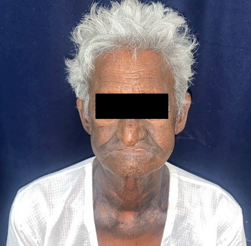
A 75-year-old male from Yavatmal, as shown in Figures 1 and 2, arrived at the Department of Oral and Maxillofacial Surgery, Sharad Pawar Dental College seeking treatment for a painful ulcer that has not healed on the posterior region of the left side of his tongue for approximately two months. The patient has a documented case of hypertension for the past ten years and has been taking 5 mg tablet of Amlodipine once daily. Additionally, the patient has a habit of chewing tobacco four to five times a day for approximately 25 years. The patient had no relevant family medical history. For about a year, the patient has experienced a burning sensation when consuming hot and spicy foods. Furthermore, the patient is having difficulties with chewing and swallowing, and has been experiencing earaches on the same side for the past two months. There is no record of any nerve-related sensations such as tingling or numbness.

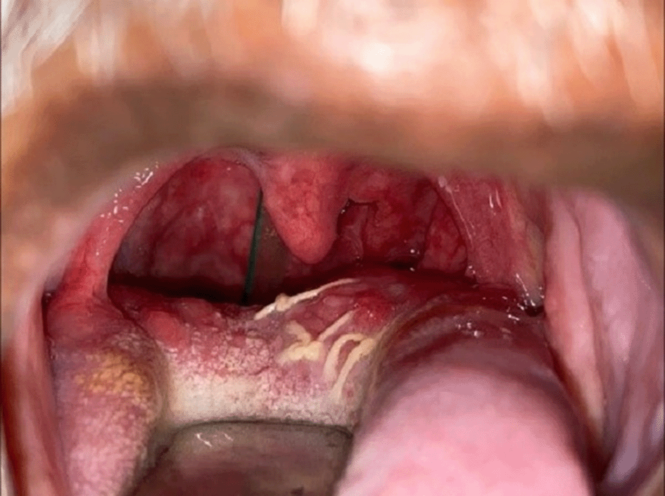
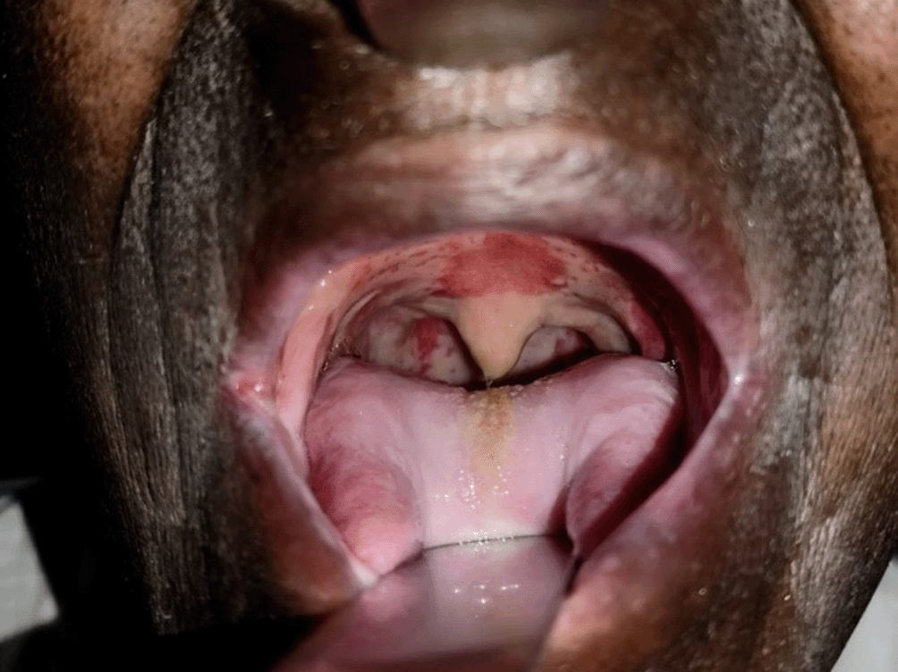
The facial appearance exhibits no noticeable imbalances, and the lips are well-formed and move smoothly and synchronously. During examination, a palpable lymph node is detected on the left side of the jaw, measuring approximately 2 x 1 cm. It has an oval shape, firm texture, and is fixed in place, but does not cause any pain. Intraoral evaluation reveals that the patient has a mouth opening of 24 mm, and both the upper and lower jaws are missing teeth. In the posterior region of the tongue on the left side, there is a single painful ulcerous and proliferative lesion. Its dimensions are approximately 3 cm in size, extending from the posterior one-third of the tongue towards the posterior end, although the full extent cannot be determined. It stretches from the midline to the lateral border of the left side of the tongue. The lesion appears reddish-pink in color, with edges that are turned outward, an irregular surface, and poorly defined borders. Palpation of the lesion reveals tenderness, firmness, and a hardened texture.
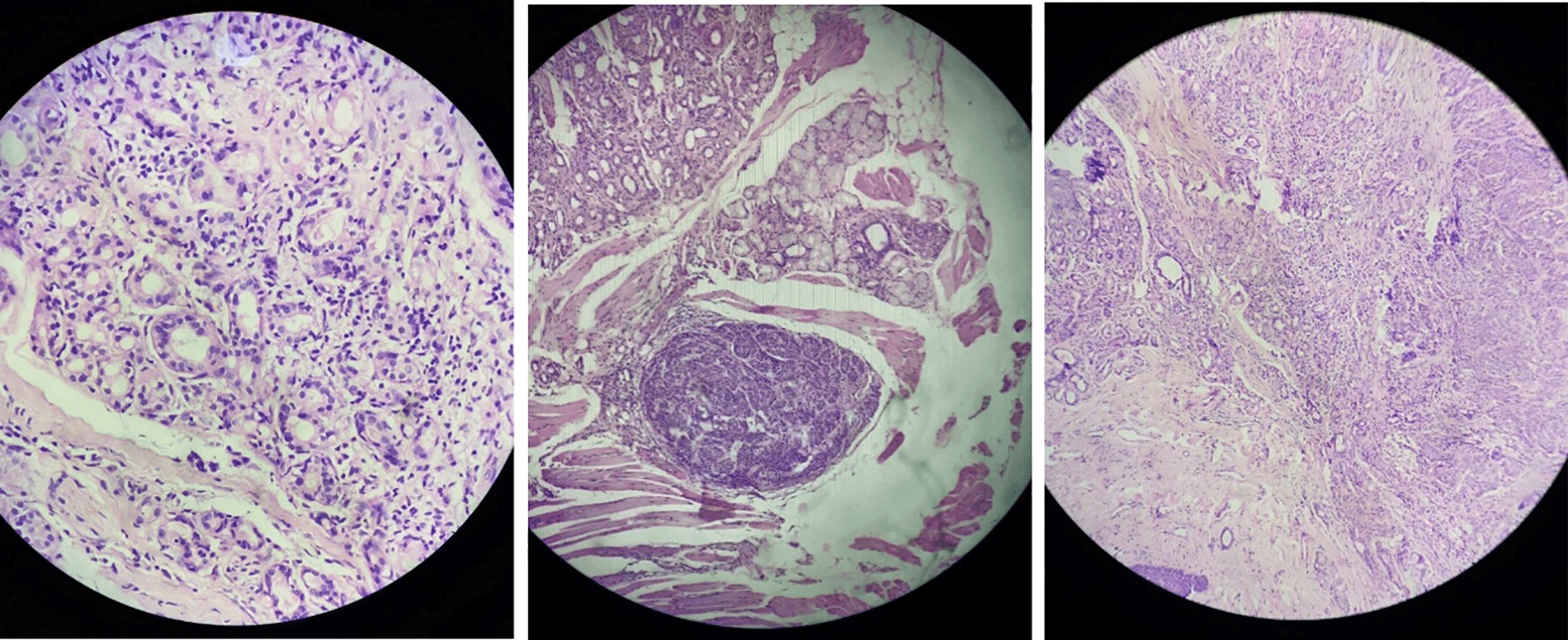
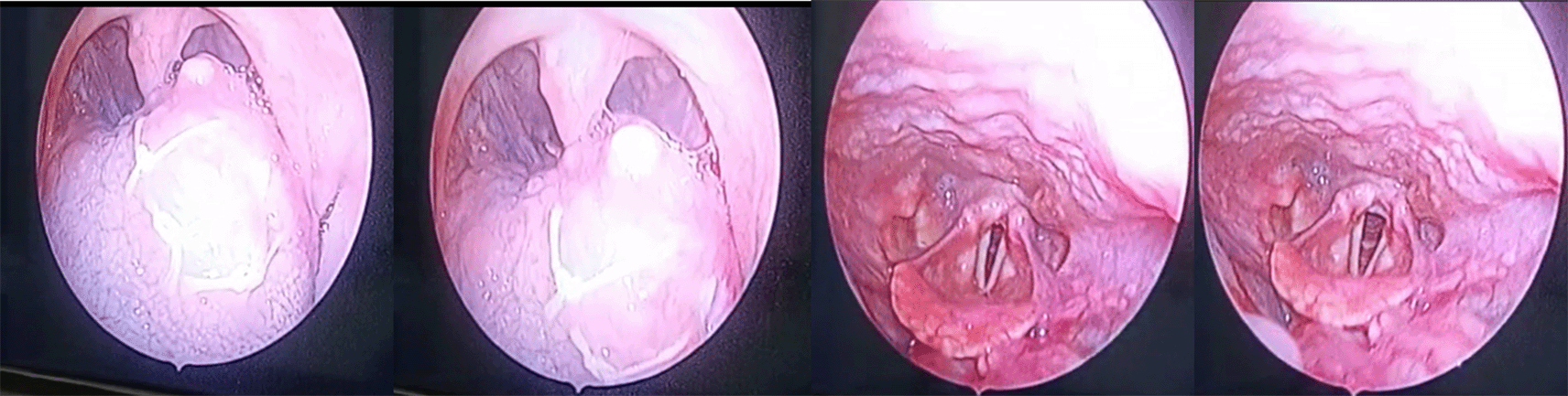
The patient has received an incisional biopsy (Figure 3) indicating adenoid cystic carcinoma with a tubular pattern. The pattern of invasion (POI) was 03, with the worst POI being 04. The depth of invasion (DOI) could not be determined due to the absence of surface epithelium. The lymphocytic response score was 03. Muscle invasion was detected, while lymph-vascular and perineural invasion were not present. Salivary gland invasion was observed.8 Direct video laryngoscopy was performed to visualize the posterior extent of the lesion (Figure 4).
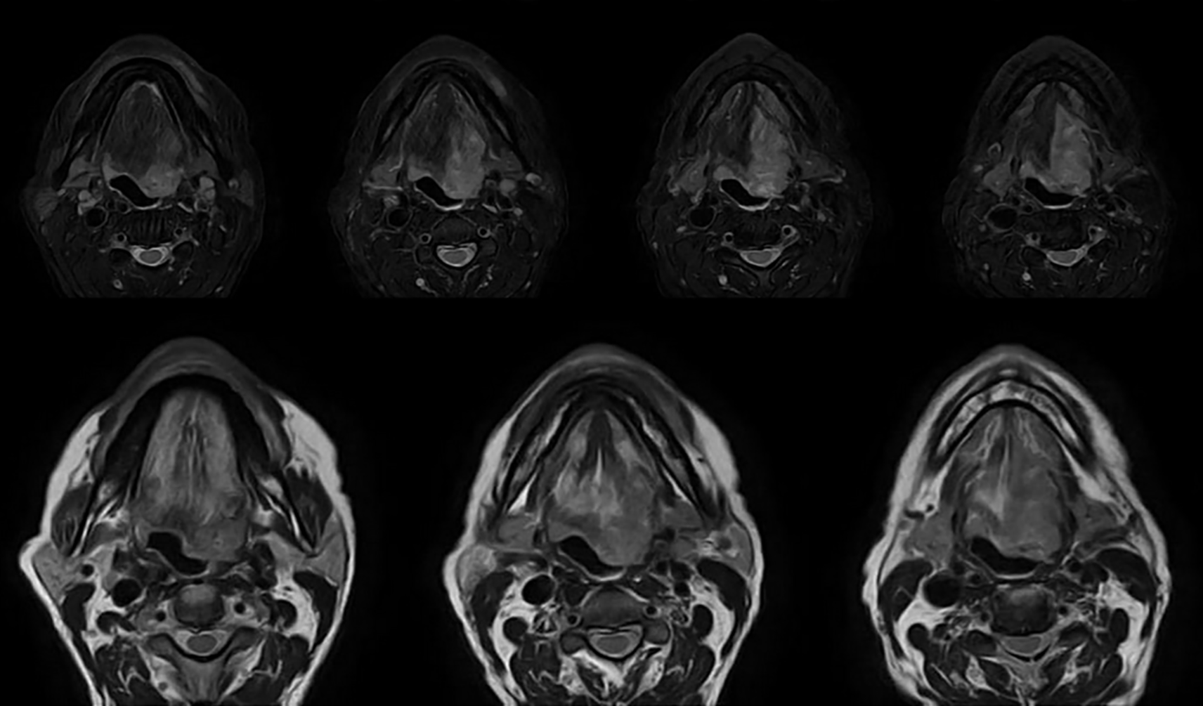
After conducting the biopsy, a magnetic resonance imaging (MRI) examination of the tongue was performed. The results revealed an area of modified signal intensity on the left half of the tongue, appearing brighter on FatSat and exhibiting similar brightness on T1-weighted and T2-weighted images. The dimensions of this area were measured to be 4.9 x 4.6 x 2.1cm, involving the intrinsic muscles of the tongue along with the genioglossus, hyoglossus, palatoglossus, and styloglossus on the left side. The lesion extended from the dorsum, reaching the alveolar ridge on the sides and crossing the midline septum posteriorly with an invasion depth of 2.9cm. The lingual tonsil and uvula were affected posteriorly, leading to the obliteration of the left side of the oropharynx. Fortunately, the mylohyoid muscle below appeared to be unaffected. Additionally, multiple lymph nodes of various sizes, ranging from sub-centimeter to centimeter, were detected in the submental, bilateral submandibular, and upper jugulodigastric regions. The largest lymph node measured 1 x 0.7cm and was observed in the right submandibular region (see Figures 5 and 6).
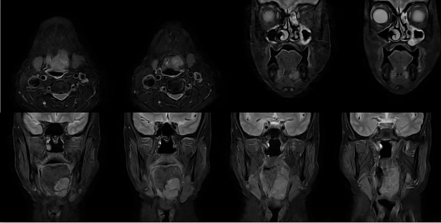
The high-resolution computed tomography (HRCT) thorax examination revealed the presence of numerous fibrotic changes beneath the pleura, accompanied by localized thickening of the pleura in both the upper and lower lobes. Additionally, a few lymph nodes exhibiting a sub-centric appearance were observed in the paratracheal, pre-vascular, and aorta-pulmonary regions. Based on these findings, the preliminary diagnosis made was carcinoma originating from the base of the tongue, categorized as pT3N2cM0, stage IV A.
The patient was recommended to undergo a surgical procedure involving the complete removal of the lesion and a significant portion of the tongue, with wide surgical margins. However, the patient expressed unwillingness to undergo any surgical intervention. As a result, the chosen treatment approach for this case was definitive chemoradiotherapy. This was planned within one month of the patient’s initial admission. This involved receiving a total dose of 70 Gy/weekly of radiation therapy, delivered in 35 fractions to both the face and neck regions using the intensity modulated radiation therapy (IMRT) technique. The patient has completed 30 fractions of radiotherapy and has also received 6 cycles of weekly injections of cisplatin, each at a dosage of 50 mg. The patient is currently undergoing treatment and has been in good health for the past month since the treatment started (refer to Figures 7 and 8).
Adenoid cystic carcinoma (ACC) is a locally aggressive tumor characterized by its infiltrative growth pattern and perineural invasion. Despite undergoing rigorous surgical resection, ACC has a high recurrence rate and tends to metastasize late. The five-year survival rate for patients with ACC is approximately 70%.9 While the lung is the most common site of distant metastasis, occurrences in the bone, liver, kidney, and prostate have also been reported.10
In a study conducted by Luna-Ortiz et al.,11 out of 68 patients with head and neck ACC, eight patients (seven females and one male) were diagnosed with ACC of the tongue, accounting for 11.7% of all head and neck ACC cases. Specifically, six cases were located at the base of the tongue, while two were in the oral tongue. Generally, the clinical history of ACC in the tongue presents as asymptomatic, with gradual submucosal tumor growth. The diagnosis is often delayed, ranging from 6 months to 8 years from the initial onset of symptoms.12 In the current study, there was an 8-month delay in diagnosing ACC in the tongue. Tumors affecting the oral tongue tend to be detected earlier due to their impact on normal functioning, whereas tumors in the base of the tongue are typically identified at later stages.
Regional lymph node metastases of adenoid cystic carcinoma (ACC) in the head and neck region are infrequent, occurring in only 6 to 10% of cases.13 One possible explanation for this rarity is that the two primary sites for ACC, namely the parotid gland and the palate/maxilla, have a low tendency to spread to the lymph nodes. A retrospective study conducted by Min et al.,9 involving 616 patients with ACC in the head and neck area, revealed that lesions located at the base of the tongue, mobile tongue, and floor of the mouth exhibited the highest rates of cervical lymph node metastasis (19.2%, 17.6%, and 15.3%, respectively).
Chen et al.14 identified several independent predictors of local recurrence in ACC. These included the absence of postoperative irradiation (P=0.007), T4 disease (P=0.0001), perineural invasion (P=0.008), and extensive involvement of nerves (P=0.02). According to Chen et al.,14 the optimal approach for successfully treating resectable ACC of the head and neck involves surgery combined with adjuvant radiation therapy at doses exceeding 60 Gy. In cases of advanced malignancies or partially resectable disease, radiotherapy alone can be employed as a standalone treatment. It is important to note that minor salivary gland ACCs located outside the major salivary glands are less amenable to surgical resection and have a lower likelihood of being cured solely through radiation therapy.15 The choice between radiotherapy alone and surgery plus radiotherapy is influenced by selection bias, as individuals with early-stage lesions are more likely to undergo surgery, while those with advanced, unresectable cancers are more inclined to receive radiotherapy as their primary treatment option.
According to Miglianico et al.,16 although there was an improvement in local-regional control, surgery alone did not significantly enhance survival rates compared to combined modality therapy. The limited survival advantage may be attributed to the high occurrence of distant metastases. Some studies propose the use of adjuvant chemotherapy alongside radiotherapy or in combination with surgery due to the high risk of hematogenous spread. However, there is insufficient convincing evidence supporting the benefits of adjuvant chemotherapy,17 and its utilization for patients with ACCs is restricted. Concurrent chemoradiotherapy can be considered as an option for individuals with advanced disease or for organ preservation purposes, but its effectiveness in treating ACC is currently under further investigation.
The ongoing RTOG 1008 clinical trial is examining whether the addition of cisplatin to standard postoperative radiotherapy improves survival in tumors of major salivary glands with high-risk factors, including pathological staging of T3-4, N1-3, or T1-2, N0, as well as a close or positive surgical margin.18 This randomized phase II/III study includes high-grade ACC (defined as >30% solid component). While awaiting these results, some limited retrospective studies suggest the use of concurrent chemo-radiotherapy in specific patients with unfavorable pathological characteristics in salivary gland cancers.19 It is possible to draw insights from the treatment approaches employed for other head and neck malignancies and consider combining radio-sensitizing chemotherapy to enhance tumor control in certain scenarios. In such cases, it is recommended to seek a multidisciplinary consultation with medical oncology.
Advanced ACC can remain dormant over an extended period, commonly exhibiting lung metastases. The tumor stage serves as a significant prognostic indicator for both overall survival and cancer recurrence. The collective five-year survival rate for ACC varies from 64% to 89%, while the ten-year survival rate ranges from 37% to 77%.20
Adenoid cystic carcinoma is an uncommon type of cancer that typically originates in the salivary glands but can also develop in other areas of the body, such as the breast, lung, or trachea. ACC is characterized by its slow growth and has the ability to spread to nearby tissues and organs, posing challenges for effective treatment. The symptoms experienced by individuals with ACC depend on the location of the tumor and may include pain, difficulty swallowing, and changes in voice or facial appearance. The standard treatment approach for ACC usually involves surgical removal of the tumor, followed by radiation therapy or chemotherapy to eliminate any remaining cancer cells. The prognosis for ACC varies depending on the cancer stage and the overall health of the patient. The patient has been showing a good response to the chosen modality and has reported a better quality of life.
In patients with ACC, the primary therapeutic objective is to achieve control over the cancer in the surrounding area, maintain normal bodily functions, and prevent the development of distant metastases. Early detection by the surgeon is crucial due to the slow-growing nature of these tumors and their tendency to invade extensively. Our aim is to enhance both prognostic outcomes and quality of life for affected individuals. For the majority of patients, concurrent radiation therapy remains the preferred treatment option. However, effective treatments for malignancies are scarce, and managing metastatic disease remains challenging. Consequently, survival rates have not shown significant improvement in recent years.
| Views | Downloads | |
|---|---|---|
| F1000Research | - | - |
|
PubMed Central
Data from PMC are received and updated monthly.
|
- | - |
Provide sufficient details of any financial or non-financial competing interests to enable users to assess whether your comments might lead a reasonable person to question your impartiality. Consider the following examples, but note that this is not an exhaustive list:
Sign up for content alerts and receive a weekly or monthly email with all newly published articles
Already registered? Sign in
The email address should be the one you originally registered with F1000.
You registered with F1000 via Google, so we cannot reset your password.
To sign in, please click here.
If you still need help with your Google account password, please click here.
You registered with F1000 via Facebook, so we cannot reset your password.
To sign in, please click here.
If you still need help with your Facebook account password, please click here.
If your email address is registered with us, we will email you instructions to reset your password.
If you think you should have received this email but it has not arrived, please check your spam filters and/or contact for further assistance.
Comments on this article Comments (0)