Keywords
Colonic perforation, Colonoscopy, Iatrogenic Disease, emergency, management, outcome
Colonoscopy is a commonly utilized procedure in gastroenterology, but it carries risks of complications, with perforation being the most dreaded. The management of colonic perforation remains a topic of debate, as it can be effectively treated through surgical or non-surgical approaches. Our objective is to detail clinical presentations, diagnostic methods, and potential therapeutic options.
For this study, we gathered clinical and radiological data from two cases of colonic perforation following colonoscopy. We examined clinical presentations, diagnostic methods employed, and the different therapeutic approaches used for each case. In both cases, patients exhibited symptoms of colonic perforation following colonoscopy. The first case was managed conservatively, with progressive clinical improvement. The second case showed signs of pneumoperitoneum, but no perforation was found during laparoscopic intervention. Both patients recovered well and experienced no complications during follow-up.
Our study highlights the importance of understanding the risks associated with colonoscopy, particularly in patients with risk factors. It also underscores the diversity of available treatment approaches for iatrogenic colonic perforation, emphasizing the significance of a multidisciplinary approach in determining the optimal therapeutic strategy.
Colonic perforation, Colonoscopy, Iatrogenic Disease, emergency, management, outcome
Colonoscopy is one of the most frequently used complementary exploration methods in gastroenterology. While it is generally considered low-risk, complications can arise, among which perforation is the most feared, even in the absence of surgical intervention. The incidence of iatrogenic colonic perforation could be as low as 0.016% of all diagnostic colonoscopy procedure1 and could reach up to 5% in interventional colonoscopies.2,3
The most commonly observed radiological translation is that of pneumoperitoneum (PNP). However, cases of pneumoperitoneum occurring without perforation following a diagnostic colonoscopy have been reported.4 The management of colonic perforation remains a controversial issue, as it can be effectively handled through both surgical and non-surgical approaches.4,5
In this study, we present two cases of colonic perforation that occurred after colonoscopy, each with distinct manifestations and managed in different ways. The aim of our work is to detail the clinical presentations, diagnostic methods, and potential therapeutic options.
The patient was a 44-year-old man of Arab descent who worked as a taxi driver and had no prior medical history of diabetes or hypertension. He was admitted to Mahmoud El Matri hospital in the gastroenterology department for a screening colonoscopy due to chronic constipation. The examination revealed moderate colon preparation using 4 liters of Polyethylene Glycol (PEG) administered orally over 4 hours before the colonoscopy. A 7 mm polyp was found in the left colon, which was surgically removed.
One day after the procedure, the patient experienced sudden, sharp, non-radiating abdominal pain. During the examination, the patient was afebrile and stable in terms of hemodynamics, respiration, and neurological status. Sensitivity was noted in the left flank without signs of peritonitis, while the rest of the abdomen was soft, depressible, and painless.
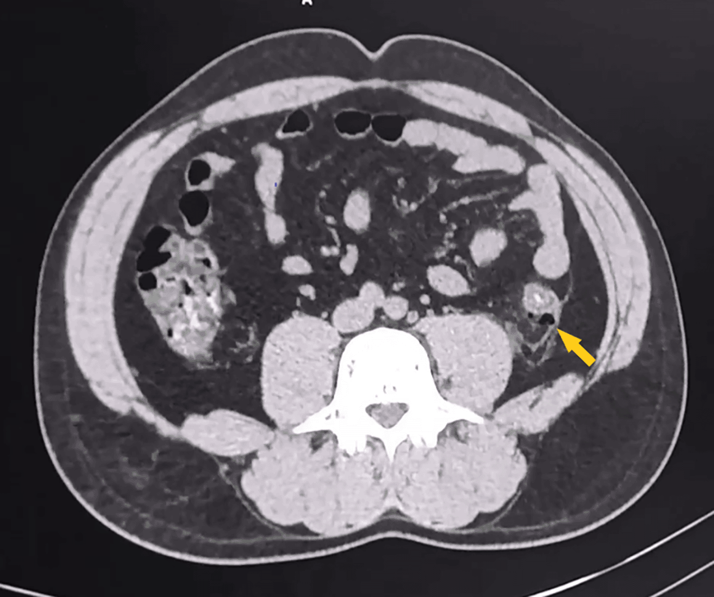
Blood tests showed a mild biological inflammatory syndrome with a c-reactive protein (CRP) level of 39 mg/ml, and no leukocytosis, a hemoglobin level of 12 g/dl, and appropriate hemostasis. As a result, an abdominal computerized tomography (CT) scan with contrast injection was performed, revealing small retroperitoneal extra-digestive air bubbles adjacent to the middle third of the descending colon (Figure 1). Thickening of the surrounding peritoneal layers and an effusion were also observed.

CT, computerized tomography.
Based on the clinical and radiological findings, an immediate surgical intervention was deemed unnecessary. Instead, the patient was treated conservatively, with a strict fasting approach, total parenteral nutrition, and close clinical and biological monitoring. Additionally, the patient was placed on empiric antibiotic therapy consisting of cefotaxime 1 g * 3/day and Metronidazole 500 mg * 3/day, administered intravenously.
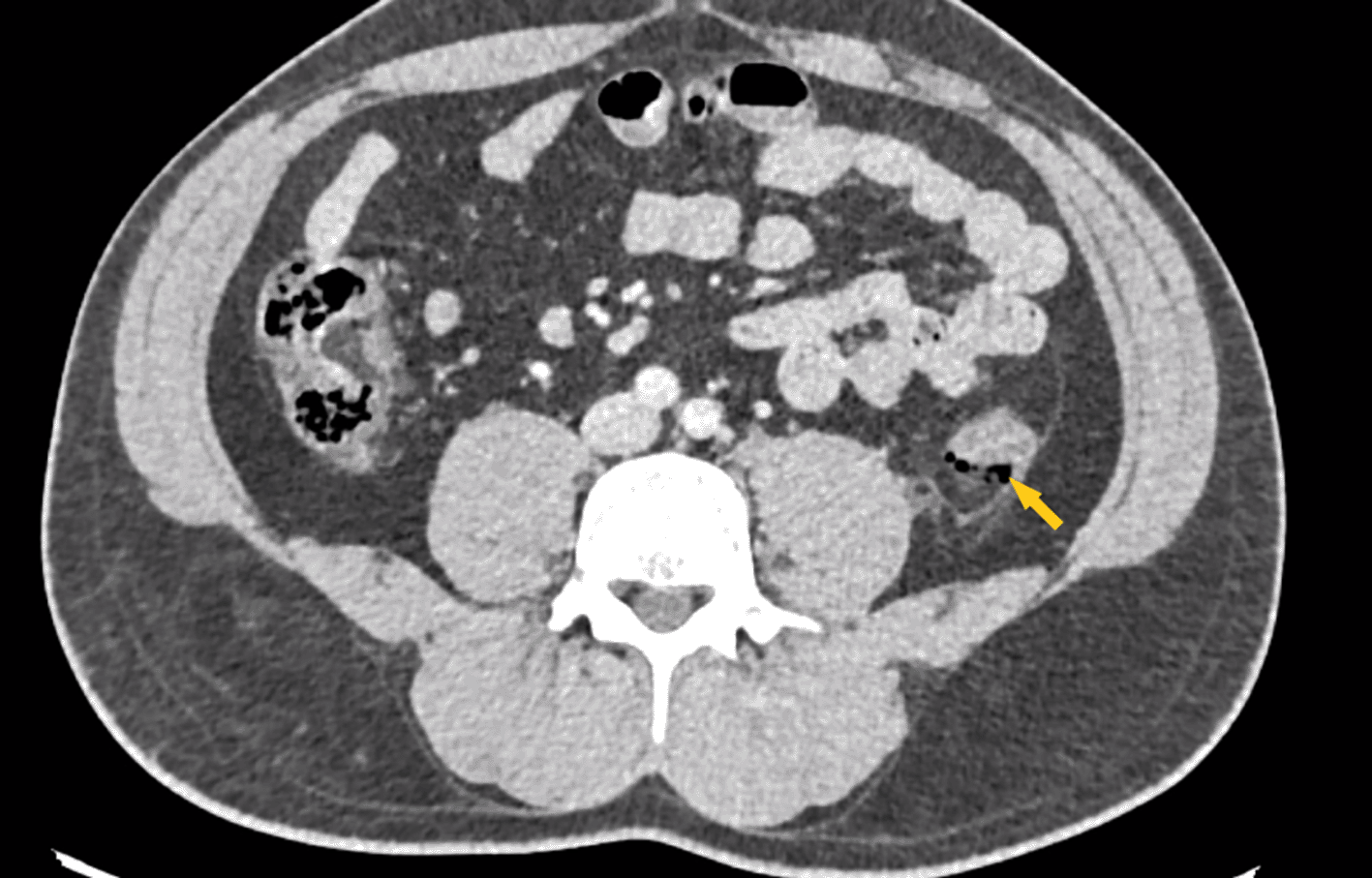
Subsequent laboratory follow-up showed regression of the biological inflammatory response. An abdominal CT scan performed on the second day of treatment revealed a stable retroperitoneal aspect adjacent to the middle third of the left colon, with an associated localized peritoneal reaction (Figure 2).

CT, computerized tomography.
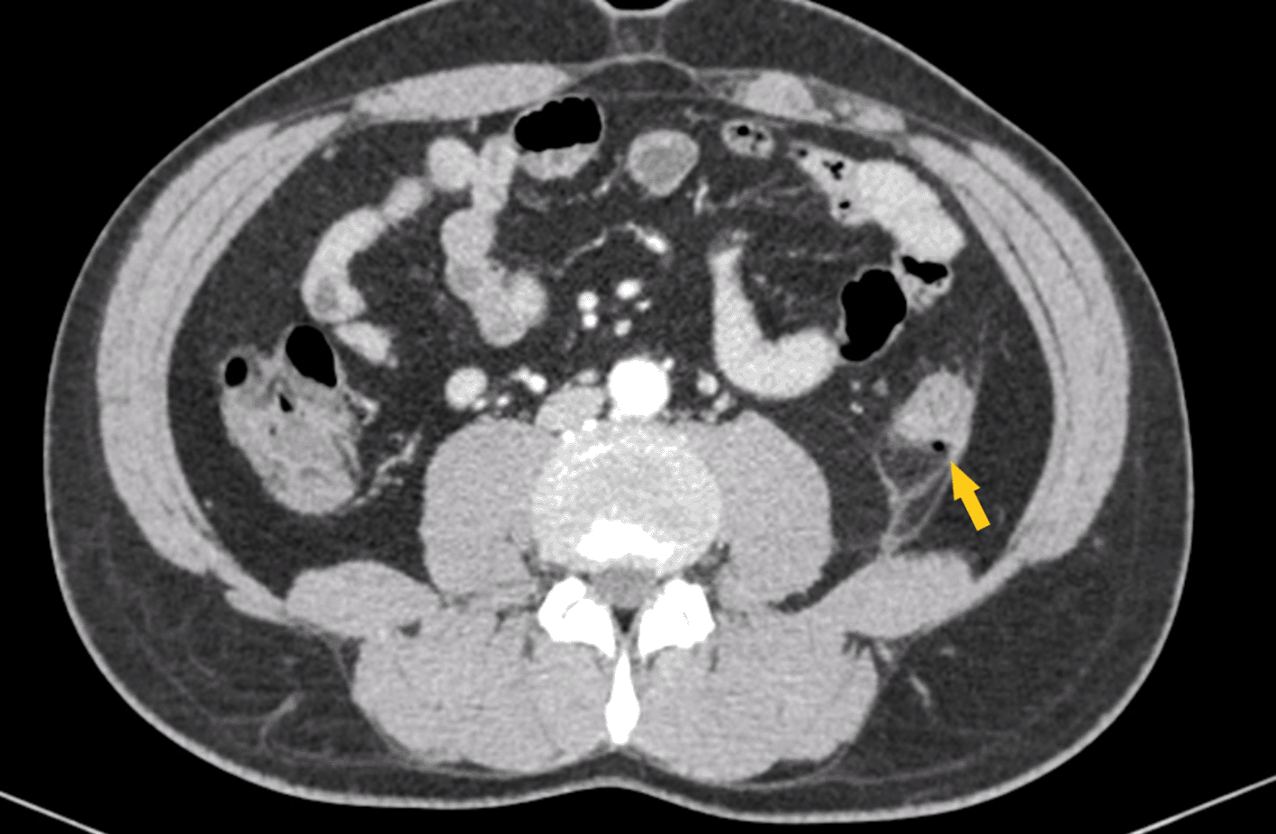
The patient’s condition evolved with clinical and biological improvement, including sustained apyrexia, disappearance of abdominal pain, and a normal abdominal examination. An abdominal CT scan performed on the fifth day of treatment demonstrated partial regression of the retroperitoneal air bubbles, as well as persistent localized peritoneal reaction (Figure 3).

CT, computerized tomography.
On the sixth day, a liquid diet via oral intake was gradually introduced and well tolerated. The patient was allowed to leave the hospital two days later.
A follow-up after two months showed the absence of complications, a normal condition. Furthermore, a two-month follow-up CT scan confirmed the complete resolution of retroperitoneal air (Figure 4).
This case concerns a 55-year-old male patient of Arab descent with no significant medical history, who worked as a fishmonger. He was admitted to Mahmoud El Matri hospital in the gastroenterology department for a screening colonoscopy.
As per the information provided by the gastroenterology consultant, despite the colonic preparation being deemed satisfactory, the colonoscope could only be advanced up to a distance of 40 cm due to technical difficulties associated with an irreducible sigmoid loop, despite multiple attempts, leading to the termination of the procedure.
In the hours following the completion of the colonoscopy, the patient began experiencing abdominal pain accompanied by vomiting. The examination revealed an afebrile patient, eupneic, hemodynamically stable with a blood pressure of 130/70 mmHg and a heart rate of 89 bpm. Palpation of the abdomen indicated painful distension. An abdominal X-ray without prior preparation was performed, revealing bilateral pneumoperitoneum associated with aerocoly (Figure 5).
Given these clinical and radiological findings, a decision was made in favor of a laparoscopic surgical intervention to explore the abdominal cavity and investigate a potential colonic perforation. However, despite a meticulous and precise exploration of the entire abdominal cavity during the procedure, no perforation, leakage, or abdominal effusion was observed. The intervention concluded with the placement of an aspiration drainage in the cul-de-sac of Douglas.
Postoperative recovery was uneventful, characterized by a reduction in abdominal pain and restoration of intestinal function. Liquid diet was gradually reintroduced, and following clinical and biological improvement, the patient was discharged from the hospital on the sixth day post-intervention. During a follow-up consultation one month later, the patient presented no complications.
The case report presents two patients who underwent colonoscopies with potential colonic perforation complications. The strengths include detailed patient information, comprehensive clinical assessments, imaging diagnostics, and the demonstration of the effectiveness of conservative management in the first case. However, the second case lacks intraoperative photos, and the duration of monitoring is short for both cases.
Colonoscopy, a commonly performed procedure and the primary diagnostic tool for colorectal cancer, also plays a role in managing specific colorectal conditions, despite its relative safety, it does entail potential complications including colonic perforation, gastrointestinal bleeding, injury to intra-abdominal organs, and cardiopulmonary instability.6
Although rare, Iatrogenic perforation carries a significant risk of morbidity and mortality.7 The optimal management approach involves a collaborative effort among endoscopists, radiologists, and surgeons, requiring their prompt availability.
The incidence of colonic perforation varies between diagnostic and therapeutic colonoscopies,8,9 with both sharing similar mechanisms such as mechanical injuries or barotrauma, but therapeutic colonoscopy presenting an additional potential risk of perforation,10 resulting in an incidence ranging from 0.03% to 0.8% for diagnostic colonoscopy and 0.15% to 3% for therapeutic colonoscopy.6
During diagnostic procedures, iatrogenic perforations are most frequently observed in the sigmoid colon and the rectosigmoid junction. This occurs because of direct mechanical injury caused by the shearing forces exerted by the colonoscope’s shaft or tip during insertion.11,12 The risk of perforation can be further elevated by pericolic adhesions, which may result from previous gynecological surgeries or abdominal inflammation, as well as by severe diverticular disease. This increased risk is particularly notable when using large-caliber instruments13,14 with excessive force.
In the realm of interventional colonoscopies, researchers have highlighted several noteworthy risk factors for iatrogenic colonic perforation. These influential factors encompass polypectomies, especially for polyps exceeding 20 mm in size, pneumatic dilatation for the management of strictures linked to inflammatory bowel diseases, the application of argon plasma coagulation, along with endoscopic mucosal resection and endoscopic submucosal dissection for colorectal neoplasms. Moreover, patient-related variables, including age, female sex, malnutrition, multiple comorbidities, a history of inflammatory bowel disease, and prior colon surgery, as well as the experience level of the endoscopist, play significant roles in the risk profile. Furthermore, it’s worth noting that the use of flexible sigmoidoscopy has also been associated with an elevated risk of perforation.13
The risk of colonic perforation exists even in the absence of interventions, whether it occurs due to direct trauma to the colonic wall by the endoscope, shearing forces, or barotrauma. Regarding frequency, the sigmoid colon is the most commonly affected site in iatrogenic colonic perforation cases (53-64%), followed by the cecum (14-24%) and the ascending colon (24%), with notably lower incidences in the transverse colon, descending colon, and rectum.6 Colonic perforations can be categorized into three types: intraperitoneal (the most prevalent scenario), extraperitoneal, or a combination of both.6
The distribution of free air within distinct anatomical regions leads to the manifestation of symptoms and clinical signs, which depend on the type of perforation.13 Typically, colonic perforation presents with acute abdominal pain, sometimes accompanied by fever. It may also exhibit signs of peritonitis such as abdominal tenderness, guarding or rigidity, abdominal distension, or, in the case of extraperitoneal perforation, subcutaneous emphysema.13
The most common clinical feature of colonic perforation is the visualization of an extra-intestinal structure during the endoscopic examination.2 However, patients with colon perforation may present with symptoms and signs of peritonitis (primarily abdominal pain and tenderness) in the hours following the completion of colonoscopy, as seen in our third reported case. When perforation is suspected, an abdominal X-ray should be performed to rule out the presence of pneumoperitoneum. Other tests, such as computed tomography (CT) scanning and magnetic resonance imaging, are also highly useful for identifying the presence of free gas.13
The use of a water-soluble contrast enema is rarely performed to detect or confirm a concealed perforation. In practice, patients can be diagnosed and treated for colonic perforation based on generalized peritonitis, even in the absence of radiological evidence of perforation.
While perforations usually occur during or within 24 hours of a colonoscopy, delayed perforations of the colon and rectum, as observed in our first case, have been reported. Therefore, physicians should consider colonic perforation if a patient presents with symptoms like fever, abdominal pain, or distension following the procedure, even if these symptoms manifest several days later. It is advisable to promptly and thoroughly assess and document any symptoms or signs indicative of iatrogenic perforation after an endoscopic procedure using a CT scan.14
Following endoscopic resection, the presence of small gas bubbles may be observed, which do not necessarily indicate a genuine iatrogenic perforation.15 Hence, it is crucial to consider radiological findings alongside endoscopic and clinical assessments. Due to the intricate nature of managing iatrogenic perforations, it is essential to have a multidisciplinary team comprising the endoscopist, radiologist, and surgeon available. The post-treatment follow-up for an iatrogenic perforation is contingent upon its type, location, and the patient’s clinical status, necessitating nearly obligatory hospitalization.
Indeed, the choice of treatment for iatrogenic perforation hinges on several factors, including the timing of diagnosis (whether intra- or post-procedure), the presence and nature of luminal contents (whether “clean” or not), the specific characteristics of the perforation (size and location), the patient’s overall health status, the expertise of the endoscopist, and the availability of closure devices. Therapeutic options encompass immediate endoscopic closure, a conservative approach, or surgical intervention. In cases where iatrogenic perforation is identified during the endoscopy, it is advisable, if feasible and reasonable, to complete the interventional procedure. Swift endoscopic closure, whenever possible, not only prevents peritonitis or mediastinitis but also reduces the necessity for surgical intervention.16–18 A variety of endoscopic clips have been applied according to the size of iatrogenic perforation.
The conservative approach entails the administration of intravenous antibiotics, withholding oral intake, continuous monitoring of hemodynamics, and maintaining close interdisciplinary follow-up.19 For malnourished patients or well-nourished individuals who won’t be able to eat for ≥ 7 days, parenteral nutrition is advised.20 If the conservative approach proves ineffective and the patient’s condition worsens, such as the development of septic or peritonitis symptoms, surgical intervention is strongly considered.21 Early surgery is typically preferred for patients with large perforations, generalized peritonitis, ongoing sepsis, deteriorating clinical conditions, or after percutaneous drainage has failed. The choice between laparoscopy and open surgery for managing iatrogenic perforations primarily depends on the perforation’s location and the surgeon’s judgment. Minimally invasive laparoscopic treatment has become the favored surgical approach for colonic iatrogenic perforations due to its superior outcomes compared to open surgery.7
Our two cases have demonstrated two different scenarios in the management and clinical presentation of colonic perforation following colonoscopy. This article has provided a comprehensive overview of iatrogenic colonic perforation, covering its incidence, risk factors, clinical presentation, diagnostic methods, and treatment options. It emphasizes the importance of a multidisciplinary approach and the consideration of various factors when determining the appropriate course of treatment. The article also highlights the increasing preference for minimally invasive laparoscopic procedures in managing colonic iatrogenic perforations.
Written informed consent for publication of their clinical details and clinical images was obtained from both patients.
All data underlying the results are available as part of the article and no additional source data are required.
| Views | Downloads | |
|---|---|---|
| F1000Research | - | - |
|
PubMed Central
Data from PMC are received and updated monthly.
|
- | - |
Is the background of the cases’ history and progression described in sufficient detail?
Yes
Are enough details provided of any physical examination and diagnostic tests, treatment given and outcomes?
Partly
Is sufficient discussion included of the importance of the findings and their relevance to future understanding of disease processes, diagnosis or treatment?
Partly
Is the conclusion balanced and justified on the basis of the findings?
Partly
References
1. Belhadj A, Touati M, Ben Othmane M, Jaouad F, et al.: Case Series: Management and outcomes of two cases of colonic perforation following colonoscopy. F1000Research. 2024; 13. Publisher Full TextCompeting Interests: No competing interests were disclosed.
Reviewer Expertise: General Surgery, Surgical endoscopy and Laparoscopy
Is the background of the cases’ history and progression described in sufficient detail?
Partly
Are enough details provided of any physical examination and diagnostic tests, treatment given and outcomes?
Partly
Is sufficient discussion included of the importance of the findings and their relevance to future understanding of disease processes, diagnosis or treatment?
Partly
Is the conclusion balanced and justified on the basis of the findings?
Partly
Competing Interests: No competing interests were disclosed.
Reviewer Expertise: Microbiome and gastrointestinal intervention therapy.
Alongside their report, reviewers assign a status to the article:
| Invited Reviewers | ||
|---|---|---|
| 1 | 2 | |
|
Version 1 08 Jan 24 |
read | read |
Provide sufficient details of any financial or non-financial competing interests to enable users to assess whether your comments might lead a reasonable person to question your impartiality. Consider the following examples, but note that this is not an exhaustive list:
Sign up for content alerts and receive a weekly or monthly email with all newly published articles
Already registered? Sign in
The email address should be the one you originally registered with F1000.
You registered with F1000 via Google, so we cannot reset your password.
To sign in, please click here.
If you still need help with your Google account password, please click here.
You registered with F1000 via Facebook, so we cannot reset your password.
To sign in, please click here.
If you still need help with your Facebook account password, please click here.
If your email address is registered with us, we will email you instructions to reset your password.
If you think you should have received this email but it has not arrived, please check your spam filters and/or contact for further assistance.
Comments on this article Comments (0)